Download presentation
Presentation is loading. Please wait.

1
Western Blotting

2
Introduction … Western blotting, also known as immunoblotting or protein blotting, is a technique used to detect the presence of a specific protein in a complex protein mixture It is a core technique in cell biology, molecular biology, virology and others. Western blots have become one of the most common analytical tools for the Detection of viral proteins.

3
Western Blot Applications for Medical Diagnosis For HIV – confirmatory HIV-test to detect anti-HIV antibody in a human serum sample For HBV – confirmatory test for Hepatitis B infection

4
The Western blotting procedure relies upon three key elements to accomplish this task: – The separation of protein mixtures by size using gel electrophoresis. – The efficient transfer of separated proteins to a solid support. – The specific detection of a target protein by appropriately matched antibodies.

5
Steps involved in western blotting 1.Sample preparation. 2.Gel Electrophoresis. 3.Blotting (or transfer). 4.Blocking. 5.Antibody Probing. 6.Detection.
. 4.Blocking. 5.Antibody Probing. 6.Detection..")
8
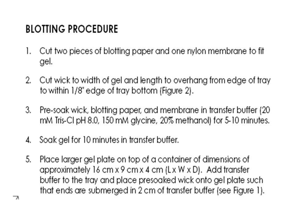
Blotting Following gel electrophoresis, the separated protein mixtures are transferred from polyacrylamide gel to membrane. Proteins are adsorbed to the membrane by hydrophobic bonds, as well as charged interactions between the membrane and protein. Membrane are much stronger and more pliable than gel and can undergo many manipulation without tearing.

9
Blotting- Blotting Membranes There are three types of membranes widely used in western blotting these are:- Nitrocellulose Membrane. Nylon Membrane. Polyvinylidene fluoride (PVDF) Membrane.
 Membrane..")
10
Transfer Methods Transfer can be performed in several ways. The goal of all methods is to move the protein from the gel to a membrane substrate for probing. Both varieties of membrane are chosen for their non-specific protein binding properties (i.e. binds all proteins equally well).
..")
11
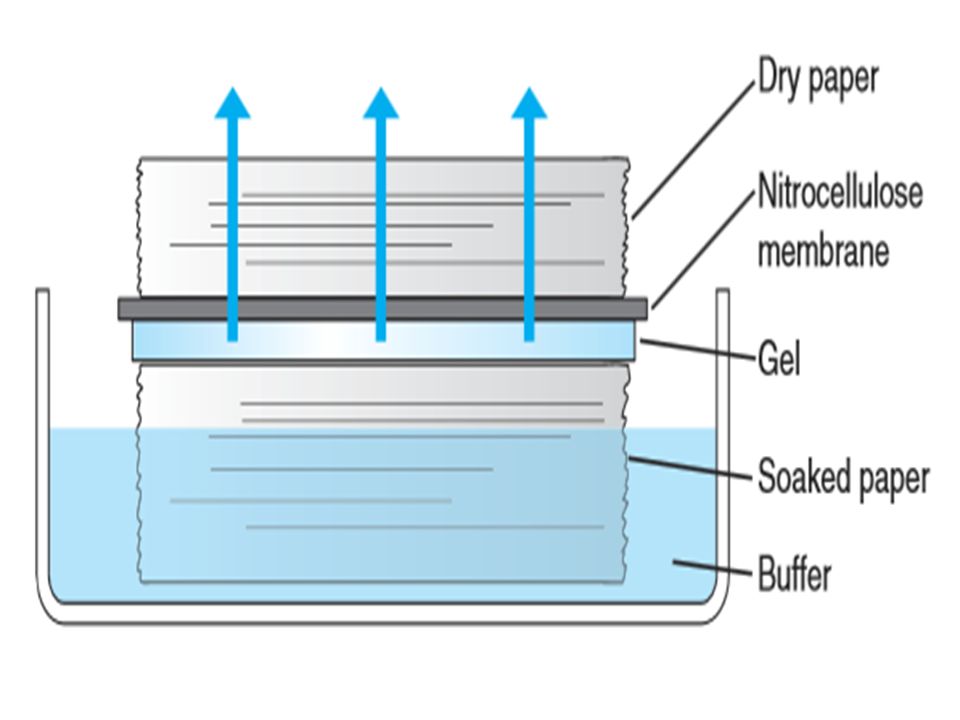
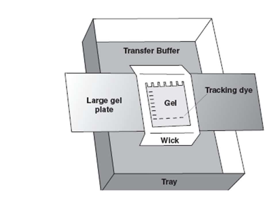
1- capillary transfer An older method of transfer For capillary transfer, the gel is placed on top of a filter papers soaked in transfer buffers. The membrane is placed directly on the gel, and dry absorbent membranes or paper towels are stacked on top of the membrane. The buffer is moved by capillary action from the lower reservoir to the dry material on top of the gel. The movement of the buffer through the gel will carry the denatured protein out of the gel.

13
When the protein contacts the nitrocellulose membrane, the protein will bind to it while the buffer will pass through to the membranes or paper towels on top. 2. A second method, called electrophoretic transfer uses electric current to move the protein from the gel to the membrane.

14
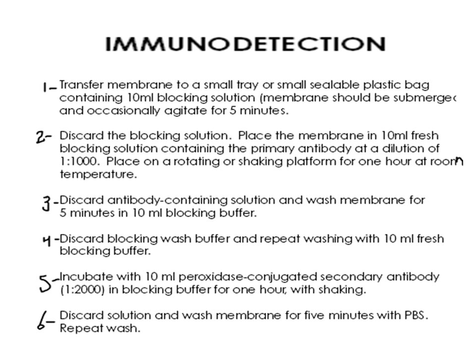
4- Blocking Since the membrane has been chosen for its ability to bind protein and as both antibodies and the target are proteins. steps must be taken to prevent the interactions between the membrane and the antibody used for detection of the target protein. Blocking of non-specific binding ( binding to all unoccupied sites on the membrane) is achieved by placing the membrane in a dilute solution of protein - typically 3-5% (BSA) or non-fat dry milk (both are inexpensive) in Tris-Buffered Saline (TBS) or PBS ( phosphate buffered saline )Tris-Buffered Saline
 is achieved by placing the membrane in a dilute solution of protein - typically 3-5% (BSA) or non-fat dry milk (both are inexpensive) in Tris-Buffered Saline (TBS) or PBS ( phosphate buffered saline )Tris-Buffered Saline.")
15
Often, a small amount of Tween 20 detergent is added to blocking and washing solutions to reduce background staining. The protein in the dilute solution attaches to the membrane in all places where the target proteins have not attached. Thus, when the antibody is added, there is no room on the membrane for it to attach other than on the binding sites of the specific target protein., this eliminates false positives

16
5- Antibody Probing Once the protein samples are separated and transferred onto a membrane, the protein of interest is detected and localized using a specific antibody. The blot will be incubated in a dilute solution of antibody, usually for a few hours at room temperature or overnight at 4°C.

17
The antibody is diluted in wash buffer (PBST or TBST) or in the blocking solution, the choice depends upon the antibody. Since antibody preparations vary in their levels of purity and specific binding properties, there will be differences in the level of dilution required The manufacturer’s datasheet should provide dilution recommendations for a particular preparation.

18
5- Antibody Probing Usually, Western blotting protocols utilize a non-labeled primary antibody directed against the target protein Wash the membrane several times in TBST while agitating, 5 minutes or more per wash, to remove residual primary antibody. A species-specific, labeled secondary antibody directed against the constant region of the primary antibody is then used.

19
The secondary antibody serves not only as a carrier of the label but is also a mechanism to amplify the emitted signals, as many secondary antibodies can theoretically bind simultaneously to the primary antibody. Secondary Ab is also diluted according to the manufacturer’s recommendations and incubated for 1 hour at RT.

20
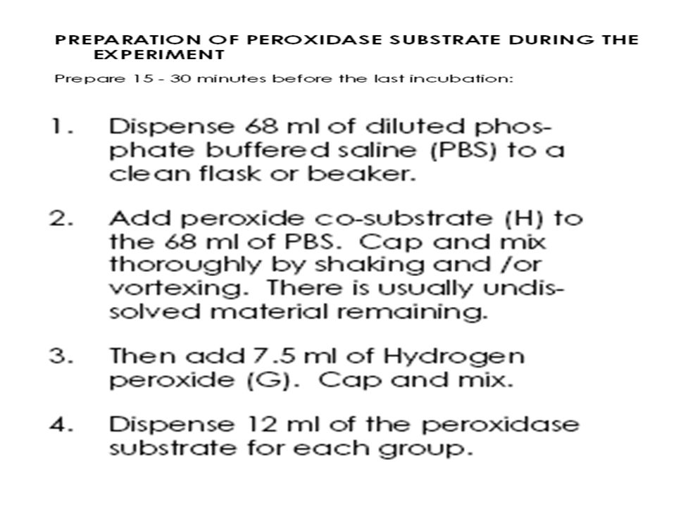
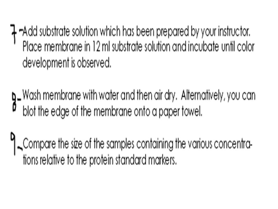
6- Detection with Substrate The most common antibody label used in Western blots is HRP, a small, stable enzyme with high specificity. The signal is detected when HRP is exposed to a substrate solution in the final step of the immunodetection procedure.

21
6- Detection with Substrate The co-substrate include numerous phenolic compounds that develop color upon oxidation. Peroxidase convert peroxide to H2O +O2 which oxidize co-substrate. The oxidized compound is colored and can be easily observed.

22
Summary: For immunological detection the membrane is placed in blocking buffer which contains detergents and blocking protein that bind to all unoccupied sites on the membrane The membrane is then incubated in buffer that contains antibody to specific antigen Subsequent, washing will remove excess,unbound antibody.

23
A secondary antibody against the first antibody,linked to an enzyme such as alkaline phosphatase or horseradish peroxidase is used for detection. The membrane is incubated in a solution of secondary antibody where it will bind selectivity to antigen-antibody complex and washed to remove excess secondary antibody. In next step,the membrane is incubated in solution containing phosphatase or peroxidase substrate. The yield chromogenic product. Area containg antigen-antibody conjugate will develop color.

24
HIV BLOT 2.2 WESTERN BLOT ASSAY

25
Introduction Screening tests are widely available for detecting antibodies to HIV. Such tests can be extremely sensitive but have a potential for being less specific, leading to false positive interpretations Independent supplemental tests of high specificity are therefore necessary to further confirm the presence of antibodies to HIV.

26
Introduction The test is intended for use as a more specific supplemental test on human serum specimens. The separated specific HIV viral antigens incorporated onto the strips via electrophoretic and blotting procedures

27
Principles of the Procedure The nitrocellulose strips are incorporated with separated, bound antigenic proteins from: Individual nitrocellulose strips are incubated with diluted serum and controls Specific antibodies to HIV if present in the specimens will bind to the HIV proteins on the strips

28
Principles of the Procedure The strips are washed to remove unbound materials Goat anti-human IgG conjugated with alkaline phosphatase is used as a secondary Ab and then the substrate is added for detection

29
Material

30
`

Similar presentations


 Lab 11. IHC IHC refers to the process of detecting antigens in cells of a tissue section by exploiting the principle of antibodies.>")
 Primary antibody Secondary antibody.>")

>")
![Lab#6 Western Blotting BCH 462[practical].](/18/6070629/big_thumb.jpg "Lab#6 Western Blotting BCH 462[practical].>")

 Northern blot (RNA) Western blot (Protein) Eastern blot (???)>")
