Download presentation
Presentation is loading. Please wait.

1
An approach to abdominal pain
Dr. Shamim Ahmad Bhat Consultant Emergency Medicine King Saud Medical City Riyadh. KSA

2
INTRODUCTION Types of pain Special Populations Assessment
History Examination Investigations Differential Diagnosis Management - overview Cases ( if time permits) Sticking to basics!
 Sticking to basics!")
3
Types Of Pain Visceral Parietal Pain
Useful to have a basic understanding of the mechanism of abdominal pain’

4
Visceral Pain Stretching of nerve fibres of walls or capsules of organs Crampy Dull Achy Often unable to lie still Bilateral innervation Caused by stretching of Unmyelinated fibres innervating the capsules or walls of organs Can also be caused by early ischaemia or inflammation Intermittent or constant Bilateral innervation – stimuli sent to both sides of spinal cord therefore felt midline regardless of the R or L sided origin of the stimulus Localization is to the approximate embryologic origin of the organ involved. E.g. T10 for appx

5
Parietal Pain Parietal peritoneum irritated
Usually anterior abdominal wall Localised to the dermatome superficial to the site of painful stimulus Irritation of Myelinated fibres in the parietal peritoneum Parietal afferents are sent from a specific area of the peritoneum and can be localized to the dermatome superficial to the painful stimulus

6
Course Visceral Non specific Parietal Localised tenderness Guarding Rigidity Rebound As the disease progresses visceral pain gives way to signs of parietal pain As localized peritonitis ( inflammation of the peritoneum) develops rigidity and rebound Patient with peritonitis lie still
 develops rigidity and rebound. Patient with peritonitis lie still.")
7
Referred Pain Examples of referred pain? Shoulder tip pain
Free fluid in abdomen eg perforated ectopic Testicular pain Renal Colic Referred pain follows the path of the embryological origin of the organ Felt distant to the site of the origin of the pain Normally felt on the SAME side as the involved organ as it is not mediated by fibres that provide bilateral innervation to the organ

8
Most Common Causes in the ED
Non-specific abd pain 34% Appendicitis 28% Biliary tract dz 10% SBO 4% Gyn disease 4% Pancreatitis 3% Renal colic % Perforated ulcer % Cancer 2% Diverticular dz 2% Other 6%

9
Special Populations WOMEN OF CHILD BEARING AGE
OLD AGE (ELDERLY PATIENTS)
")
10
Elderly May lack physical findings despite having serious pathology
As patients age increases diagnostic accuracy declines Risk of Vascular Catastrophes Assume surgical cause until proven otherwise 30-40% of geris with abdo pain need surgery Biliary tract Disease is the commonest cause Age > 65 need to think of reasons not to CT! Mortality is 7% in the over 80’s - equivalent to AMI!

11
Elderly Patient think Nasties!
AAA Ischaemic Gut Bowel Obstruction Diverticulitis Perforated Peptic Ulcer Cholecystitis Appendicitis Constipation is a diagnosis of exclusion – never make until all serious pathology has been excluded!

12
Women of Childbearing Age
Must Ascertain whether PREGNANT ALL WOMEN OF CHILDBEARING AGE WITH ABDO PAIN NEED BHCG Gravid uterus displaces intra-abdominal organs making presentations atypical Pregnant women still get common surgical abdominal conditions Needs to prove that they are not pregnant Can affect nature of presentation – ie atypical presentation. Pregnancy related condition- ectopic/ abruption etc The pregnant patient presenting with nontraumatic abdominal pain adds dozens of potential diagnoses to the equation. It is also common for typical illness- es (like appendicitis) to present in atypical fashion due to the anatomic changes brought about by an enlarging uterus
 to present in atypical fashion due to the anatomic changes brought about by an enlarging uterus.")
13
History What are the key points of the abdominal pain history?
For Symptoms associated with the pain think from the top down Nausea Vomiting Dyspepsia Change in bowels Urinary Symptoms PV loss or bleeding

14
History HPC Pain Associated Symptoms – Consultations/ Presentations
Provocative Palliative Quality Radiation Symptoms associated with Timing Taken for the pain Consultations/ Presentations Associated Symptoms – Gastro – intestinal Genito-urinary Gynaecologic For Symptoms associated with the pain think from the top down Nausea Dyspepsia Vomiting Change in bowels Urinary Symptoms PV loss or bleeding

15
History PMH PSH DM HT Liver Disease Renal Disease
Sexually Transmitted Infections PSH Abdominal Surgery Pregnancies Deliveries/ Abortions/ Ectopics Trauma

16
History Meds ALLS NSAIDs Steroids OCP/ Fertility Drugs Narcotics
Immunosuppressant Chemotherapy agent ALLS Contrast Analgesic

17
High Yield Questions Which came first – pain or vomiting?
How long have you had the pain? Constant or intermittent? History of cancer, diverticulosis, gall stones, Inflammatory BD? Vascular history, HT, heart disease or AF? Advanced age = increased risk of nasty Pain first is worst –more likely to be caused by surgical disease Pain for less than 48 hours more likely to have a surgical cause Constant pain more likely to be serious than intermittent First episode more likely to be something bad Abs and steroids mask infection Vascular history etc – consider mesenteric ischaemia or AAA

18
Examination Lots of information from the end of the bed Vital Signs
Distressed vs. non distressed Lying still - peritonitis Writhing – Renal Colic Vital Signs NEVER ignore abnormal vital signs! Always document as part of your assessment Absent or diminished BS add little clinically significant info Hyperacive or obstructed BS are slightly more useful Can have normal BS in SBO Palpaption How do you diminish voluntary guarding PR PV All women with lower abdo pain looking for cervical excitation, adnexal tenderness, discharge – dont forget to take genital swabs when you perform the PV – blue and pinswabs for general and STD culture

19
ABDOMINAL EXAMINATION
This video shows an a thorough GI examination. Once you have perfected the full examination in the emergency department setting you need to perform a slick focused examination.

20
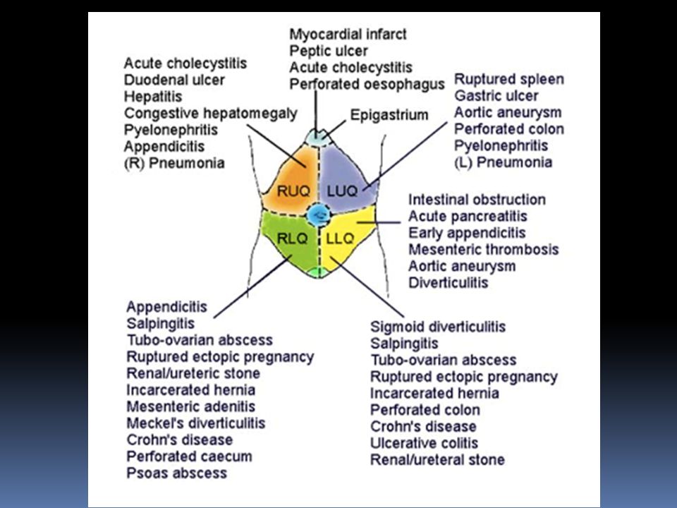
Whiteboard Think Medical Surgical Female ( Don’t forget non-intra abdominals) Stress AAA Male Female Differentials according to quadrant of pain

22
Investigations Bedside Bloods UE
Leucocyte Esterase and nitrites Urine HCG ECG – anyone with upper abdominal pain or elderly Bloods ALL WOMEN OF CHILDBEARING AGE NEED BHCG What are your differentials? Avoid machine gun approach! CXR looking for perforation, referred pathology eg pneumonia, associated problems eg effusion with pancreatitis

23
Radiology CXR –?perforation ?Extra abdominal pathology
?Complications of intra-abdominal disease Abdominal XR is overutilised.

24
Tracking Renal Calculi Foreign Body
Which of the following is NOT an indication for plain abdominal imaging? Bowel Obstruction Constipation Tracking Renal Calculi Foreign Body As other diagnostic modalities have become more readily available over the past decade, the role of plain film abdominal radiography has come under question
![]()
25
SOME INTRESTING AXRs

26
What would you do with this?
If passed through the oesophagus into stomach will be OK If vomiting Abdo pain Toxic – e.g. Pb or will need endoscopy. Iron Safety pins Heavy metals

27
Patient brought in from the airport after getting off flight from South America

28
Large L renal Calculus & stones radio opaque CT is the primary investigation of choice AXR will identify sufficiently large radiopaque stones such as calcium, struvite, and cystine stones Miss small stones or stones overlying bony structures, and will not detect obstruction. Used to tract progress of radiopaque stones to avoid multiple CTs

29
Other imaging ULTRASOUND Biliary Disease Good for gynae complaints
Rule out Ectopic pregnancy Appendicitis in children No radiation If negative for appx does not exclude

30
CT ABDOMEN CT is accurate for diagnosis of Avoid repeated CT scans
Renal colic Appendicitis Diverticulitis AAA Intra abdominal Abscesses Mesenteric Ischaemia Bowel Obstruction Avoid repeated CT scans Limit use in younger patients Avoid where possible in pregnant females CT use has double in the last decade Excellent diagnostic modality but radiation dose associated with it

31
Imaging Dose (mSV) CXR equivalents Pelvic XR 0.6 6 Abdominal XR 0.7 7
CT abdo-pelvis 14 140 CT aortogram 24 240 Mettler, FA, Huda, W, Yoshizumi, TT, Mahesh, M. Effective doses in radiology and diagnostic nuclear medicine: a catalog. Radiology 2008; 248:254. Sievert (symbol: Sv) is the International System of Units (SI) SI derived unit of dose equivalent radiation.
 is the International System of Units (SI) SI derived unit of dose equivalent radiation.")
32
Management Resuscitate with ABC APPROACH
Large bore access N Saline bolus 20ml/kg x 2 if shocked If bleeding think hypotensive resuscitation All should be NBM until provisional diagnosis Ensure normothermia Maintenance fluids and fluid balance Analgesia doesn’t mask signs Use a the pain scale Morphine titrated to pain. Normally 0.1mg/Kg Paracetamol adjunct NSAIDs for renal colic Correct Electrolytes Thromboprophylaxis

33
Cases

34
Case 1 21 year old female 24 hour history of vague peri-umbilical abdominal pain. Moved down to the RIF. Now constant and sharp. Associated with 2x vomits and feels flushed No appetite Normal Bowels

35
What clinical signs may lead you to a diagnosis of appendicitis?
Lie still RIF tenderness Rebound Rovsig’s sign Psoas Sign Lie still RIF tender Rebound Weak cough or grabs abdomen whilst coughing Rovsigs sign – highly sensitive for acute appx – gentle pressure on L then release - pain on the R PR May get referred tenderness but seldom changes management Psoas sign – lie patient L lateral and extend the hip – if increased pain of appx sitting on psoas – 95% specific

36
Imaging? AXR rarely useful USS Not as good as CT
Good for female to exclude gynae pathology If appendix is visualised is useful CT Only if there is doubt about diagnosis Sensitivity up to 98% High radiation dose Diagnose other pathology if no appendicitis Elderly AXR rarely useful – may show appx faecolith, appendicieal gas, localised ileus, blurred R psoas muscle, free air if perforated CT greater sensitivity, accuracy and negative predictive value than US. High radiation dose – women and children Provides alternative diagnoses CT does not change management in men but can prevent unnecessary appendectomies in women

37
CT examination of the right lower quadrant after the administration of intravenous and enteric contrast material shows a dilated, fluid-filled appendix with a thickened wall (arrows). There are inflammatory changes in the adjacent fat tissue (arrowheads). Laparotomy confirmed the diagnosis of acute appendicitis, and an appendectomy was performed. The patient had an uneventful recovery.
. Laparotomy confirmed the diagnosis of acute appendicitis, and an appendectomy was performed. The patient had an uneventful recovery..")
38
Management NPO Analgesia Anti-emetic if necessary Maintenance fluids
IVABs – e.g. Ceftriaxone, Gentamicin and Metronidazole Surgical Referral Delay may lead to perforation and peritonitis IV abs reduce the incidence of wound infection and abdominal wall abscess OT either laparoscopy – reduced inpatient stay and analgesia requirements but takes longer WCC sensitive but not specific

39
Case 2 40 yr old obese female RUQ pain Pain is constant
nausea, vomiting fevers and chills PMH Asthma MEDS OCP SH Drinks 2 std / week Smokes 20/day Nil drugs

40
On Examination Looks distressed. Not jaundiced T 38 C P 120 BP 100/60
RR 20 Sats 98% RA Tender in the RUQ and Murphy’s positive. What is Murphy’s sign?

41
What blood Tests will you order on this patient?

42
HB 13.8 WCC 16.0 Neuts 12.4 Lymph 1.6 EUC Normal Bil 9 (<18) ALP 450 (30-130) GGT 320 (<60) ALT 41 (5-55) AST 30 (5-55) Amylase 28 (<120) Lipase 40 (<60)
 AST 30 (5-55) Amylase 28 (<120) Lipase 40 (<60)")
43
Gall bladder here is 74mm ie greater than 5cm = enlarged

44
GB wall 7mm= > 5mm = thickened

45
Management NPO IVF IV abs –Ampicillin + Gentamicin
Analgesia +- anti emetic Refer to surgeons CT if diagnosis unclear or USS unavailable USS is the investigation of choice

46
Case 3 52 yr old alcoholic Constant epigastric pain radiating to the back. Worsening over the past 2 days Improved with sitting up and forwards Nausea and vomiting Bowels OK PMH Chronic Airways Limitation Alcoholic Gastritis MEDS Thiamine 100 mg daily SH Boarding house resident Drinks 4 litres wine/day Smokes 20/day 52 yr old alcoholic from Bigge Park

47
Looks unwell and dehydrated T38.4C P105 BP 130/70 RR 18 Sats 93% RA
Cullen’s Sign Blue discolouration around periumbilicus due to haemoperitoneum – necrotic pancreatitis or intraabdominal bleeding Grey Turner’s sign Grey discolouration to the flanks due to haemaglobin catbolism his sign takes 24–48 hours. It can predict a severe attack of acute pancreatitis,[1] with mortality rising from 8-10% to 40%

48
Tender Epigastrium and RUQ
Reduced AE L base Tender Epigastrium and RUQ No guarding/ rebound Turners sign – bluish discoloration of flank due to pancreatitis Neither sign is specific to pancreatitis – can occur in other causes of retroperitoneal haemorrhage

49
What blood tests will you order?

50
Blood Results Glucose 15 Alb 23 Ca (Corr) 2.0 Haem HB 114 WCC 17
Biochem Na 129 K 4.0 Cr 62 Ur 8.0 Amylase 1080 (<120) Lipase 950 (<60) Bil 11 ( 18) GGT 900 (<60) ALP 200 ( < 140) AST 300 (5-55) ALT 250 (5-55) LDH 800( ) Glucose 15 Alb 23 Ca (Corr) 2.0 Haem HB 114 WCC 17 Coags Normal NB Lipase is more sensitive than amylase for pancreatitis. If lipase is 3x upper limit of normal this is almost 100 % sensitive for pancreatitis Rises within 4-8 hours in acute pancreatitis. Peaks at 24 hours. Normal after 2 weeks Amylase - > 3x upper limit of normal 80% sensitive, 95 % specific. Once > % sensitiveEUC Normal Bil 9 (<18) ALP 450 (30-130) GGT 320 (<60) ALT 41 (5-55) AST 30 (5-55) Amylase 28 (<120) Lipase 40 (<60)
 Lipase 950 (<60) Bil 11 ( 18) GGT 900 (<60) ALP 200 ( < 140) AST 300 (5-55) ALT 250 (5-55) LDH 800( ) Glucose 15. Alb 23. Ca (Corr) 2.0. Haem. HB 114. WCC 17. Coags Normal. NB Lipase is more sensitive than amylase for pancreatitis. If lipase is 3x upper limit of normal this is almost 100 % sensitive for pancreatitis. Rises within 4-8 hours in acute pancreatitis. Peaks at 24 hours. Normal after 2 weeks. Amylase - > 3x upper limit of normal 80% sensitive, 95 % specific. Once > % sensitiveEUC Normal. Bil 9 (<18) ALP 450 (30-130) GGT 320 (<60) ALT 41 (5-55) AST 30 (5-55) Amylase 28 (<120) Lipase 40 (<60)")
51
What imaging will you perform ( if any)?
")
52
CXR

53
Imaging CT USS CXR Look for complications
Confirms diagnosis Identifies complications Help’s grade severity Not always necessary in ED USS Poor visualisation of pancreas Good for looking at gall stones/ biliary tree dilatation CXR Look for complications Pleural Effusion, Atelectasis, ARDS AXR – not specific for pancreatitis – may see sentinal loop . Good for looking for other diagnoses such as obstruction

54
Management O2 NBM IVF Analgesia +-Antibiotics (controversial)
Correct Electrolytes Thromboprophylaxis ICD/Art-line/CVC depending on severity Surgical Admit +_ ICU review Several trials show that antibiotic prophylaxis reduce the development of sepsis Large volumes of fluid may be needed due to ileus, effusions and oedema Correct electrolytes such as hypocalcaemia Antibiotics indicated if proven pancreatic necrosis on CT Prophylactic abs may decrease septic complications in severe necrotising pancreatitis

55
Causes G all stones E toh T rauma S teroids M umps A utoimmune
S corpion Bites H yperlidaemia/ hypercalcaemia/hypothermia E RCP D rugs Gall stones commonest cause in women Alcohol – normally after 10 years of heavy drinking. Commonest cause in men Alcohol and gall stomes account for 90% cases Drugs Azathiprine, Sulphonamides, thiazides, frusemide, oestrogens,tetracylcines,valproate

56
Case 4 27 yr old female 6/40 LIF constant severe sharp pain
Radiating to the back Light bright red PV spotting Feels light headed PMH IVF Previous D+C x 2 Ovarian Cysts MEDS Nil SH Lives with partner Non-smoker Non-Drinker

57
On Examination Looks unwell. Pale, diaphoretic, restless P 150
BP 70/40 RR 26 Sats 98% RA Tender and guarding in the LIF PV Bright red blood spotting L adnexal tenderness ++

58
How do you manage this patient?
Panic! ( don’t!) Call for senior help Large bore IV access x 2 (16 G or larger) Urgent Cross Match Fluid resuscitation Call O+G urgently Needs OR immediately Cautious volume resus
 Call for senior help. Large bore IV access x 2 (16 G or larger) Urgent Cross Match. Fluid resuscitation. Call O+G urgently. Needs OR immediately. Cautious volume resus.")
59
Case 5 88 yr old female. Peri-umbilical, colicky abdominal pain for 2 days Abdominal distension Vomits x 10 Reduced flatus for 2 days. PMH Cholecystectomy appendectomy TAH BSO Hypertension

60
On examination Looks distressed Lying Still T 37.5 P 110 sinus
BP 150/80 RR 18 Sats 98% RA Abdomen Distended Generally tender No guarding rebound or rigidity High pitched bowel sounds

61
Investigations CT good at differentiating mechanical obstruction from an ileus or for the differentiation of simple SBO from stangulation

62
Investigations Labs AXR CXR CT

63
Dilated loops of small Bowel
Multiple Air fluid levels 6 Adhesions Neoplasm Sms Hernias-external, internal Crohns \

64
Fee air under the diaphrag – small bowel obstruction and perforation

65
Management NPO Fluid resuscitation
Monitor volume status – may have large volume shifts Correct Electrolytes Analgesia NG if vomiting IV Abs – Ceftriaxone, metronidazole Urgent Surgical consult for OR IV abs for all people with mechanical obstruction as risk of infection and septicaemia is high Discontinue medications that limit gut motility

66
Small Bowel Adhesions Hernias Polyps Lymphoma Adenocarcinoma
Gall Stones Inflammatory BD Bezoar – vegetable matter or pulp form persimmons – concretion in the small intestine UPPER LIMIT OF NORMAL DIAM FOR SMALL BOWEL IS 2.5CM Small intesitne has linear densities EXTENDING ACROSS the bowel lumen ( plicae circulares)
")
67
Large Bowel Almost never adhesions or hernia CARCINOMA Diverticulitis
Sigmoid Volvulus Faecal Impaction UPPER LIMIT OF NORMAL FOR LARGE BOWEL IS 5.5 CM Colon - larger diameter. Thick densities HAUSTRA EXTEND PARTIALLY into the lumen

68
Case 6 73 yr old male presents with sudden onset of central abdominal pain radiating to the back. He also reports weakness to both legs PMH HT Hypercholesterolemia Current smoker 30/day MEDS Aspirin 100mg Daily Perindopril 5 mg Daily Atorvastatin 10 mg Daily SH Lives Alone Fully independent with ALS Occasional alcohol

69
Examination Distressed P 130 BP 80/60 RR 26 Sats 99% RA Abdomen
Non-distended Generally tender.

70
Bedside Ultrasound 9cm Abdominal Aortic Aneurysm (AAA):
The diagnosis of AAA (defined as a maximal aortic diameter of greater than 3 cm) and ruptured AAA has increased over the last several years. majority of patients with rup- tured AAA do not present with the classic triad of abdominal pain, shock, and pulsatile abdominal mass. Nearly 30% of patients who present to the ED with ruptured AAA are initially misdiagnosed. Early detection of a ruptured AAA with prompt surgical intervention can decrease mortality from 75 to 35%
 and ruptured AAA has increased over the last several years. majority of patients with rup- tured AAA do not present with the classic triad of abdominal pain, shock, and pulsatile abdominal mass. Nearly 30% of patients who present to the ED with ruptured AAA are initially misdiagnosed. Early detection of a ruptured AAA with prompt surgical intervention can decrease mortality from 75 to 35%")
71
Management of ruptured AAA
Senior help ABC Large Bore IV Access x 2 Hypotensive resuscitation Analgesia Ensure O neg available Ensure normothermia Urgent Vascular Consult To OR Cautious fluids – if give rapid volume expansion may cause loss of retroperitoneal tamponade and accelerated bleeding If patient is perfusing vital organs this can be delayed Target SBP of Obviously if pre arrest need volume resuscitation

72
Last Case! 85 yr old male. Nursing home resident
Central Abdominal Pain Sudden onset. Severe PMH Dementia MI MEDS Clopidogrel 75 mg Daily Metoprolol 25 mg BD Perindopril 5 mg daily SH Mild dementia Forgetful Requires some assistance with bathing and toileting Feeds Self Walks with frame Non-smoker Non-drinker

73
Examination Looks dry and emaciated P 120- 140 BP 110/70 RR 30
Sats 96% RA T 37.4 C Abdomen Generally tender No guarding rigidity or rebound

74
ECG

75
Differential?

76
ABG pH 7.10 pCO2 15 P02 80 Bic 8 BE -15 Lactate 10.2

77
Management ABC NPO IV access IVF Analgesia IV abs
Urgent Surgical Consult Urgent CT mesenteric angiogram OR Surgical Emergency Small bowel has warm ischaemic time of 2-3 hours Rapidly progresses to gangrene, septic shock and death Need high index of suspicion to diagnose it Severe pain but little tenderness on examination

78
Mesenteric Ischaemia Surgical Emergency
Small bowel has warm ischaemic time of 2-3 hours Rapidly progresses to gangrene, septic shock and death Need high index of suspicion to diagnose it Severe pain but little tenderness on examination

79
Case 7 40 yr old male presents with sudden onset of severe R loin to groin pain. Excruciating pain. Coming in waves. Feels nauseated and has vomited x 2. Patient is agitated, pacing around the room, unable to sit still. Screaming in pain. P 120 sinus BP 160/80 T 37.0 C RR 18 Sats 99% RA R renal angle tender

80
Differential Diagnosis?
Renal Colic Pancreatitis Cholecystitis Appendicitis Ruptured/leaking AAA

81
UA Erythrocytes ++++ No leucocytes No nitrites

82
KUB XR Ray Where do the ureters run on this XR? Medial to the tips of the transverse processes of the lumbar vertebrae Crosses the pelvic brim at the sacro-iliac joint Shadow then passes to the ischial spine and to the pubic tubercle

83
Investigations UA EUC FBC (other bloods if diagnosis unclear) CT KUB
Beware phleboliths and calcified mesenteric lLM Trace the path of the ureters carefully on the fil

84
Management Analgesia NSAID Morphine IV titrated to pain
IV fluids – maintenance only Observe

85
Who should we CT CT On going pain Impaired renal function Fever
Diagnosis not clear

86
Indications for admission
Infection Impaired Renal Function Pain ongoing– needing IV opiates Stone > 5mm Obstruction/hydronephrosis on CT Stag horn Calculus on CT

87
Take Home Message Exclude life threatening pathology
BHCG in female of child bearing age Be mindful of radiation exposure Beware of Abdominal pain in the Elderly Never ignore abnormal vital signs Ask for help if not sure about diagnosis/Rx

88
Thanks for your patience!

Similar presentations