Download presentation
Presentation is loading. Please wait.

1
Pharmacology – III PHL-418
Pancreatic Hormones & Antidiabetic Drugs Dr. Hassan Madkhali Assistant Professor Department of Pharmacology E mail: 1

2
Content Layout with List
OVERVIEW: PANCREASE and PANCREATIC HORMONES INSULIN AND GLUCAGON ACTIONS OF INSULIN DIABETES MELLITUS DRUGS FOR THE TREATMENT OF DIABETES MELLITUS INSULIN ORAL ANTI-DIABETIC DRUGS

3
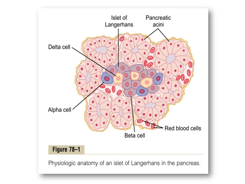
Pancreas Physiologic Anatomy of the Pancreas Insulin Glucagon
Digestive functions Secretes two important hormones Insulin Glucagon Secretes other hormones, such as amylin, somatostatin, and pancreatic polypeptide Physiologic Anatomy of the Pancreas Two major types of tissues The acini,which secrete digestive juices into duodenum The islets of Langerhans, which secrete insulin and glucagon into blood. The islets contain three major types of cells alpha, beta, delta cell The beta cells 60 % of all the cells of the islets, lie mainly in the middle of each islet and secrete insulin and amylin, The alpha cells, 25 % of the total, secrete glucagon The delta cells, about 10 %, secrete somatostatin.

5
Hormones Insulin is a polypeptide containing two amino acid chains (21 and 30 amino acids, respectively) connected by disulfide bridges. Glucagon is a straight-chain polypeptide of 29 amino acid residues. Both insulin and glucagon circulate unbound to carrier proteins and have short half-lives of 6 minutes. Approximately 50% of the insulin and glucagon in blood is metabolized in the liver; most of the remaining hormone is metabolized by the kidneys.

6
Insulin Is a Hormone Associated with Energy Abundance
When there is great abundance of energy-giving foods in the diet, especially excess amounts of carbohydrates, insulin is secreted in great quantity. Insulin plays an important role in storing the excess energy. In the case of excess carbohydrates, it causes them to be stored as glycogen mainly in the liver and muscles. Excess carbohydrates is also converted under the stimulus of insulin into fats and stored in the adipose tissue. Insulin has a direct effect in promoting amino acid uptake by cells and conversion of these amino acids into protein. In addition, it inhibits the breakdown of the proteins that are already in the cells.

7
Actions of Insulin To initiate its effects on target cells, insulin first binds with and activates a membrane receptor protein The insulin receptor is a tetramer made up of two α-subunits that lie outside the cell membrane and two β-subunits that penetrate the cell membrane and protrude into the cytoplasm When insulin binds with the alpha subunits on the outside of the cell, portions of the beta subunits protruding into the cell become autophosphorylated. Thus, the insulin receptor is an example of an enzyme-linked receptor Autophosphorylation of the beta subunits of the receptor activates a local tyrosine kinase, which in turn causes phosphorylation of multiple other intracellular enzymes including a group called insulin-receptor substrates (IRS). The net effect is to activate some of these enzymes while inactivating others. In this way, insulin directs the intracellular metabolic machinery to produce the desired effects on carbohydrate, fat, and protein metabolism.
. The net effect is to activate some of these enzymes while inactivating others. In this way, insulin directs the intracellular metabolic machinery to produce the desired effects on carbohydrate, fat, and protein metabolism.")
9
Effect on Carbohydrate Metabolism
Immediately after a high-carbohydrate meal, glucose that is absorbed into the blood causes rapid secretion of insulin Insulin causes rapid uptake, storage, and use of glucose by almost all tissues of the body, but especially by the muscles, adipose tissue, and liver.

10
In Muscle, Insulin Promotes the Uptake and Metabolism of Glucose
Mostly muscle tissue depends not on glucose for its energy but on fatty acids, because normal resting muscle membrane is only slightly permeable to glucose, except when the muscle fiber is stimulated by insulin. Under two conditions the muscles do use large amounts of glucose. During moderate or heavy exercise: because exercising muscle fibers become more permeable to glucose even in the absence of insulin During few hours after a meal: At this time the blood glucose concentration is high and the pancreas is secreting large quantities of insulin. The extra insulin causes rapid transport of glucose into the muscle cells. Abundant glucose transported into the muscle cells is stored in the form of muscle glycogen

11
In the Liver, Insulin Promotes Glucose Uptake and Storage, and Use
Insulin causes most of the glucose absorbed after a meal to be stored almost immediately in the liver in the form of glycogen. The mechanism of glucose uptake and storage in the liver : Insulin inactivates liver phosphorylase, which normally causes liver glycogen to split into glucose. Insulin causes enhanced uptake of glucose from blood by liver by increasing the activity of the enzyme glucokinase, causes the initial phosphorylation of glucose after it diffuses into liver Insulin also increases the activities of the enzymes that promote glycogen synthesis, glycogen synthase

12
Glucose Is Released from the Liver Between Meals
When the blood glucose level begins to fall to a low level between meals, several events cause the liver to release glucose back into the circulating blood: The decreasing blood glucose causes the pancreas to decrease its insulin secretion. The lack of insulin then reverses all the effects for glycogen storage The lack of insulin activates the enzyme phosphorylase, causes the splitting of glycogen into glucose phosphate. The enzyme glucose phosphatase, now becomes activated by the insulin lack and causes the phosphate radical to split away from the glucose Thus, the liver removes glucose from the blood when it is present in excess after a meal and returns it to the blood when the blood glucose concentration falls between meals

13
Insulin Promotes Conversion of Excess Glucose into Fatty Acids and Inhibits Gluconeogenesis in Liver. When the quantity of glucose entering the liver cells is more, insulin promotes the conversion of all this excess glucose into fatty acids. These packaged as triglycerides in VLDL and transported by blood to the adipose tissue and deposited as fat. Insulin also inhibits gluconeogenesis.

14
Lack of Effect of Insulin on Glucose Uptake and Usage by the Brain
Insulin has little effect on uptake or use of glucose in brain Instead, the brain cells are permeable to glucose The brain cells are also quite different from most other cells of the body in that they normally use only glucose for energy and can use other energy substrates, such as fats, only with difficulty. It is essential that the blood glucose level always be maintained above a critical level When the blood glucose falls too low, symptoms of hypoglycemic shock develop, characterized by progressive nervous irritability that leads to fainting, seizures, and even coma.

15
Effect of Insulin on Carbohydrate Metabolism in Other Cells
Insulin increases glucose transport into and glucose usage by most other cells of the body The transport of glucose into adipose cells mainly provides substrate for the glycerol portion of the fat molecule. Therefore, in this indirect way, insulin promotes deposition of fat in these cells.

16
Effect of Insulin on Fat Metabolism
Insulin Promotes Fat Synthesis and Storage Insulin has several effects that lead to fat storage in adipose tissue. Insulin increases the utilization of glucose by body Insulin promotes fatty acid synthesis, in liver cells Fatty acids are then transported from the liver by way of the blood lipoproteins to the adipose cells to be stored

17
Role of Insulin in Storage of Fat in the Adipose Cells
Insulin has two other essential effects that are required for fat storage in adipose cells: Insulin inhibits the action of hormone-sensitive lipase. This is the enzyme that causes hydrolysis of the triglycerides already stored in the fat cells. Insulin promotes glucose transport through the cell membrane into the fat cells. Some of this glucose is then used to synthesize minute amounts of fatty acids, but forms large quantities of a-glycerol phosphate. This substance supplies the glycerol that combines with fatty acids to form the triglycerides that are the storage form of fat

18
Insulin Deficiency Increases Use of Fat for Energy
All aspects of fat breakdown and use for providing energy are greatly enhanced in the absence of insulin. This occurs even normally between meals when secretion of insulin is minimal, but it becomes extreme in diabetes mellitus . Insulin Deficiency Causes Lipolysis of Storage Fat and Release of Free Fatty Acids. Consequently, the plasma concentration of free fatty acids begins to rise within minutes. This free fatty acid then becomes the main energy substrate used by essentially all tissues of the body besides the brain.

19
Insulin Deficiency Increases Plasma Cholesterol and Phospholipid Concentrations
The excess of fatty acids in the plasma associated with insulin deficiency also promotes liver conversion of some of the fatty acids into phospholipids and cholesterol, two of the major products of fat metabolism. These two substances, along with excess triglycerides formed at the same time in the liver, are then discharged into the blood in the lipoproteins, so the plasma lipoproteins increase This high lipid concentration—especially the high concentration of cholesterol—promotes the development of atherosclerosis in people with serious diabetes.

20
Excess Usage of Fats During Insulin Lack Causes Ketosis and Acidosis
Insulin lack also causes excessive amounts of acetoacetic acid to be formed in the liver cells. At the same time, the absence of insulin also depresses the utilization of acetoacetic acid in the peripheral tissues. Thus, so much acetoacetic acid is released from the liver Some of the acetoacetic acid is also converted into b-hydroxybutyric acid and acetone. These two substances, along with the acetoacetic acid, are called ketone bodies, and their presence in large quantities in the body fluids is called ketosis. In severe diabetes the acetoacetic acid and the b-hydroxybutyric acid can cause severe acidosis and coma, which often leads to death.

21
Effect of Insulin on Protein Metabolism and on Growth
Insulin Promotes Protein Synthesis and Storage During the few hours after a meal proteins are also stored in the tissues by insulin Insulin stimulates transport of many of amino acids into the cells, eg valine, leucine, isoleucine, tyrosine, and phenylalanine. Insulin increases the translation of mRNA, thus forming new proteins Over a longer period of time, insulin also increases the rate of transcription of selected DNA, forming increased quantities of RNA and still more protein synthesis Insulin inhibits the catabolism of proteins In the liver, insulin depresses the rate of gluconeogenesis, this suppression of gluconeogenesis conserves the amino acids in the protein stores of the body.

22
Summary The major effects of insulin on muscle and adipose tissue are:
(1) Carbohydrate metabolism: (2) Lipid metabolism: (3) Protein metabolism: it increases the rate of glucose transport across the cell membrane. it increases the rate of glycolysis by increasing hexokinase (glucokinase) and 6-phosphofructokinase activity. it stimulates the rate of glycogen synthesis and decreases the rate of glycogen breakdown. it decreases the rate of lipolysis in adipose tissue and hence lowers the plasma fatty acid level. it stimulates fatty acid and triacylglycerol synthesis in tissues. it increases the uptake of triglycerides from the blood into adipose tissue and muscle. it decreases the rate of fatty acid oxidation in muscle and liver. it increases the rate of transport of some amino acids into tissues. it increases the rate of protein synthesis in muscle, adipose tissue, liver, and other tissues. it decreases the rate of protein degradation in muscle (and perhaps other tissues).
 Carbohydrate metabolism: (2) Lipid metabolism: (3) Protein metabolism: it increases the rate of glucose transport across the cell membrane. it increases the rate of glycolysis by increasing hexokinase (glucokinase) and. 6-phosphofructokinase activity. it stimulates the rate of glycogen synthesis and decreases the rate of glycogen breakdown. it decreases the rate of lipolysis in adipose tissue and hence lowers the plasma fatty acid level. it stimulates fatty acid and triacylglycerol synthesis in tissues. it increases the uptake of triglycerides from the blood into adipose tissue and muscle. it decreases the rate of fatty acid oxidation in muscle and liver. it increases the rate of transport of some amino acids into tissues. it increases the rate of protein synthesis in muscle, adipose tissue, liver, and other. tissues. it decreases the rate of protein degradation in muscle (and perhaps other tissues).")
23
Insulin and Growth Hormone Interact Synergistically to Promote Growth
Because insulin is required for the synthesis of proteins, it is as essential for growth of an animal as growth hormone is. A combination of these hormones causes dramatic growth. Thus, it appears that the two hormones function synergistically to promote growth.

24
Mechanisms of Insulin Secretion
Insulin vesicles or granules Beta-cell Glycolysis

26
Glucagon -Glucagon is also called the hyperglycemic hormone
-The binding of glucagon to hepatic receptors results in activation of adenylyl cyclase and generation of the second messenger cyclic AMP, which in turn activates protein kinase, leading to phosphorylation that results in the activation or deactivation of a number of enzymes.

27
Effects on Glucose Metabolism
Glucagon Promotes Hyperglycemia Greatly enhance the availability of glucose to the organs of the body Glucagon stimulates glycogenolysis: Glucagon has immediate and pronounced effects on the liver to increase glycogenolysis and the release of glucose into the blood. This effect is achieved through activation of liver phosphorylase and simultaneous inhibition of glycogen synthase. Glucagon stimulates gluconeogenesis: Glucagon increases the hepatic extraction of amino acids from the plasma and increases the activities of key gluconeogenic enzymes.

28
Other Effects of Glucagon
Occurs only when its concentration rises well above the maximum normally found in the blood. Activates adipose cell lipase, making increased quantities of fatty acids available to the energy systems of the body. Glucagon also inhibits the storage of triglycerides in the liver, which prevents the liver from removing fatty acids from the blood. Enhances the strength of the heart Increases blood flow in some tissues, especially the kidneys Enhances bile secretion Inhibits gastric acid secretion.

29
Regulation of Glucagon Secretion
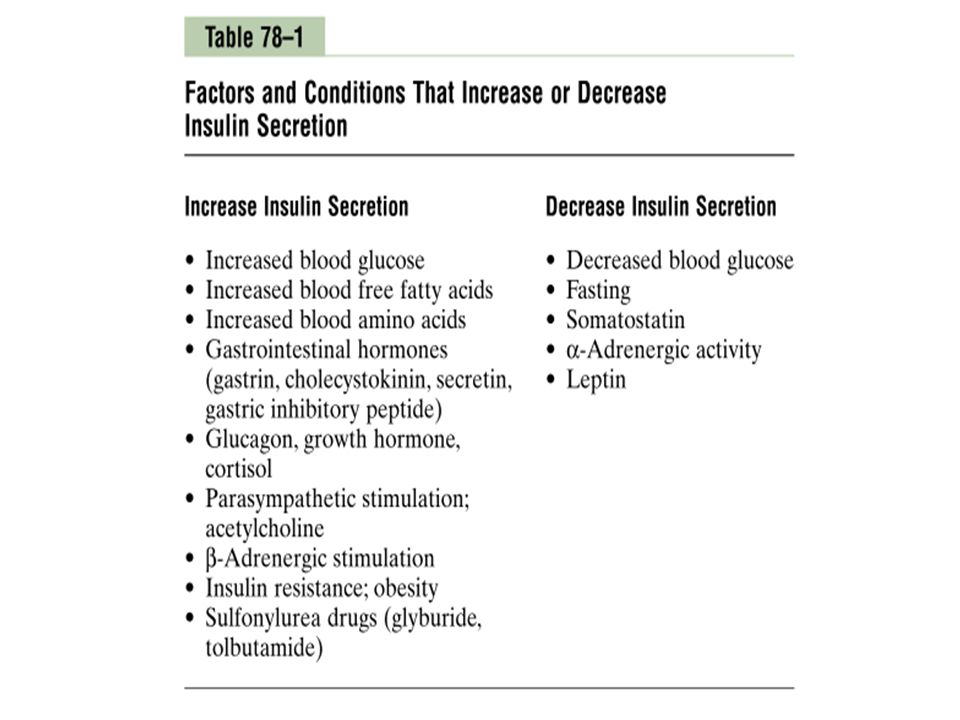
Increased Blood Glucose Inhibits Glucagon Secretion Increased Blood Amino Acids Stimulate Glucagon Secretion Exercise Stimulates Glucagon Secretion Somatostatin Inhibits Glucagon and Insulin Secretion

30
Somatostatin Somatostatin is a peptide hormone secreted by δ cells of the pancreatic islets (also produced in the hypothalamus) in response to: - blood glucose - plasma amino acids - fatty acids Somatostatin decreases gastrointestinal functions by: - motility - secretion - absorption Somatostatin splanchnic blood flow Somatostatin release of: - insulin - glucagon

31
Diabetes Mellitus (DM)
Diabetes mellitus is a syndrome of impaired carbohydrate, fat, and protein metabolism caused by either lack of insulin secretion or decreased sensitivity of the tissues to insulin. Two major forms of diabetes mellitus Type I diabetes mellitus, also called insulin-dependent diabetes mellitus (IDDM), is caused by impaired secretion of insulin. Type II diabetes mellitus, also called non–insulin-dependent diabetes mellitus (NIDDM), is caused by resistance to the metabolic effects of insulin in target tissues. Other forms include: -Gestational DM, GDM (triggered by pregnancy) -DM can also result rarely from diseases of the pancreas, and medications (Drug-induced diabetes: thiazide diuretics, beta-blockers and statins).
, is caused by impaired secretion of insulin. Type II diabetes mellitus, also called non–insulin-dependent diabetes mellitus (NIDDM), is caused by resistance to the metabolic effects of insulin in target tissues. Other forms include: -Gestational DM, GDM (triggered by pregnancy) -DM can also result rarely from diseases of the pancreas, and medications (Drug-induced diabetes: thiazide diuretics, beta-blockers and statins).")
32
DM in KSA The yearly total number of registered cases of diabetes according to gender (G) and type (T) of diabetes from the start of registry in 2000 to 2012. -Impaired glucose tolerance (IGT) is a pre-diabetic state of hyperglycemia -Secondary diabetes is diabetes that results as a consequence of another medical condition. The yearly total number of registered cases of diabetes according to gender (G) and type (T) of diabetes from the start of registry in 2000 to 2012. Ref: Khalid Al-Rubeaan et al. A Web-Based Interactive Diabetes Registry for Health Care Management and Planning in Saudi Arabia. J Med Internet Res 2013;15(9):e202.
 and type (T) of diabetes from the start of registry in 2000 to Impaired glucose tolerance (IGT) is a pre-diabetic state of hyperglycemia. -Secondary diabetes is diabetes that results as a consequence of another medical condition. The yearly total number of registered cases of diabetes according to gender (G) and type (T) of diabetes from the start of registry in 2000 to Ref: Khalid Al-Rubeaan et al. A Web-Based Interactive Diabetes Registry for Health Care Management and Planning in Saudi Arabia. J Med Internet Res 2013;15(9):e202.")
33
DM in KSA Source: International Diabetes Federation
Middle East & North Africa (MENA)
")
34
TREATMENT OF DM Anti-diabetic drugs: Insulin Incretin mimetics
Oral Anti-diabetics

35
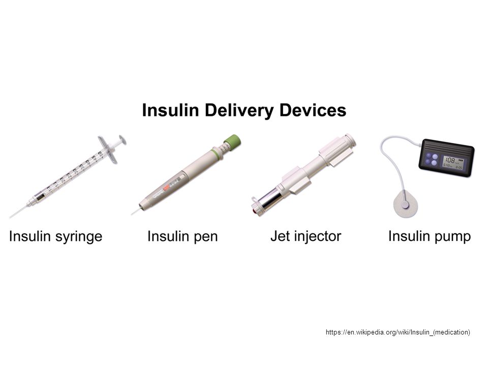
Insulin Insulin is a 51 AA peptide Not active orally.
Insulin is inactivated by insulinase found mainly in liver and kidney. Dose reduced in renal insufficiency Sources of Insulin : Bovine pancreas / Porcine pancreas Human insulin: Recombinant DNA origin

36
Insulin preparations :
Rapid acting insulin : Lispro, Aspart and Glulisine Short acting insulin: Regular (crystalline) Intermediate acting insulin: NPH (isophane) and Lente (insulin zinc) Long acting insulin: Ultralente, Detimir and Glargine Insulin Duration Route Features Lispro 3 – 5 hrs I.V or S.C Onset within 15 minutes Regular (crystalline) 7 – 10 hrs common NPH (Neutral protamine hagedorn) 16 – 20 hrs S.C NPH can mix with regular Ultralente 24 – 30 hrs Basal level Insulin glargine (Lantus), an insulin analog which is suitable for once-daily dosing.
 Intermediate acting insulin: NPH (isophane) and Lente (insulin zinc) Long acting insulin: Ultralente, Detimir and Glargine. Insulin. Duration. Route. Features. Lispro. 3 – 5 hrs. I.V or S.C. Onset within 15 minutes. Regular (crystalline) 7 – 10 hrs. common. NPH (Neutral protamine hagedorn) 16 – 20 hrs. S.C. NPH can mix with regular. Ultralente. 24 – 30 hrs. Basal level. Insulin glargine (Lantus), an insulin analog which is suitable for once-daily dosing.")
37
https://en.wikipedia.org/wiki/Insulin_(medication)
38
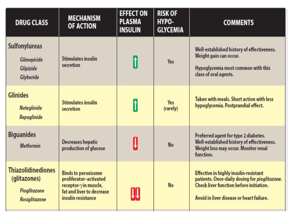
Oral Anti-diabetic drugs
A. Sulfonylureas: B. Meglitinides (Glinides): Repaglinide (Prandin), Nateglinide (Starlix) C. Biguanines: Metformin (Glucophage) D. Thiozolidonediones (glitazones): Pioglitazone (Actos), rosiglitazone (Avandia) E. α-Glucosidase inhibitors: Acarbose (Precose), Miglitol (Glycet) F. Dipeptidyl peptidase IV inhibitors (DPP-4 inhibitors): Sitagliptin (Januvia) G. Sodium-glucose cotransporter 2 (SGLT2) inhibitors: Canagliflozin (Invokana) 1st generation 2nd generation Acetohexamide (Dymelor) Chlorpropamide (Diabinese) Tolazamide (Tolinase) Tolbutamide (Orinase) Glyburide (Micronase) Glipizide (Glucotrol) Glimepiride (Amaryl) Repaglinide Nateglinide
: Repaglinide (Prandin), Nateglinide (Starlix) C. Biguanines: Metformin (Glucophage) D. Thiozolidonediones (glitazones): Pioglitazone (Actos), rosiglitazone (Avandia) E. α-Glucosidase inhibitors: Acarbose (Precose), Miglitol (Glycet) F. Dipeptidyl peptidase IV inhibitors (DPP-4 inhibitors): Sitagliptin (Januvia) G. Sodium-glucose cotransporter 2 (SGLT2) inhibitors: Canagliflozin (Invokana) 1st generation. 2nd generation. Acetohexamide (Dymelor) Chlorpropamide (Diabinese) Tolazamide (Tolinase) Tolbutamide (Orinase) Glyburide (Micronase) Glipizide (Glucotrol) Glimepiride (Amaryl) Repaglinide. Nateglinide.")
39
(RPG = random plasma glucose; FPG = fasting plasma glucose; OGTT = oral glucose tolerance test; PG = plasma glucose; HbA1c = glycosylated hemoglobin A1c; HBGM = home blood glucose monitoring; DM = diabetes mellitus; GI = gastrointestinal; PPG = postprandial glucose) Diabetic rages: Fasting plasma glucose: Greater than 125 mg/dL Random plasma glucose: Greater than 200 mg/dL Postprandial glucose at 2 hours: Greater than 200 mg/dL Impaired fasting glucose: Fasting glucose of mg/dL Impaired glucose tolerance testing: Postprandial glucose at 2 hours of mg/dL The value for hypoglycemia is as follows: Value less than 60 mg/dL Normal ranges: Fasting plasma glucose: mg/dL Postprandial plasma glucose at 2 hours: Less than 140 mg/dL Random plasma glucose: Less than 140 mg/dL
 Diabetic rages: Fasting plasma glucose: Greater than 125 mg/dL. Random plasma glucose: Greater than 200 mg/dL. Postprandial glucose at 2 hours: Greater than 200 mg/dL. Impaired fasting glucose: Fasting glucose of mg/dL. Impaired glucose tolerance testing: Postprandial glucose at 2 hours of mg/dL. The value for hypoglycemia is as follows: Value less than 60 mg/dL. Normal ranges: Fasting plasma glucose: mg/dL. Postprandial plasma glucose at 2 hours: Less than 140 mg/dL. Random plasma glucose: Less than 140 mg/dL.")
40
GI side effects such as nausea and diarrhea

41
Triple combination include metformin, Sulfonylurea and pioglitazone
Note: Metformin is considered to be one of the most effective therapeutics for treating type 2 diabetes, why? because it specifically reduces hepatic gluconeogenesis without increasing insulin secretion, inducing weight gain or posing a risk of hypoglycaemia. (This is explain why it is included in the most of combination therapies) Ref: BEATRIZ LUNA and MARK N. FEINGLOS. Oral Agents in the Management of Type 2 Diabetes Mellitus. AMERICAN FAMILY PHYSICIAN. MAY 1, 2001 / VOLUME 63, NUMBER 9
 Ref: BEATRIZ LUNA and MARK N. FEINGLOS. Oral Agents in the Management of Type 2 Diabetes Mellitus. AMERICAN FAMILY PHYSICIAN. MAY 1, 2001 / VOLUME 63, NUMBER 9.")
42
Combinations: Pioglitazone & metformin (Actoplus Met)
Glyburide & metformin (Glucovance) Glipizide & metformin (Metaglip) Sitagliptin & metformin (Janumet) Saxagliptin & metformin (kombiglyze) Repaglinide & metformin (Prandimet) Pioglitazone & glimepiride (Duetact)
 Glipizide & metformin (Metaglip) Sitagliptin & metformin (Janumet) Saxagliptin & metformin (kombiglyze) Repaglinide & metformin (Prandimet) Pioglitazone & glimepiride (Duetact)")
45
Metformin MOA

46
MOA PPARs: peroxisome proliferator-activated receptors

47
Sulfonylureas & Meglitinides MOA
inhibit the efflux of K+

48
α-Glucosidase inhibitors MOA

49
Dipeptidyl peptidase IV inhibitors and Incretin mimetics MOA
Incretin mimitics bind to GLP-1 receptors GLP-1: glucagon-like peptide-1

50
SGLT2 inhibitors MOA

51
Source

52
Pharmacist roles and responsibilities in diabetes care and management
Inform the diabetic patients that health education could make a significant difference and it is needed to give the diabetic patients a certain understanding of the disease Inform them about the importance of compliance with medication, diet and exercise, weight control and the use of herbal preparations. Encourage them to do self blood glucose monitoring regularly. Monitor and promote patient adherence to recommended treatment regimens Identify and resolving drug-related problems Provide education Remind them about the importance of doing regular exams Inform and convince them that unhealthy diet and physical inactivity are the most important risk factors of DM. Encourage them to participate actively in managing and monitoring their condition.

53
Thank you ? Animated open book effect (Difficult)
Tip: You will need to use drawing guides and the ruler to position the objects on this slide. To display the drawing guides and the ruler, do the following: On the Home tab, in the Slides group, click Layout, and then click Blank. Right-click the slide background area, and then click Grid and Guides. In the Grid and Guides dialog box, under Guide settings, select Display drawing guides on screen. (Note: One horizontal and one vertical guide will display on the slide at 0.00, the default position. The spine of the book will be aligned to the vertical drawing guide.) On the View tab, in the Show/Hide group, select Ruler. To reproduce the first shape in the Book cover group on this slide, do the following: On the Home tab, in the Drawing group, click Shapes, and then under Rectangles click Rounded Rectangle (second option from the left). On the slide, drag to draw a rounded rectangle. Select the rounded rectangle. Under Drawing Tools, on the Format tab, in the Size group, do the following: In the Shape Height box, enter 4.5”. In the Shape Width box, enter 3.33”. On the rounded rectangle, drag the yellow diamond adjustment handle to the left to decrease the amount of rounding on the corners. On the Home tab, in the bottom right corner of the Drawing group, click the Format Shape dialog box launcher. In the Format Shape dialog box, click Fill in the left pane, select Gradient fill in the Fill pane, and then do the following: In the Type list, select Linear. Click the button next to Direction, and then click Linear Right (first row, fourth option from the left). In the Angle box, enter 0°. Under Gradient stops, click Add gradient stops or Remove gradient stops until two stops appear in the slider. Also under Gradient stops, customize the gradient stops that you added as follows: Select the first stop in the slider, and then do the following: In the Position box, enter 0%. Click the button next to Color, and then under Theme Colors, click Red, Accent 2, Darker 50% (sixth row, sixth option from the left). Select the second stop in the slider, and then do the following: In the Position box, enter 100%. Click the button next to Color, and then under Theme Colors, click Red, Accent 2, Darker 25% (fifth row, sixth option from the left). Also in the Format Shape dialog box, click Line Color in the left pane. In the Line Color pane, select No line. Also in the Format Shape dialog box, click 3-D Format in the left pane, and then in the 3-D Format pane, do the following: Under Bevel, click the button next to Top, and then under Bevel click Circle (first row, first option from the left). Next to Top, in the Width box, enter 4 pt, and in the Height box, enter 4 pt. Under Surface, click the button next to Material, and then under Standard click Warm Matte (second option from the left). Also under Surface, click the button next to Lighting, and then under Neutral, click Three Point (first row, first option from the left). On the slide, drag the rounded rectangle until the left edge is against the vertical drawing guide. On the Home tab, in the Drawing group, click Arrange, point to Align, and then do the following: Click Align to Slide. Click Align Middle. To reproduce the second shape in the Book cover group on this slide, do the following: On the Home tab, in the Clipboard group, click the arrow under Paste, and then click Duplicate. On the slide, drag the duplicate rectangle until the left edge is against the vertical drawing guide. Under Drawing Tools, on the Format tab, in the Size group, in the Shape Width box, enter 0.73”. Click the button next to Direction, and then click Linear Left (first row, fifth option from the left). In the Angle box, enter 180°. Click the button next to Color, and then under Theme Colors, click Black, Text 1 (first row, second option from the left). In the Transparency box, enter 50%. In the Transparency box, enter 100%. Also in the Format Shape dialog box, click 3-D Format in the left pane. In the 3-D Format pane, under Bevel, click the button next to Top, and then under No Bevel, click None. To reproduce the third shape (first small rectangle on the book spine) in the Book cover group on this slide, do the following: Select the first, larger rectangle on the slide. On the Home tab, in the Clipboard group, click the arrow to the right of Copy, and then click Duplicate. Select the third, duplicate rectangle. Under Drawing tools, on the Format tab, in the Size group, do the following: In the Shape Height box, enter 0.08”. In the Shape Width box, enter 0.73”. Click the button next to Direction, and then click Linear Diagonal (first row, third option from the left). In the Angle box, enter 135°. Under Bevel, next to Top, in the Width box, enter 3 pt, and in the Height box, enter 3 pt. Under Surface, click the button next to Lighting, and then under Neutral click Soft (first row, third option from the left). To reproduce the rest of the shapes (other small rectangles on the book spine) in the Book cover group on this slide, do the following: Select the third, smaller rectangle. On the Home tab, in the Clipboard group, click the arrow to the right of Copy, and then click Duplicate. Repeat this process for a total of four thin, rounded rectangles. To position the four thin, rounded rectangles on the book spine, do the following: Drag the first rectangle 1.75” above the horizontal drawing guide, with the left edge touching the vertical drawing guide. Drag the second rectangle 0.75” above the horizontal drawing guide, with the left edge touching the vertical drawing guide. Drag the third rectangle 0.75” below the horizontal drawing guide, with the left edge touching the vertical drawing guide. Drag the fourth rectangle 1.75” below the horizontal drawing guide, with the left edge touching the vertical drawing guide. On the Home tab, in the Editing group, click Select, and then click Select All. On the Home tab, in the Drawing group, click Arrange, and then click Group. On the Home tab, in the Editing group, click Select, and then click Selection Pane. On the Selection and Visibility pane, double-click the group to edit the name, and then enter Book cover. To reproduce the first shape in the Inside-left pages group on this slide, do the following: Under Drawing Tools, on the Format tab, in the Size group, do the following: Click the button next to Color, and then under Theme Colors, click Red Accent 2, Darker 25% (fifth row, sixth option from the left). Also in the Format Shape dialog box, click Line Color in the left pane, and then in the Line Color pane, select No line. On the slide, drag the rectangle until the right edge is against the vertical guideline. On the Home, tab, in the Drawing group, click Arrange, point to Align, and then do the following: To reproduce the second shape in the Inside-left pages group on this slide, do the following: On the Home tab, in the Drawing group, click Shapes, and then under Rectangles click Rectangle (first option from the left). On the slide, drag to draw a rectangle. In the Shape Height box, enter 4.33”. In the Shape Width box, enter 3.15”. Under Gradient stops, click Add gradient stops or Remove gradient stops until five stops appear in the slider. Click the button next to Color, and then under Theme Colors, click White, Background 1, Darker 35% (fifth row, first option from the left). In the Position box, enter 5%. Click the button next to Color, and then under Theme Colors, click White, Background 1 (first row, first option from the left). Select the third stop in the slider, and then do the following: In the Position box, enter 18%. Click the button next to Color, and then under Theme Colors, click White, Background 1, Darker 5% (second row, first option from the left). Select the fourth stop in the slider, and then do the following: In the Position box, enter 38%. Select the fifth stop in the slider, and then do the following: In the Position box, enter 93%. Click the button next to Color, and then under Theme Color,s click White, Background 1 (first row, first option from the left). Also in the Format Shape dialog box, click Shadow in the left pane. In the Shadow pane, click the button next to Presets, under Outer click Offset Right (second row, first option from the left), and then do the following: In the Transparency box, enter 60%. In the Size box, enter 100%. In the Blur box, enter 4 pt. In the Distance box, enter 3 pt. On the slide, drag the rectangle until the right edge touches the vertical drawing guide. Press and hold CTRL, and then in the Selection and Visibility task pane, select the rectangle and the rounded rectangle to the left of the vertical drawing guide. In the Selection and Visibility task pane, double-click the new group to edit the name, and then enter Inside-left pages. To reproduce the first shape in the Inside-right pages with text group, do the following: In the Selection and Visibility task pane, select the Inside-left pages group. On the Home tab, in the Clipboard group, click the arrow to the right of Copy, and then click Duplicate. On the Home tab, in the Drawing group, click Arrange, point to Rotate, and then click More Rotation Options. In the Size and Position dialog box, on the Size tab, under Size and rotation, in the Rotation box, enter 180°. In the Selection and Visibility task pane, double-click the new group to edit the name, and then enter Inside-right pages. On the slide, drag the rectangle until the left edge is against the vertical drawing guide. To reproduce the text effects in the Inside-right pages with text group, do the following: On the Insert tab, in the Text group, click Text Box, and then on the slide, drag to draw a text box. Enter text in the text box, and then select the text. (Note: To reproduce the example above, enter Introduction.) On the Home tab, in the Font group, do the following: In the Font list, select Vivaldi. In the Font Size list, select 18. On the Home tab, in the Paragraph group, click Center to center the text in the text box. On the slide, drag the text box until the left edge of the text is 1” to the right of the vertical drawing guide and the bottom edge of the text is 0.5” above the horizontal drawing guide. To reproduce the page edges in the Inside-right pages with text group, do the following: On the Home tab, in the Drawing group, click Shapes, and then under Lines click Line (first option from the left). On the slide, press and hold SHIFT, and then drag to draw a straight, vertical line. Select the line. Under Drawing Tools, on the Format tab, in the Size group, in the Shape Width box, enter 4.32”. Under Drawing Tools, on the Format tab, in the Shape Styles group, click the arrow next to Shape Outline, and then under Theme Colors, click White, Background 1, Darker 15% (third row, first option from the left). On the Home tab, in the Clipboard group, click the arrow to the right of Copy, and then click Duplicate. Repeat this process for a total of six lines. On the slide, drag the six lines until they are bunched together in a dense group, no wider than 0.5”. In the Selection and Visibility task pane, press and hold CTRL, and then select all six straight connectors (lines). Point to Align, and then click Align Selected Objects. Point to Align, and then click Distribute Horizontally. Point to Align, and then click Align Middle. Click Group. On the slide, drag the group of lines until the right edge of the group of lines is touching the right edge of the white rectangle to the right of the vertical drawing guide. In the Selection and Visibility task pane, press and hold CTRL, and then select the group of lines, the text box, and the Inside-right pages group. On the Home tab, in the Drawing group, click the arrow under Arrange, and then click Group. In the Selection and Visibility task pane, double-click the new group to edit the name, and then enter Inside-right pages with text. To reproduce the animation effects on this slide, do the following: In the Selection and Visibility pane, select the Book cover group. On the Home tab, in the Drawing group, click Arrange, and then click Bring to Front. In the Selection and Visibility pane, select the Inside-left pages group. On the Home tab, in the Drawing group, click Arrange, and then click Bring Forward. In the Selection and Visibility pane, select the Book cover group. On the Animations tab, in the Advanced Animation group, click Add Animation, point to Exit, and then click More Exit Effects. In the Add Exit Effect dialog box, under Basic, click Wipe. On the Animations tab, in the Timing group, do the following: In the Start list, select With Previous. In the Duration box, enter 1.00 second. Also on the Animations tab, in the Animation group, click Effect Options, and then click From Right. In the Selection and Visibility pane, select the Inside-left pages group. On the Animations tab, in the Advanced Animation group, lick Add Animation, point to Entrance, and then click More Entrance Effects. In the Add Entrance Effect dialog box, under Basic, click Wipe. In the Start list, select After Previous. In the Duration box, select 1.00 seconds. To reproduce the background effects on this slide, do the following: Right-click the slide background area, and then click Format Background. In the Format Background dialog box, click Fill in the left pane, select Gradient fill in the Fill pane, and then do the following: Click the button next to Direction, and then click Linear Down (first row, second option from the left). In the Position box, enter 63%. Click the button next to Color list, and then under Theme Colors, click Black, Text 1, Lighter 50% (second row, second option from the left). Thank you ?
 On the View tab, in the Show/Hide group, select Ruler. To reproduce the first shape in the Book cover group on this slide, do the following: On the Home tab, in the Drawing group, click Shapes, and then under Rectangles click Rounded Rectangle (second option from the left). On the slide, drag to draw a rounded rectangle. Select the rounded rectangle. Under Drawing Tools, on the Format tab, in the Size group, do the following: In the Shape Height box, enter In the Shape Width box, enter On the rounded rectangle, drag the yellow diamond adjustment handle to the left to decrease the amount of rounding on the corners. On the Home tab, in the bottom right corner of the Drawing group, click the Format Shape dialog box launcher. In the Format Shape dialog box, click Fill in the left pane, select Gradient fill in the Fill pane, and then do the following: In the Type list, select Linear. Click the button next to Direction, and then click Linear Right (first row, fourth option from the left). In the Angle box, enter 0°. Under Gradient stops, click Add gradient stops or Remove gradient stops until two stops appear in the slider. Also under Gradient stops, customize the gradient stops that you added as follows: Select the first stop in the slider, and then do the following: In the Position box, enter 0%. Click the button next to Color, and then under Theme Colors, click Red, Accent 2, Darker 50% (sixth row, sixth option from the left). Select the second stop in the slider, and then do the following: In the Position box, enter 100%. Click the button next to Color, and then under Theme Colors, click Red, Accent 2, Darker 25% (fifth row, sixth option from the left). Also in the Format Shape dialog box, click Line Color in the left pane. In the Line Color pane, select No line. Also in the Format Shape dialog box, click 3-D Format in the left pane, and then in the 3-D Format pane, do the following: Under Bevel, click the button next to Top, and then under Bevel click Circle (first row, first option from the left). Next to Top, in the Width box, enter 4 pt, and in the Height box, enter 4 pt. Under Surface, click the button next to Material, and then under Standard click Warm Matte (second option from the left). Also under Surface, click the button next to Lighting, and then under Neutral, click Three Point (first row, first option from the left). On the slide, drag the rounded rectangle until the left edge is against the vertical drawing guide. On the Home tab, in the Drawing group, click Arrange, point to Align, and then do the following: Click Align to Slide. Click Align Middle. To reproduce the second shape in the Book cover group on this slide, do the following: On the Home tab, in the Clipboard group, click the arrow under Paste, and then click Duplicate. On the slide, drag the duplicate rectangle until the left edge is against the vertical drawing guide. Under Drawing Tools, on the Format tab, in the Size group, in the Shape Width box, enter Click the button next to Direction, and then click Linear Left (first row, fifth option from the left). In the Angle box, enter 180°. Click the button next to Color, and then under Theme Colors, click Black, Text 1 (first row, second option from the left). In the Transparency box, enter 50%. In the Transparency box, enter 100%. Also in the Format Shape dialog box, click 3-D Format in the left pane. In the 3-D Format pane, under Bevel, click the button next to Top, and then under No Bevel, click None. To reproduce the third shape (first small rectangle on the book spine) in the Book cover group on this slide, do the following: Select the first, larger rectangle on the slide. On the Home tab, in the Clipboard group, click the arrow to the right of Copy, and then click Duplicate. Select the third, duplicate rectangle. Under Drawing tools, on the Format tab, in the Size group, do the following: In the Shape Height box, enter In the Shape Width box, enter Click the button next to Direction, and then click Linear Diagonal (first row, third option from the left). In the Angle box, enter 135°. Under Bevel, next to Top, in the Width box, enter 3 pt, and in the Height box, enter 3 pt. Under Surface, click the button next to Lighting, and then under Neutral click Soft (first row, third option from the left). To reproduce the rest of the shapes (other small rectangles on the book spine) in the Book cover group on this slide, do the following: Select the third, smaller rectangle. On the Home tab, in the Clipboard group, click the arrow to the right of Copy, and then click Duplicate. Repeat this process for a total of four thin, rounded rectangles. To position the four thin, rounded rectangles on the book spine, do the following: Drag the first rectangle 1.75 above the horizontal drawing guide, with the left edge touching the vertical drawing guide. Drag the second rectangle 0.75 above the horizontal drawing guide, with the left edge touching the vertical drawing guide. Drag the third rectangle 0.75 below the horizontal drawing guide, with the left edge touching the vertical drawing guide. Drag the fourth rectangle 1.75 below the horizontal drawing guide, with the left edge touching the vertical drawing guide. On the Home tab, in the Editing group, click Select, and then click Select All. On the Home tab, in the Drawing group, click Arrange, and then click Group. On the Home tab, in the Editing group, click Select, and then click Selection Pane. On the Selection and Visibility pane, double-click the group to edit the name, and then enter Book cover. To reproduce the first shape in the Inside-left pages group on this slide, do the following: Under Drawing Tools, on the Format tab, in the Size group, do the following: Click the button next to Color, and then under Theme Colors, click Red Accent 2, Darker 25% (fifth row, sixth option from the left). Also in the Format Shape dialog box, click Line Color in the left pane, and then in the Line Color pane, select No line. On the slide, drag the rectangle until the right edge is against the vertical guideline. On the Home, tab, in the Drawing group, click Arrange, point to Align, and then do the following: To reproduce the second shape in the Inside-left pages group on this slide, do the following: On the Home tab, in the Drawing group, click Shapes, and then under Rectangles click Rectangle (first option from the left). On the slide, drag to draw a rectangle. In the Shape Height box, enter In the Shape Width box, enter Under Gradient stops, click Add gradient stops or Remove gradient stops until five stops appear in the slider. Click the button next to Color, and then under Theme Colors, click White, Background 1, Darker 35% (fifth row, first option from the left). In the Position box, enter 5%. Click the button next to Color, and then under Theme Colors, click White, Background 1 (first row, first option from the left). Select the third stop in the slider, and then do the following: In the Position box, enter 18%. Click the button next to Color, and then under Theme Colors, click White, Background 1, Darker 5% (second row, first option from the left). Select the fourth stop in the slider, and then do the following: In the Position box, enter 38%. Select the fifth stop in the slider, and then do the following: In the Position box, enter 93%. Click the button next to Color, and then under Theme Color,s click White, Background 1 (first row, first option from the left). Also in the Format Shape dialog box, click Shadow in the left pane. In the Shadow pane, click the button next to Presets, under Outer click Offset Right (second row, first option from the left), and then do the following: In the Transparency box, enter 60%. In the Size box, enter 100%. In the Blur box, enter 4 pt. In the Distance box, enter 3 pt. On the slide, drag the rectangle until the right edge touches the vertical drawing guide. Press and hold CTRL, and then in the Selection and Visibility task pane, select the rectangle and the rounded rectangle to the left of the vertical drawing guide. In the Selection and Visibility task pane, double-click the new group to edit the name, and then enter Inside-left pages. To reproduce the first shape in the Inside-right pages with text group, do the following: In the Selection and Visibility task pane, select the Inside-left pages group. On the Home tab, in the Clipboard group, click the arrow to the right of Copy, and then click Duplicate. On the Home tab, in the Drawing group, click Arrange, point to Rotate, and then click More Rotation Options. In the Size and Position dialog box, on the Size tab, under Size and rotation, in the Rotation box, enter 180°. In the Selection and Visibility task pane, double-click the new group to edit the name, and then enter Inside-right pages. On the slide, drag the rectangle until the left edge is against the vertical drawing guide. To reproduce the text effects in the Inside-right pages with text group, do the following: On the Insert tab, in the Text group, click Text Box, and then on the slide, drag to draw a text box. Enter text in the text box, and then select the text. (Note: To reproduce the example above, enter Introduction.) On the Home tab, in the Font group, do the following: In the Font list, select Vivaldi. In the Font Size list, select 18. On the Home tab, in the Paragraph group, click Center to center the text in the text box. On the slide, drag the text box until the left edge of the text is 1 to the right of the vertical drawing guide and the bottom edge of the text is 0.5 above the horizontal drawing guide. To reproduce the page edges in the Inside-right pages with text group, do the following: On the Home tab, in the Drawing group, click Shapes, and then under Lines click Line (first option from the left). On the slide, press and hold SHIFT, and then drag to draw a straight, vertical line. Select the line. Under Drawing Tools, on the Format tab, in the Size group, in the Shape Width box, enter Under Drawing Tools, on the Format tab, in the Shape Styles group, click the arrow next to Shape Outline, and then under Theme Colors, click White, Background 1, Darker 15% (third row, first option from the left). On the Home tab, in the Clipboard group, click the arrow to the right of Copy, and then click Duplicate. Repeat this process for a total of six lines. On the slide, drag the six lines until they are bunched together in a dense group, no wider than In the Selection and Visibility task pane, press and hold CTRL, and then select all six straight connectors (lines). Point to Align, and then click Align Selected Objects. Point to Align, and then click Distribute Horizontally. Point to Align, and then click Align Middle. Click Group. On the slide, drag the group of lines until the right edge of the group of lines is touching the right edge of the white rectangle to the right of the vertical drawing guide. In the Selection and Visibility task pane, press and hold CTRL, and then select the group of lines, the text box, and the Inside-right pages group. On the Home tab, in the Drawing group, click the arrow under Arrange, and then click Group. In the Selection and Visibility task pane, double-click the new group to edit the name, and then enter Inside-right pages with text. To reproduce the animation effects on this slide, do the following: In the Selection and Visibility pane, select the Book cover group. On the Home tab, in the Drawing group, click Arrange, and then click Bring to Front. In the Selection and Visibility pane, select the Inside-left pages group. On the Home tab, in the Drawing group, click Arrange, and then click Bring Forward. In the Selection and Visibility pane, select the Book cover group. On the Animations tab, in the Advanced Animation group, click Add Animation, point to Exit, and then click More Exit Effects. In the Add Exit Effect dialog box, under Basic, click Wipe. On the Animations tab, in the Timing group, do the following: In the Start list, select With Previous. In the Duration box, enter 1.00 second. Also on the Animations tab, in the Animation group, click Effect Options, and then click From Right. In the Selection and Visibility pane, select the Inside-left pages group. On the Animations tab, in the Advanced Animation group, lick Add Animation, point to Entrance, and then click More Entrance Effects. In the Add Entrance Effect dialog box, under Basic, click Wipe. In the Start list, select After Previous. In the Duration box, select 1.00 seconds. To reproduce the background effects on this slide, do the following: Right-click the slide background area, and then click Format Background. In the Format Background dialog box, click Fill in the left pane, select Gradient fill in the Fill pane, and then do the following: Click the button next to Direction, and then click Linear Down (first row, second option from the left). In the Position box, enter 63%. Click the button next to Color list, and then under Theme Colors, click Black, Text 1, Lighter 50% (second row, second option from the left). Thank you.")
Similar presentations


![Islets of Langerhan. Prof. K. Sivapalan. 08-01-14Islets of Langerhan2 Histology. A cells 20 % [glucogon] B cells 50% [Insulin] D cells 8% [somatostatin]](/15/4663650/big_thumb.jpg "Islets of Langerhan. Prof. K. Sivapalan. 08-01-14Islets of Langerhan2 Histology. A cells 20 % [glucogon] B cells 50% [Insulin] D cells 8% [somatostatin]>")


 muscle proteins liver glycogen fat lipids glucose.>")

 muscle proteins liver glycogen fat lipids glucose.>")


 state>")
>")

