Download presentation
Presentation is loading. Please wait.

7
CT Scan coronal reconstruction of the cervical spine illustrating a fracture of the bodies of C4 and C5. These are two reformatted CT images of the cervical spine. The green arrows point to a transverse fracture of the base of the dens (odontoid). Sagittal Reconstruction of a CT Scan Showing a Cervical Fracture with Dislocation at the Level of C6/7
. Sagittal Reconstruction of a CT Scan Showing a Cervical Fracture with Dislocation at the Level of C6/7.")
8
CT scan - Sagittal (T4 fracture dislocation and L4 fracture ).
.")
9
Sagittal reformatted CT- image (1) shows Fracture of D8 over D9. (2) Coronal images show lateral translation at the same level. (3) Axial CT- image 3 shows “Double vertebral body” at the level of fracture.
 Coronal images show lateral translation at the same level. (3) Axial CT- image 3 shows Double vertebral body at the level of fracture..")
10
Computed Tomography (CT) Scan of the Lumbar Spine (Shown by Compression Fracture of the Spine). A CT Scan taken from the Side of a Burst Fracture in the Lumbar Spine.

11
CT scan was performed and showed a fracture-dislocation centered on L4 vertebra. CT scan shows a collapsed, compressed osteoporotic fracture in L3.

12
Initial sagittal computed tomography scan showing herniation of the C5-C6 calcified disk located at the central and posterior border (arrow).
.")
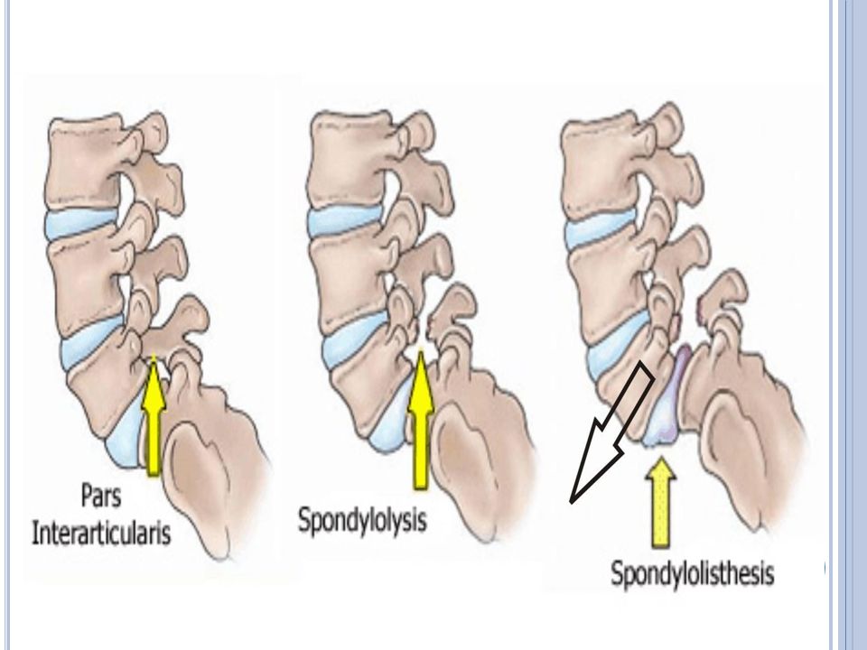
13
Axial and Sagittal CT scans of T6-T7. There is a calcified disc protruding into the spinal canal, extending from the midline and more prominent to the left.

14
This sagittal reconstruction of the lumbar spine obtained following a lumbar discogram shows contrast, (white), within the L4/5 disc which has a normal appearance. The appearance of contrast within the L5/S1 disc is abnormal demonstrating a degenerated disc with a mild posterior disc protrusion/herniation.

16
CT of lumbar spine: seen is an incomplete closure of the spine's lamina (spina bifida) Magnetic resonance imaging (MRI) scan of a sagittal section - Spina bifida.
 Magnetic resonance imaging (MRI) scan of a sagittal section - Spina bifida.")
17
Sagittal CT through the lumbar spine revealing an additional lucent line in the posterior elements of the L4 vertebral body (black arrow).
.")
18
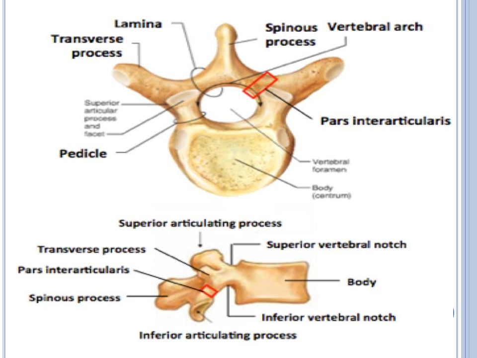
The CT scan shows the relevant anatomy of a normal pars interarticularis and one that exhibits spondylolysis.

19
Midline sagittal CT scan again demonstrates the L5-S1 spondylolisthesis. Preoperative sagittal CT scan showing spondylolisthesis.

21
CT scan – C5 spondylolisthesis. CT scan – C5 spondylolisthesis postoperative.

22
Non- contrast CT Scan of the entire spine shows diffuse multiple osteolytic lesions.

23
CT scan, Tumor at C2-C3 C2 C3

24
Bone metastases: Sagittal CT of the thorax in bone windows shows multiple lesions. Reconstruction images of the CT chest showing multiple osteoblastic lesions in thoraco- lumbar vertebrae. Skeletal changes in myeloma, thoracic spine.

25
Sagittal (A) and axial (B) computed tomography scans showing an osteolytic lesion of the L4 vertebral body. Sagittal CT reformation of the lumbar spine (a) shows a large sclerotic lesion nearly completely involving the L5 vertebral body.
 shows a large sclerotic lesion nearly completely involving the L5 vertebral body..")
26
Post-operative CT scan showing screws and plate in cross-section.

28
Post-op axial CT scans showing cage and pedicle screw placement. Post-op sagittal CT scan (left) and coronal CT scan (right) showing adequate spondylolisthesis reduction.
 and coronal CT scan (right) showing adequate spondylolisthesis reduction..")
Similar presentations


:e4.>")
