Download presentation
Presentation is loading. Please wait.

1
Protons (hydrogen nuclei act like little magnets) MRI Collective Magnetic Moment of Protons (M 0 ) Each pixel is a glass of protons B 0 = 3T (not to scale) Energy antiparallel parallel
 MRI Collective Magnetic Moment of Protons (M 0 ) Each pixel is a glass of protons B 0 = 3T (not to scale) Energy antiparallel parallel")
2
B0B0 Model of Head Coil collective spins from one voxel (M 0 ) for example
 for example")
3
3 Tesla magnet field B0B0 Protons (hydrogen nuclei act like little magnets) Not all the protons line up – thermal energy MRI Collective Magnetic Moment of Protons (M 0 ) from one voxel Classical picture of Quantum Phenomenon
 Not all the protons line up – thermal energy MRI Collective Magnetic Moment of Protons (M 0 ) from one voxel Classical picture of Quantum Phenomenon")
4
B0B0 Model of Head Coil Each voxel is a “cup of hydrogen nuclei” (Avogadro numbers)
")
5
B0B0 Model of Head Coil ExciteListen Collective Magnetic Moment (M 0 ) of Protons Flipped 90 ° start end Radio Waves (B 1 ) Larmor Frequency υ L =γ’B 0
 of Protons Flipped 90 ° start end Radio Waves (B 1 ) Larmor Frequency υ L =γ’B 0")
6
M 0 excitation in lab reference frame (x, y, z) and rotating coordinate frame (x’,y’,z’) Thanks Donald Plewes “Spin Gymnatics” Larmor Frequency υ L =γ’B 0
 and rotating coordinate frame (x’,y’,z’) Thanks Donald Plewes Spin Gymnatics Larmor Frequency υ L =γ’B 0")
7
MRI excite B0B0 Radio Waves start end M0M0 Collective Magnetic Moment (M 0 ) of protons flipped 90 ° Larmor Frequency υ L =γ’B 0
 of protons flipped 90 ° Larmor Frequency υ L =γ’B 0")
8
3.1 T 127 MHz 3.0 T 123 MHz 2.9 T 119 MHz excite Like a swing. Got one of the 3 orthogonal spatial dimensions when we excite. Z gradient on Larmor Frequency υ L =γ’B 0

9
3.1 T 127 MHz 3.0 T 123 MHz 2.9 T 119 MHz better to LISTEN like this Got second of the 3 orthogonal spatial dimensions when we listen. X gradient on fast slow regular Model of Head Coil Larmor Frequency υ L =γ’B 0

10
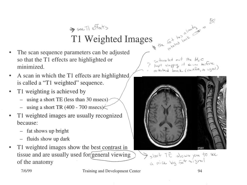
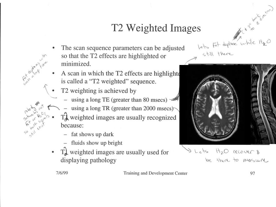
MRI listen start end Make image based on Protons loosing energy Make image based on Protons dephasing Slow Precession Fast Precession Fat and water loose energy and dephase at different rates T1 (energy lose time constant) T2 (dephasing time constant) Axial MRI Head Water Fat bright dark
 T2 (dephasing time constant) Axial MRI Head Water Fat bright dark")
13
T1 decay – “spins back down” Collective Magnetic Moment of Protons end start B0B0 signal we “hear” V Time T1 Recovery MR Signal Time Typical T1 Graph 1 s

14
T2 decay – separation (dephasing) of “collective magnetic moment” sometime after RF excitation Immediately after RF excitation = collective magnectic moment individual spins separation (dephasing) a little time later T2 Decay MR Signal Typical T2 Graph Time 50 ms
 of collective magnetic moment sometime after RF excitation Immediately after RF excitation = collective magnectic moment individual spins separation (dephasing) a little time later T2 Decay MR Signal Typical T2 Graph Time 50 ms")
15
T2 Decay MR Signal T1 Recovery MR Signal 50 ms 1 s T1 Contrast time TE – echo time TR – repeat time

16
T2 Decay MR Signa l T1 Recovery MR Signal 50 ms 1 s T2* and T2 Contrast TE – echo time TR – repeat time

17
Spin echo (most common sequence) signal strength equation (Density of protons contributing to signal) (signal loss due to T1) (Signal loss due to T2) Intensity of a voxel
 signal strength equation (Density of protons contributing to signal) (signal loss due to T1) (Signal loss due to T2) Intensity of a voxel")
18
Properties of Body Tissues MRI has high contrast for different tissue types!

Similar presentations


 T2 weighted – (fmri)>")


>")


 Medical imaging systems: MRI.>")


 Imaging>")

 Magnetic Resonance Imaging (MRI)>")