Download presentation
Presentation is loading. Please wait.

1
Radiography of the Shoulder Jennifer Nicol PGY-1 August 6, 2009

2
Objectives BRIEF Anatomy Review Standard shoulder views Radiographs of shoulder injuries NOT: ◦ Treatment ◦ Other imaging modalities ◦ Pediatric imaging

4
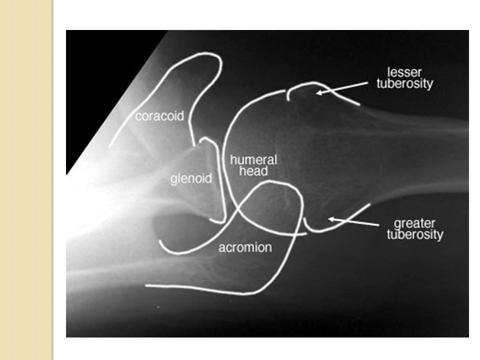
Anatomy

5
Shoulder Views Over 15 views of shoulder described Trauma series: ◦ 3 views: AP Trans-scapular “Y-view” Axillary Modified axillary

6
AP view True AP - 45˚tilt ◦ Glenohumeral joint with no bony overlap ◦ Preferred in trauma AP int/ext rotation ◦ Highlight tuberosities ◦ Soft tissue injuries Clavicle and AC joint

8
Transcapular view Projects along long axis scapula Simple, reproducible Good for visualising anterior, posterior dislocations

9
Acromion Coracoid Body

10
Axillary View Glenohumeral joint in cephalocaudal plane Lesions of glenoid rim, humeral head, caracoid Axial view of shoulder

12
Modified Axillary View Reverse axillary when pt can’t abduct

13
Retrospective 1690 shoulder exams ◦ Mod axillary view used 104 times ◦ Identified additional pathology in 30 cases No comparison b/t standard and modified axillary

14
Approach to Shoulder XR AP: ◦ If ext/int rotation look at tuberosity orientation ◦ Glenohumeral region Alignment Distance b/t humeral head and glenoid Bones ◦ AC region ◦ Other regions (clavicle, ribs, scapular spine,lungs)
")
15
Approach to Shoulder XR Other views: ◦ Humeral head to glenoid ◦ Prox humerus ◦ Glenoid rim ◦ Scapula ◦ Carocoid ◦ Acromion

16
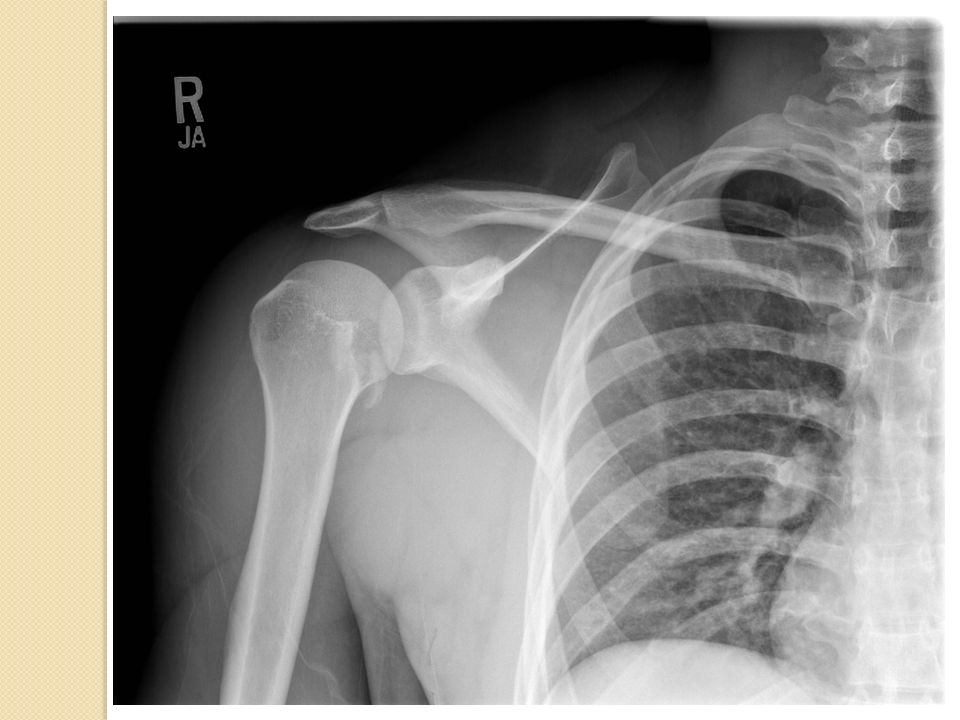
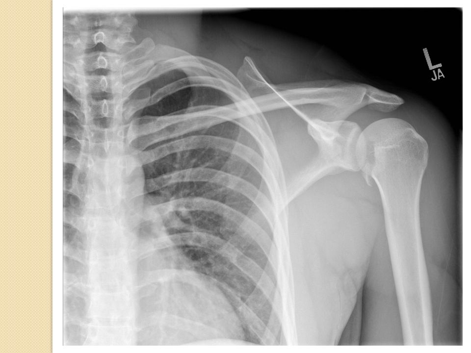
1)Glenoid Alignment Distance bones 2)AC Alignment Carocoid-clavicle space 3)Other Lungs, scapula, ribs, clavicle Type I AC injury
Glenoid Alignment Distance bones 2)AC Alignment Carocoid-clavicle space 3)Other Lungs, scapula, ribs, clavicle Type I AC injury")
17
Type III AC injury

18
Posterior Dislocation

19
Positive Rim Sign

20
Trough Sign

21
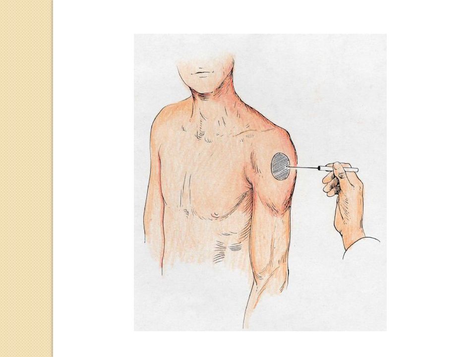
No Mercedes!!!!

22
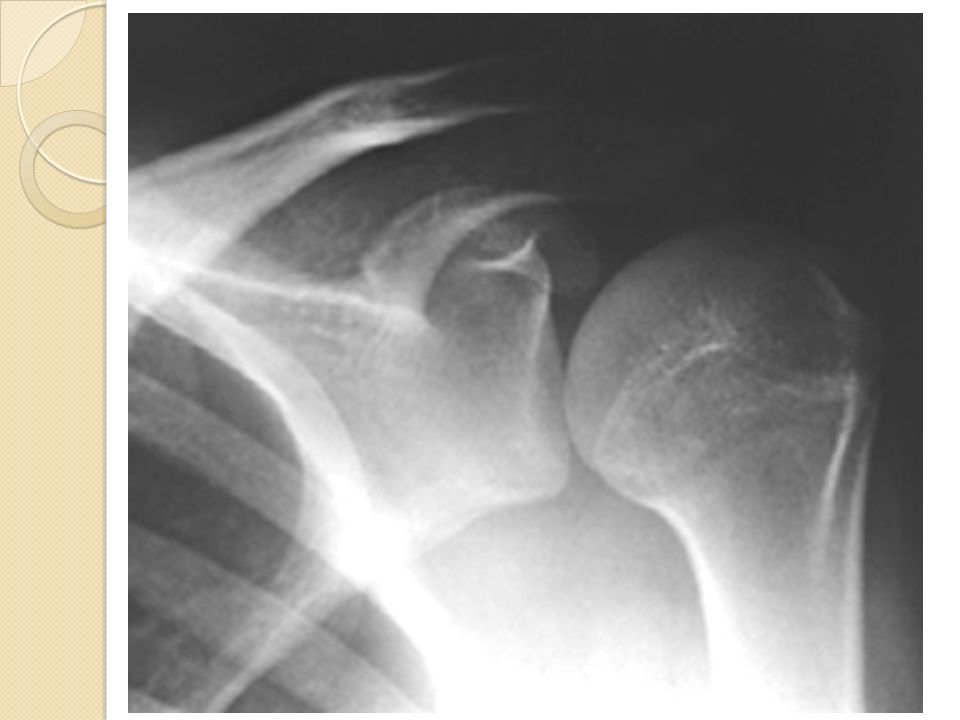
Avulsion Lesser Tuberosity

25
Bilateral shoulder dislocation spontaneously reduced with bilateral reverse Hill-Sachs lesions

26
Posterior Dislocations Have high suspicion with correct mechanism Don’t miss – clinical exam important Look for associated fractures Types: ◦ Subacromial (98%) ◦ Subglenoid ◦ subspinosus
 ◦ Subglenoid ◦ subspinosus")
27
Anterior Dislocation

29
Scapular View: Anterior Dislocation

30
Hill-Sachs deformity

31
AP Bankhart Injury Axillary

32
Greater Tuberosity Fracture

33
Anterior Dislocations 4 Types ◦ Subcoracoid ◦ Subglenoid ◦ Subclavicular ◦ Intrathoracic

34
Anterior Dislocations Check Neurovascular exam pre-post reduction Don’t delay reduction – NV injury increases with time Recurrence high – 80% <30

35
Inferior Dislocation

36
Subglenoid anterior dislocation

37
Pseudodislocation

Similar presentations


>")


 JOINT>")


