Download presentation
Presentation is loading. Please wait.

1
1 The Heart

2
Heart Anatomy & Basic Function

3
(1) Cardiovascular Function Cardiovascular = Heart, Arteries, Veins, Blood Function:Function: –Transportation –Blood = transport vehicle –Carries oxygen, nutrients, wastes, and hormones –Movement provided by pumping of heart
 Cardiovascular Function Cardiovascular = Heart, Arteries, Veins, Blood Function:Function: –Transportation –Blood = transport vehicle –Carries oxygen, nutrients, wastes, and hormones –Movement provided by pumping of heart")
4
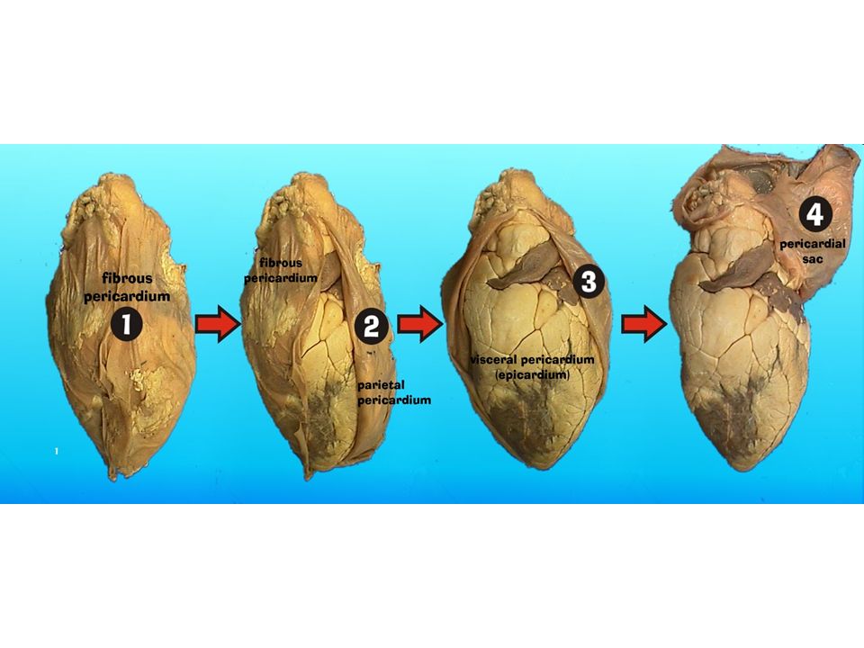
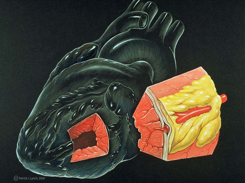
(2) Cardiac Tissues Outermost = Pericardium & Epicardium –Pericardium is a membrane anchoring heart to diaphragm and sternum –Pericardium secretes lubricant (serous fluid) –Epicardium is outermost muscle tissue Middle = Myocardium –Contains contractile muscle fibers Innermost = Endocardium –Lines Cardiac Chambers
 Cardiac Tissues Outermost = Pericardium & Epicardium –Pericardium is a membrane anchoring heart to diaphragm and sternum –Pericardium secretes lubricant (serous fluid) –Epicardium is outermost muscle tissue Middle = Myocardium –Contains contractile muscle fibers Innermost = Endocardium –Lines Cardiac Chambers")
5
5 Pericardium (see next slide) Starting from the outside… Without most of pericardial layers
 Starting from the outside… Without most of pericardial layers")
6
6 Heart’s position in thorax

7
7 In mediastinum – behind sternum and pointing left, lying on the diaphragm It weighs 250-350 gm (about 1 pound) Feel your heart beat at apex (this is of a person lying down)
 Feel your heart beat at apex (this is of a person lying down)")
10
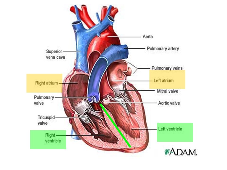
(3) Cardiac Chambers Human heart has 4 chambers –2 Atria Superior = primary receiving chambers, do not actually pump Blood flows into atria –2 Ventricles Pump blood Contraction = blood sent out of heart + circulated Chambers are separated by septum… –Due to separate chambers, heart functions as double pump
 Cardiac Chambers Human heart has 4 chambers –2 Atria Superior = primary receiving chambers, do not actually pump Blood flows into atria –2 Ventricles Pump blood Contraction = blood sent out of heart + circulated Chambers are separated by septum… –Due to separate chambers, heart functions as double pump")
12
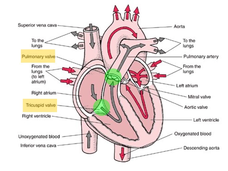
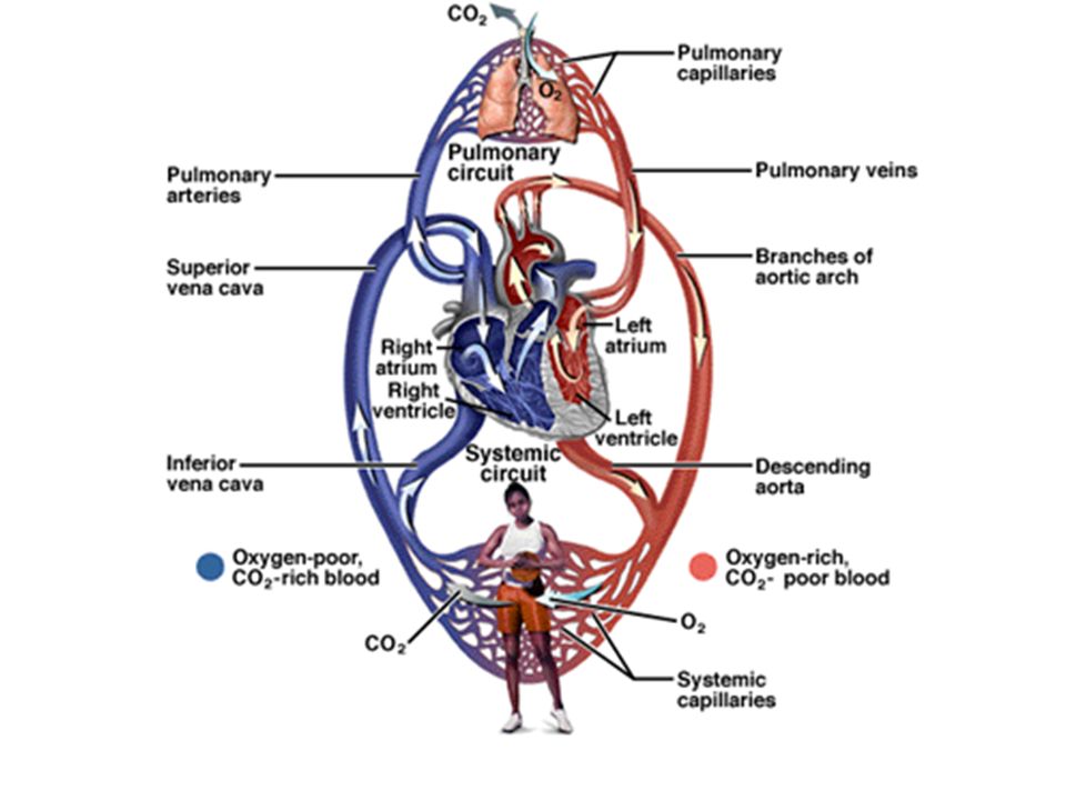
Deoxygenated Blood …To the lungs Oxygenated Blood …To the rest of the body

13
(4) Pulmonary Circulation Pulmonary = Deoxygenated Blood Involves Right Side of Heart Pathway:Pathway: 1.Superior / Inferior Vena Cava 2.Right Atrium Tricuspid Valve 3.Right Ventricle Pulmonary Semilunar Valve 4.Left Pulmonary Artery 5.Lungs
 Pulmonary Circulation Pulmonary = Deoxygenated Blood Involves Right Side of Heart Pathway:Pathway: 1.Superior / Inferior Vena Cava 2.Right Atrium Tricuspid Valve 3.Right Ventricle Pulmonary Semilunar Valve 4.Left Pulmonary Artery 5.Lungs")
15
(5) Systemic Circulation Systemic = Oxygenated Blood Involves Left Side of Heart Pathway:Pathway: 1.Left Pulmonary Vein 2.Left Atrium Bicuspid Valve 3.Left Ventricle Aortic Semilunar Valve 4.Aorta 5.All Other Tissues
 Systemic Circulation Systemic = Oxygenated Blood Involves Left Side of Heart Pathway:Pathway: 1.Left Pulmonary Vein 2.Left Atrium Bicuspid Valve 3.Left Ventricle Aortic Semilunar Valve 4.Aorta 5.All Other Tissues")
18
18 Pattern of flow (simple to more detailed) Body RA RV Lungs LA LV Boby Body to right heart to lungs to left heart to body Body, then via vena cavas and coronary sinus to RA, to RV, then to lungs via pulmonary arteries, then to LA via pulmonary veins, to LV, then to body via aorta From body via SVC, IVC & coronary sinus to RA; then to RV through tricuspid valve; to lungs through pulmonic valve and via pulmonary arteries; to LA via pulmonary veins; to LV through mitral valve; to body via aortic valve then aorta LEARN THIS
 Body RA RV Lungs LA LV Boby Body to right heart to lungs to left heart to body Body, then via vena cavas and coronary sinus to RA, to RV, then to lungs via pulmonary arteries, then to LA via pulmonary veins, to LV, then to body via aorta From body via SVC, IVC & coronary sinus to RA; then to RV through tricuspid valve; to lungs through pulmonic valve and via pulmonary arteries; to LA via pulmonary veins; to LV through mitral valve; to body via aortic valve then aorta LEARN THIS")
19
19 In the fetus, the RA received oxygenated blood from mom through umbilical cord, so blood R to L through the foramen ovale: fossa ovalis is left after it closes The pulmonary trunk had high resistance (because lungs not functioning yet) & ductus arteriosus shunted blood to aorta; becomes ligamentum arteriosum after birth
 & ductus arteriosus shunted blood to aorta; becomes ligamentum arteriosum after birth")
20
(6) Cardiac Valves [4 main valves] When the heart is relaxed… –Blood passively fills atrium –Flows right past tricuspid / bicuspid valves –Semilunar Valves remain shut When the heart contracts (pumps)… –Tricuspid / Bicuspid valves swing up and shut –Blood ejected out of ventricle –Semilunar Valves open up
![(6) Cardiac Valves [4 main valves] When the heart is relaxed… –Blood passively fills atrium –Flows right past tricuspid / bicuspid valves –Semilunar Valves remain shut When the heart contracts (pumps)… –Tricuspid / Bicuspid valves swing up and shut –Blood ejected out of ventricle –Semilunar Valves open up](http://images.slideplayer.com/27/8940010/slides/slide_20.jpg "(6) Cardiac Valves [4 main valves] When the heart is relaxed… –Blood passively fills atrium –Flows right past tricuspid / bicuspid valves –Semilunar Valves remain shut When the heart contracts (pumps)… –Tricuspid / Bicuspid valves swing up and shut –Blood ejected out of ventricle –Semilunar Valves open up")
22
22 Function of AV valves

23
23 Function of semilunar valves (Aortic and pulmonic valves)
")
24
24 Note positions of valves Valves open and close in response to pressure differences Trabeculae carnae Note papillary muscles, chordae tendinae (heart strings): keep valves from prolapsing (purpose of valve = 1 way flow)
: keep valves from prolapsing (purpose of valve = 1 way flow)")
25
25

26
26 Heartbeat Systole: contraction Diastole: filling Normal rate: 60-100 Slow: bradycardia Fast: tachycardia ***Note: blood goes to RA, then RV, then lungs, then LA, then LV, then body; but the fact that a given drop of blood passes through the heart chambers sequentially does not mean that the four chambers contract in that order; the 2 atria always contract together, followed by the simultaneous contraction of the 2 ventricles Definition: a single sequence of atrial contraction followed by ventricular contraction See http://www.geocities.com/Athens/Forum/6100/1heart.html

27
27 Heart sounds Called S1 and S2 S1 is the closing of AV (Mitral and Tricuspid) valves at the start of ventricular systole S2 is the closing of the semilunar (Aortic and Pulmonic) valves at the end of ventricular systole –Separation easy to hear on inspiration therefore S2 referred to as A2 and P2 Murmurs: the sound of flow –Can be normal –Can be abnormal
 valves at the start of ventricular systole S2 is the closing of the semilunar (Aortic and Pulmonic) valves at the end of ventricular systole –Separation easy to hear on inspiration therefore S2 referred to as A2 and P2 Murmurs: the sound of flow –Can be normal –Can be abnormal")
28
28 “EKG” ( or ECG, electrocardiogram) Electrical depolarization is recorded on the body surface by up to 12 leads Pattern analyzed in each lead P wave=atrial depolarization QRS=ventricular depolarization T wave=ventricular repolarization
 Electrical depolarization is recorded on the body surface by up to 12 leads Pattern analyzed in each lead P wave=atrial depolarization QRS=ventricular depolarization T wave=ventricular repolarization")
29
29 Electrical conduction system: (Explanation in next slides) specialized cardiac muscle cells that carry impulses throughout the heart musculature, signaling the chambers to contract in the proper sequence
 specialized cardiac muscle cells that carry impulses throughout the heart musculature, signaling the chambers to contract in the proper sequence")
30
30 Conduction system SA node (sinoatrial) –In wall of RA –Sets basic rate: 70-80 –Is the normal pacemaker Impulse from SA to atria Impulse also to AV node via internodal pathway AV node –In interatrial septum
 –In wall of RA –Sets basic rate: –Is the normal pacemaker Impulse from SA to atria Impulse also to AV node via internodal pathway AV node –In interatrial septum")
31
31 Conduction continued SA node through AV bundle (bundle of His) –Into interventricular septum –Divides R and L bundle branches become subendocardial branches (“Purkinje fibers”) Contraction begins at apex
 –Into interventricular septum –Divides R and L bundle branches become subendocardial branches ( Purkinje fibers ) Contraction begins at apex")
32
32

33
33 Artificial Pacemaker

34
34 Autonomic innervation Sympathetic –Increases rate and force of contractions Parasympathetic (branches of Vagus n.) –Slows the heart rate http://education.med.nyu.edu/courses/old/physiology/courseware/ekg_pt1/EKGseq.html For a show on depolarization:
 –Slows the heart rate For a show on depolarization:")
35
35 Blood supply to the heart (there’s a lot of variation) A: Right Coronary Artery; B: Left Main Coronary Artery; C: Left Anterior Descending (LAD, or Left Anterior Interventricular); D: Left Circumflex Coronary Artery; G: Marginal Artery; H: Great Cardiac Vein; I: Coronary sinus, Anterior Cardiac Veins.
 A: Right Coronary Artery; B: Left Main Coronary Artery; C: Left Anterior Descending (LAD, or Left Anterior Interventricular); D: Left Circumflex Coronary Artery; G: Marginal Artery; H: Great Cardiac Vein; I: Coronary sinus, Anterior Cardiac Veins.")
36
36 Anterior view L main coronary artery arises from the left side of the aorta and has 2 branches: LAD and circumflex R coronary artery emerges from right side of aorta

37
37 Note that the usual name for “anterior interventricular artery” is the LAD (left anterior descending)
")
38
38 A lot of stuff from anterior view Each atrium has an “auricle,” an ear-like flap

39
39

Similar presentations


 in thorax, in inferior mediastinum>")


