Download presentation
Presentation is loading. Please wait.

1
PROJECT#3(b) Astrocyte Analysis
BY Bhimanathini Venkatsai Sai Kumar Maddula

2
Contents Astrocyte Image Segmentation Thresholding Astrocyte Analysis
Convert Stack file to 8-bit Apply Threshold Segmentation by 3D Viewer Selection of Seed Point Overlapping/Touching cell Structure Apply Singletonize 3D

3
Astrocyte Astrocyte , are characteristic star-shaped Glial cells in the brain and spinal cord. They are the most abundant cell of the human brain.

4
IMAGE SEGMENTATION Image segmentation is the process of assigning a label to every pixel in an image such that pixels with the same label share certain visual characteristics like colour, texture etc. The result of image segmentation is a set of segments that collectively cover the entire image, or a set of contours extracted from the image

5
Thresholding This is method of Image segmentation, where grayscale image thresholding can be used to create binary image. There are few thresholding methods based on the information the algorithm manipulates. Such as: Clustering based methods Entropy based methods Histogram shape based methods Etc…

6
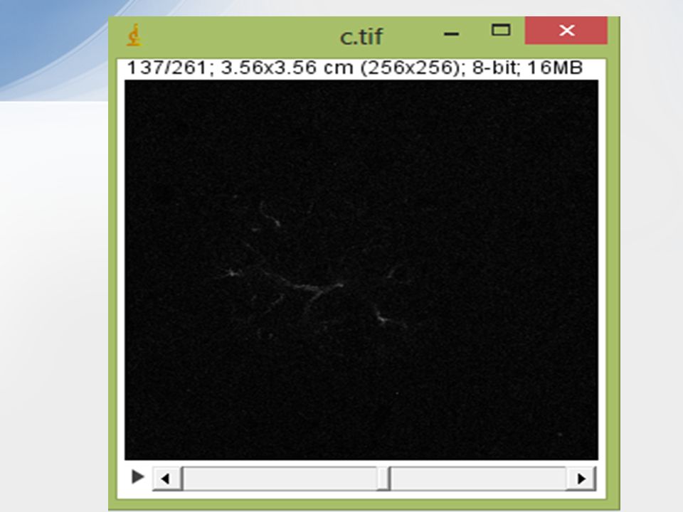
Astrocyte Analysis: Load the stack File into ImageJ
File Open Browse Stack File

7
Convert the Stack File to 8-bit image
If Stack file is not in 8-bit image format. Convert it into 8-bit image format. Image Type click 8-bit Where: Scale 0= Black Scale 128=Medium Gray Scale 255= White

9
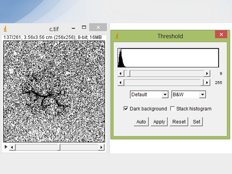
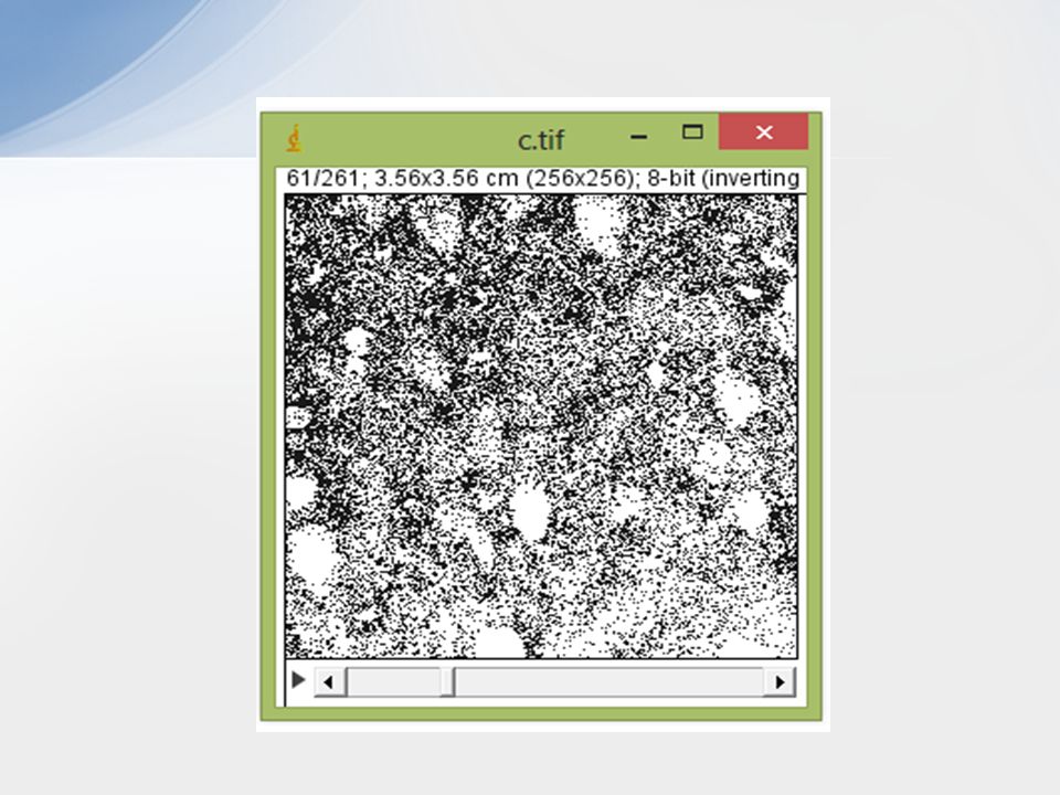
Apply Threshold: We use Thresholding for detecting edges , counting particals or measuring areas. Image Adjust select ‘Threshold’ Note: Check ‘Dark Background’ in the Threshold pop-up window and Apply.

11
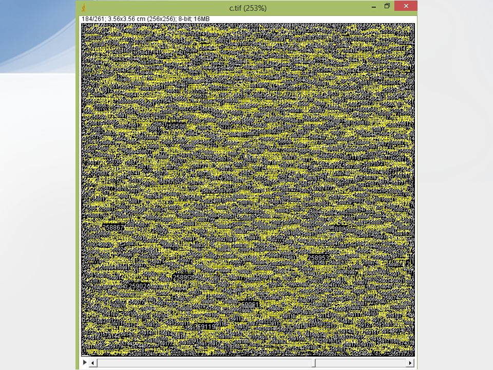
Note: ‘calculate threshold for each image’ should be checked.

12
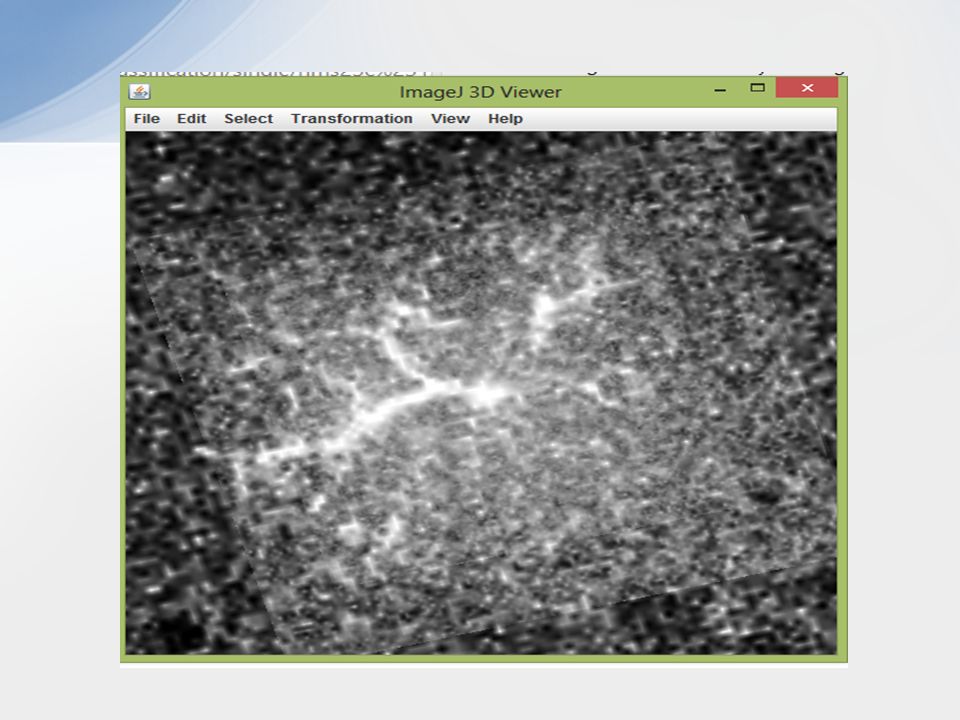
Segmentation by 3D Viewer
Here 3D Viewer uses Java 3D to provide hardware-accelerated 3D visualization of image stacks as volumes, surfaces and orthoslices. Plugins Segmentation Click ‘Segment blob in 3D Viewer’

15
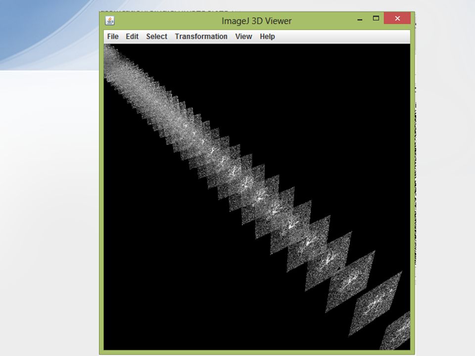
Selection of Seed Point
Select a seed point as a start (using ‘Point Selections’ Modern image segmentation techniques are based on PDE (Partial differential equations. The Fiji Plugin provides two PDE based methods i) Fast Marching ii) Level Sets.
 Fast Marching. ii) Level Sets.")
17
Overlapping/Touching Cell Structures
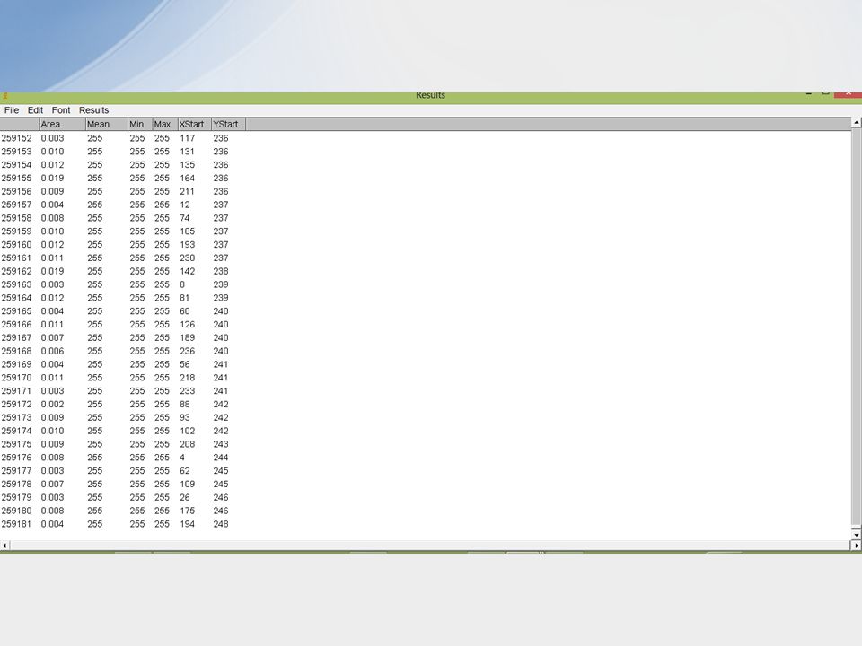
Here we apply Thresholding on binary image stack file then we follow the below steps.. 1) Process Binary Watershed 2) Analyze Analyze Particles Outlines Display Result
 Process Binary Watershed. 2) Analyze Analyze Particles Outlines. Display Result.")
20
Thank you..!!

Similar presentations