Download presentation
Presentation is loading. Please wait.

1
Venous thromboembolism

2
Learning objectives Gain organised knowledge in the subject area VTE
Be able to correctly interpret diagnostic information in suspected VTE Know and apply the relevant evidence and/or guidelines to different clinical presentations of VTE Be aware of common cognitive biases in the diagnosis and management of VTE Learning objectives always the same for each topic …

3
Scenarios

4
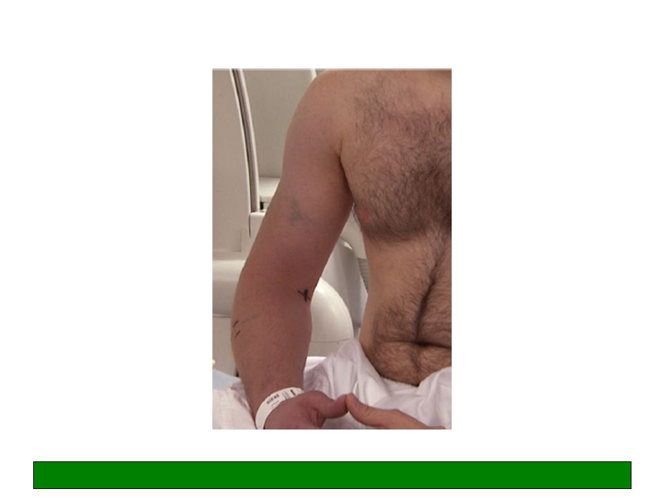
Scenario 1 A 34-year-old man was admitted with acute onset of heaviness, pain and functional impairment of his right arm. The arm was cyanotic and swollen. For the past few weeks, he reported transient paraesthesia of his right arm during weightlifting. He had a fracture of the right clavicle after a ski accident 5 years previously which was managed conservatively. There was no personal or family history of thrombosis.

6
Doppler USS right arm showed axillary and subclavian vein thrombosis
In small groups answer the following Qs: What are the treatment options in this case? What further investigations should be performed? What follow-up is required? Treatment options – Anticoagulation (LMWH plus warfarin – stop LMWH when after min 5 days or two therapeutic INRs whichever is sooner) If severe symptoms/signs and low bleeding risk/good functional status, refer to vascular for catheter directed thrombolysis or pharmaco-mechanical thrombectomy Further investigations – Imaging of the thorax a) CT or MRI of the thoracic inlet to look for cause of the compression; imaging of the thorax to look for cancer (lung/lymphoma) – discuss protocol with radiologist. Follow-up – Referral to vascular surgery Any cancer referrals Lymphoedema clinic (compression not recommended acutely but may be needed for PTS) Wells Score for DVT not validated for upper limb cases. D-dimer not validated/not recommended. No validated diagnostic or treatment strategy or national guideline. Management based on consensus statements e.g. American College Chest Physicians 2012.
 If severe symptoms/signs and low bleeding risk/good functional status, refer to vascular for catheter directed thrombolysis or pharmaco-mechanical thrombectomy. Further investigations – Imaging of the thorax a) CT or MRI of the thoracic inlet to look for cause of the compression; imaging of the thorax to look for cancer (lung/lymphoma) – discuss protocol with radiologist. Follow-up – Referral to vascular surgery. Any cancer referrals. Lymphoedema clinic (compression not recommended acutely but may be needed for PTS) Wells Score for DVT not validated for upper limb cases. D-dimer not validated/not recommended. No validated diagnostic or treatment strategy or national guideline. Management based on consensus statements e.g. American College Chest Physicians")
7
Upper limb DVT: summary
Increasing incidence 5-10% all DVTs Primary Effort-related thrombosis (Paget-Schroetter Syndrome). Weights, tennis, repeated overhead activities (swimming, decorating). Most have underlying VTOS Idiopathic* Secondary most common Indwelling central venous catheters/ports Cancers, mainly lung and lymphoma Surgery/trauma/immobilisation of arm Pregnancy and ovarian hyperstimulation syndrome Effort-related usually good history and most common cause of primary upper limb DVT VTOS = venous thoracic outlet syndrome Idiopathic require thrombophilia screen and further Ix – up to one quarter thought to be idiopathic have a cancer* Idiopathic = no obvious underlying risk factor or VTOS can be identified. More likely to have abnormal thrombophilia screen. Secondary most common cause of upper limb DVT overall
. Weights, tennis, repeated overhead activities (swimming, decorating). Most have underlying VTOS. Idiopathic* Secondary most common. Indwelling central venous catheters/ports. Cancers, mainly lung and lymphoma. Surgery/trauma/immobilisation of arm. Pregnancy and ovarian hyperstimulation syndrome. Effort-related usually good history and most common cause of primary upper limb DVT. VTOS = venous thoracic outlet syndrome. Idiopathic require thrombophilia screen and further Ix – up to one quarter thought to be idiopathic have a cancer* Idiopathic = no obvious underlying risk factor or VTOS can be identified. More likely to have abnormal thrombophilia screen. Secondary most common cause of upper limb DVT overall.")
8
Upper limb DVT: summary
Initial management Anticoagulation: 3 months for idiopathic and catheter related (if catheter removed); indefinite if on-going risk factors Catheter directed thrombolysis / pharmaco-mechanical thrombectomy in selected patients Investigations/follow-up Doppler USS initial investigation of choice If primary upper limb DVT: imaging of the thorax +/- thrombophilia screen If secondary: address underlying cause (e.g. catheter) OP vascular surgery referral Complications Post-thrombotic syndrome (in one study, 53% pts with upper limb VTE due to VTOS) VTOS = venous thoracic outlet syndrome Patients can be started on initial treatment and have urgent OP Ix and FU – imaging of the thorax should be discussed with a radiologist on a case by case basis so studies are protocolled correctly. OP ref to vascular surgery is for treatment of VTOS in selected cases. There are some cancer cases when stenting may be considered acutely (e.g. SVC syndrome) – usually by a vascular radiologist. Stenting in cases of VTOS is not advised without decompression surgery first, as the stent just fractures/re-occludes.
; indefinite if on-going risk factors. Catheter directed thrombolysis / pharmaco-mechanical thrombectomy in selected patients. Investigations/follow-up. Doppler USS initial investigation of choice. If primary upper limb DVT: imaging of the thorax +/- thrombophilia screen. If secondary: address underlying cause (e.g. catheter) OP vascular surgery referral. Complications. Post-thrombotic syndrome (in one study, 53% pts with upper limb VTE due to VTOS) VTOS = venous thoracic outlet syndrome. Patients can be started on initial treatment and have urgent OP Ix and FU – imaging of the thorax should be discussed with a radiologist on a case by case basis so studies are protocolled correctly. OP ref to vascular surgery is for treatment of VTOS in selected cases. There are some cancer cases when stenting may be considered acutely (e.g. SVC syndrome) – usually by a vascular radiologist. Stenting in cases of VTOS is not advised without decompression surgery first, as the stent just fractures/re-occludes.")
9
Knowledge gaps application of the wrong heuristic
Common cognitive biases in the diagnosis and management of upper limb DVT Knowledge gaps application of the wrong heuristic i.e. treating upper limb DVT the same as lower limb DVT and applying inappropriate diagnostic, management and further investigation strategies Heuristic = “rule of thumb” Patterns we look for and how we respond to familiar situations

10
From the 2012 consensus guidelines of the American College of Chest Physicians
See further reading – summary of ACCP recommendations.

11
Any questions at this point?

12
Scenario 2 A 50-year-old woman was admitted with pain, redness and swelling of her thigh that had developed over the preceding 48 hours ?cellulitis. Her only past medical history was gastro-oesophageal reflux disease and varicose veins.

14
She was started on iv flucloxacillin by the FY1 doctor
In small groups answer the following Qs: What is the diagnosis? What further investigations should be performed? What are the treatment options in this case? Superficial thrombophlebitis of the proximal thigh (?near sapheno-femoral junction) Doppler USS of the affected leg to exclude occult DVT Anti-embolism stockings In this case, probably LMWH prophylactic dose 30 days (minimum) General advice – elevation, hot towel, do not restrict mobility, watch for signs of DVT, ?topical NSAIDs
 Doppler USS of the affected leg to exclude occult DVT. Anti-embolism stockings. In this case, probably LMWH prophylactic dose 30 days (minimum) General advice – elevation, hot towel, do not restrict mobility, watch for signs of DVT, topical NSAIDs.")
15
Revision of anatomy: deep vs superficial veins of the leg and the sapheno-femoral junction

16
Superficial thrombophlebitis: summary
Inflammation of the superficial veins Swelling, pain, redness Scanty evidence base re: management SIGN guidelines and NICE ‘clinical knowledge summary’ broadly agree

17
Superficial thrombophlebitis: summary
SIGN Guideline 122 10-21% of pts have a DVT, more likely if >5cm inflammation or within 10cm of sapheno-femoral junction Topical treatments only alleviate symptoms Oral NSAIDs prevent extension, recurrence and progression to DVT, so does LMWH Not clear if LMWH (prophylactic or therapeutic doses) is better than NSAIDs – but in high risk cases LMWH recommended* NICE CKS The risk of DVT should be considered Treat with oral NSAIDs and paracetamol, class 1 AES and advice. Topical NSAIDs can be used for symptomatic relief LMWH not routinely recommended, but experts advise it should be used in high risk cases (no dose recommendation) Treat super-added infection with antibiotics So what does SIGN say? More clear and I recommend this: Patients with clinical signs of superficial thrombophlebitis affecting the proximal long saphenous vein should have an ultrasound scan to exclude concurrent DVT. Patients with superficial thrombophlebitis should have anti-embolism stockings and can be considered for treatment with prophylactic doses of LMWH for up to 30 days or fondaparinux for 45 days. If LMWH is contraindicated, 8-12 days of oral NSAIDs should be offered. Patients with superficial thrombophlebitis at, or extending towards, the sapheno-femoral junction can be considered for therapeutic anticoagulation for 6-12 weeks.
 is better than NSAIDs – but in high risk cases LMWH recommended* NICE CKS. The risk of DVT should be considered. Treat with oral NSAIDs and paracetamol, class 1 AES and advice. Topical NSAIDs can be used for symptomatic relief. LMWH not routinely recommended, but experts advise it should be used in high risk cases (no dose recommendation) Treat super-added infection with antibiotics. So what does SIGN say More clear and I recommend this: Patients with clinical signs of superficial thrombophlebitis affecting the proximal long saphenous vein should have an ultrasound scan to exclude concurrent DVT. Patients with superficial thrombophlebitis should have anti-embolism stockings and can be considered for treatment with prophylactic doses of LMWH for up to 30 days or fondaparinux for 45 days. If LMWH is contraindicated, 8-12 days of oral NSAIDs should be offered. Patients with superficial thrombophlebitis at, or extending towards, the sapheno-femoral junction can be considered for therapeutic anticoagulation for 6-12 weeks.")
18
Confirmation bias if the patient has been seen first by someone else
Common cognitive biases in the diagnosis and management of superficial thrombophlebitis Knowledge gaps application of the wrong heuristic (i.e. treating as ‘cellulitis’) not knowing the risk of occult DVT at presentation Confirmation bias if the patient has been seen first by someone else Heuristic = “rule of thumb” Patterns we look for and how we respond to familiar situations
 not knowing the risk of occult DVT at presentation. Confirmation bias if the patient has been seen first by someone else. Heuristic = rule of thumb Patterns we look for and how we respond to familiar situations.")
19
Any questions at this point?

20
Scenario 3 A 45 year old man presented to Ambulatory Care with right leg swelling and pain, which had occurred spontaneously over the last 3 days On examination, his right calf was 5cm wider than the left His only past medical history was hypertension

21
What should be done at this point?
Would you do a d dimer? – no. High Wells Score – straight to imaging. Bloods for FBC, U&E, LFT, baseline clotting (all related to proposed treatment with LMWH/warfarin)
")
22
Doppler USS right leg showed a femoral vein thrombosis
In small groups answer the following Qs: What is the management in this case? (pharmacological and non-pharmacological) What is the duration of anti-coagulation? What further investigations should be performed? What follow-up is required? Treatment options – Anticoagulation (LMWH plus warfarin – stop LMWH after a min of 5 days treatment or two therapeutic INRs - whichever is sooner) Below knee graduated compression stockings for the affected leg during day for up to two years (TEDs) Counselling re anticoagulant therapy and written information /card (v important) Decision re duration of anti-coagulation: In ALL cases of unprovoked DVT they need screening for cancer: history and examination, CXR, bloods (incl FBC, calcium/LFTs), urinalysis. In patients aged over 40 who do not have signs/symptoms of cancer based on this initial screen, consider abdo/pelvis CT and mammogram for women. How many of your departments do this in every case? Studies have suggested that among patients with VTE, a large number of the cancers most commonly found are those in the abdomen and pelvic areas (ovary, pancreas, liver, kidney colon). Adopting an intensive testing strategy which involves ultrasound scans, antigen tests, tumour markers, FOB, PSA or colonoscopy increases false positives without significant increases in sensitivity. 9.4% of patients with first episode unprovoked VTE who underwent the baseline routine tests had cancer detected. Over the course of 2 years, an additional 24/201(11.9%) developed cancer. These overall rates of an underlying cancer in patients with an unproved VTE are consistent with data from multiple large published registry studies. There were no cancers detected in the above study in pts under the age of 40. The recommendation is limited to patients with an apparently unprovoked VTE (as in the study), which represents only 20% of patients with a VTE, and further restricting it to patients over the age of 40years, in whom a cancer has not been detected by routine investigations, will significantly limit the workload and capacity impact on the NHS, of this recommendation while potentially maximising benefit. Test for thrombophilia in patients with unprovoked DVT in whom it is planned to stop treatment. Don’t test people on treatment.
 What is the duration of anti-coagulation What further investigations should be performed What follow-up is required Treatment options – Anticoagulation (LMWH plus warfarin – stop LMWH after a min of 5 days treatment or two therapeutic INRs - whichever is sooner) Below knee graduated compression stockings for the affected leg during day for up to two years (TEDs) Counselling re anticoagulant therapy and written information /card (v important) Decision re duration of anti-coagulation: In ALL cases of unprovoked DVT they need screening for cancer: history and examination, CXR, bloods (incl FBC, calcium/LFTs), urinalysis. In patients aged over 40 who do not have signs/symptoms of cancer based on this initial screen, consider abdo/pelvis CT and mammogram for women. How many of your departments do this in every case Studies have suggested that among patients with VTE, a large number of the cancers most commonly found are those in the abdomen and pelvic areas (ovary, pancreas, liver, kidney colon). Adopting an intensive testing strategy which involves ultrasound scans, antigen tests, tumour markers, FOB, PSA or colonoscopy increases false positives without significant increases in sensitivity. 9.4% of patients with first episode unprovoked VTE who underwent the baseline routine tests had cancer detected. Over the course of 2 years, an additional 24/201(11.9%) developed cancer. These overall rates of an underlying cancer in patients with an unproved VTE are consistent with data from multiple large published registry studies. There were no cancers detected in the above study in pts under the age of 40. The recommendation is limited to patients with an apparently unprovoked VTE (as in the study), which represents only 20% of patients with a VTE, and further restricting it to patients over the age of 40years, in whom a cancer has not been detected by routine investigations, will significantly limit the workload and capacity impact on the NHS, of this recommendation while potentially maximising benefit. Test for thrombophilia in patients with unprovoked DVT in whom it is planned to stop treatment. Don’t test people on treatment.")
23
Duration of treatment? Question: What is the risk of a recurrent DVT in a 30 year old man who presents with a single unprovoked DVT? Cancer patients 6 months then review risks/benefits LMWH only Provoked DVT/PE 3 months then review risks/benefits Unprovoked DVT/PE What’s the risk of a recurrent DVT in a 30 year old man who presents with a single unprovoked DVT?? Answer: When a major reversible risk factor such as surgery can be identified as the sole explanation for VTE, then the risk of recurrence is relatively low (i.e. ≈3% in the first year). In contrast, the risk is high (≈10% in the first year) in patients with unprovoked (“idiopathic”) VTE and in those with persistent, irreversible, or other risk factors. Recurrence of thrombosis is 31.6% in the first 6 months of IVDU pts Patients should be counselled re this. What about IVDUs?? – 6 months LMWH only then review risks/benefits. Look out for other complications at presentation: infected DVT, bacteraemia, groin abscess etc. Some reports of Vit K deficiency, chaotic lifestyle and interaction with methadone/HIV meds means warfarin inappropriate. Who is following DVT patients up in your hospital??
. In contrast, the risk is high (≈10% in the first year) in patients with unprovoked ( idiopathic ) VTE and in those with persistent, irreversible, or other risk factors. Recurrence of thrombosis is 31.6% in the first 6 months of IVDU pts. Patients should be counselled re this. What about IVDUs – 6 months LMWH only then review risks/benefits. Look out for other complications at presentation: infected DVT, bacteraemia, groin abscess etc. Some reports of Vit K deficiency, chaotic lifestyle and interaction with methadone/HIV meds means warfarin inappropriate. Who is following DVT patients up in your hospital")
24
Lower limb DVT: summary
?DVT history, exam and Wells Score If DVT suspected plus Wells = likely then do a proximal Doppler USS (whole leg not recommended) NICE recommends doing a d dimer in likely cases so that if the proximal scan is negative they can be offered a repeat scan in one week if their d dimer is high … This is because proximal scans may miss calf DVTs that could extend – you don’t need a repeat scan if a whole leg USS was performed first If DVT suspected plus Wells = unlikely then get a d dimer, if positive get a scan, if negative DVT is virtually excluded For all cases, if you can’t get a Doppler within 4 hours then treat and get a scan within 24 hours Consider and communicate alternative explanations for symptoms in patients with negative scans Don’t forget to arrange further investigations/FU for new DVT cases History and examination is KEY! If the history is clearly not a DVT at all, don’t even go here – for example trauma/ ruptured head of gastrocnemius while running for the bus, calf tears etc. How many of your depts follow this guideline? D-dimer tests have relatively high sensitivity but low specificity (false positive results common). When the sensitivity of a d-dimer test increase, its specificity decreases. To be useful in the diagnosis of DVT, a D-dimer test has high sensitivity and high negative value - fewer people with DVT will be missed. Therefore, a negative D-dimer may be useful in excluding DVT but a positive D-dimer is of no diagnostic value, it merely mandates further testing. Whilst a negative D-dimer test is good enough to exclude the diagnosis of DVT in a patient with an “unlikely” pre-test clinical probability it is not good enough in those with a “likely” pre-test probability. The Guideline Development Group had recommended proximal leg vein ultrasound scans as the clinical importance of picking up extra calf vein blood clots by scanning the whole leg is uncertain. Moreover, the evidence review suggested that ultrasound scan of calf veins are not very sensitive in picking up calf vein DVT. A repeat proximal leg vein scan is recommended to ensure that any clots propagating to the proximal veins are not missed. The GDG also modelled that an above-knee ultrasound with a repeat if negative is more cost-effective than a single above-knee or full-leg ultrasound scan.
 NICE recommends doing a d dimer in likely cases so that if the proximal scan is negative they can be offered a repeat scan in one week if their d dimer is high … This is because proximal scans may miss calf DVTs that could extend – you don’t need a repeat scan if a whole leg USS was performed first. If DVT suspected plus Wells = unlikely then get a d dimer, if positive get a scan, if negative DVT is virtually excluded. For all cases, if you can’t get a Doppler within 4 hours then treat and get a scan within 24 hours. Consider and communicate alternative explanations for symptoms in patients with negative scans. Don’t forget to arrange further investigations/FU for new DVT cases. History and examination is KEY! If the history is clearly not a DVT at all, don’t even go here – for example trauma/ ruptured head of gastrocnemius while running for the bus, calf tears etc. How many of your depts follow this guideline D-dimer tests have relatively high sensitivity but low specificity (false positive results common). When the sensitivity of a d-dimer test increase, its specificity decreases. To be useful in the diagnosis of DVT, a D-dimer test has high sensitivity and high negative value - fewer people with DVT will be missed. Therefore, a negative D-dimer may be useful in excluding DVT but a positive D-dimer is of no diagnostic value, it merely mandates further testing. Whilst a negative D-dimer test is good enough to exclude the diagnosis of DVT in a patient with an unlikely pre-test clinical probability it is not good enough in those with a likely pre-test probability. The Guideline Development Group had recommended proximal leg vein ultrasound scans as the clinical importance of picking up extra calf vein blood clots by scanning the whole leg is uncertain. Moreover, the evidence review suggested that ultrasound scan of calf veins are not very sensitive in picking up calf vein DVT. A repeat proximal leg vein scan is recommended to ensure that any clots propagating to the proximal veins are not missed. The GDG also modelled that an above-knee ultrasound with a repeat if negative is more cost-effective than a single above-knee or full-leg ultrasound scan.")
25
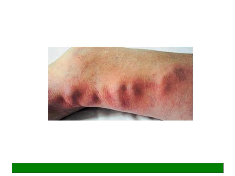
What’s this called? Phlegmasia cerulea dolens
(Phlegmasia alba dolens = white leg, more severe) Massive DVT causing obstruction and severe pain. Call vascular surgery. NICE: Consider catheter-directed thrombolytic therapy /embolectomy for patients with symptomatic iliofemoral DVT who have: symptoms of less than 14 days’ duration and good functional status and a life expectancy of 1 year or more and a low risk of bleeding. Phlegmasia stems from a Greek term (phlegma) meaning inflammation. It has been used in the medical literature in reference to extreme cases of lower-extremity deep venous thrombosis (DVT) that cause critical limb ischemia and possible limb loss. Phlegmasia alba dolens (PAD) describes the patient with swollen and white leg because of early compromise of arterial flow secondary to extensive DVT. This condition is also known as “milk leg,” especially as it affects women in the third trimester of pregnancy or post partum. Phlegmasia cerulea dolens (PCD) is more advanced and considered a precursor of frank venous gangrene. It is characterised by severe swelling and cyanosis and blue discoloration of the extremity. It was first described by Hildanus in the 16th century. Later, the term PCD was first used by Gregoire in Its rarity notwithstanding, phlegmasia is a life-threatening condition. It is crucial for nonvascular specialists to be able to recognise this condition promptly and accurately; treatment is time-sensitive.
 Massive DVT causing obstruction and severe pain. Call vascular surgery. NICE: Consider catheter-directed thrombolytic therapy /embolectomy for patients with symptomatic iliofemoral DVT who have: symptoms of less than 14 days’ duration and. good functional status and. a life expectancy of 1 year or more and. a low risk of bleeding. Phlegmasia stems from a Greek term (phlegma) meaning inflammation. It has been used in the medical literature in reference to extreme cases of lower-extremity deep venous thrombosis (DVT) that cause critical limb ischemia and possible limb loss. Phlegmasia alba dolens (PAD) describes the patient with swollen and white leg because of early compromise of arterial flow secondary to extensive DVT. This condition is also known as milk leg, especially as it affects women in the third trimester of pregnancy or post partum. Phlegmasia cerulea dolens (PCD) is more advanced and considered a precursor of frank venous gangrene. It is characterised by severe swelling and cyanosis and blue discoloration of the extremity. It was first described by Hildanus in the 16th century. Later, the term PCD was first used by Gregoire in Its rarity notwithstanding, phlegmasia is a life-threatening condition. It is crucial for nonvascular specialists to be able to recognise this condition promptly and accurately; treatment is time-sensitive.")
26
Groupthink (‘that’s the way we do things around here’)
Common cognitive biases in the diagnosis and management of lower limb DVT Groupthink (‘that’s the way we do things around here’) Psych-out error and visceral bias (IVDUs) Omission bias (the tendency towards inaction, rooted in the principle of ‘first do no harm’ e.g. radiation, anxiety/harm from further Ix, explanation and treatment) … Many depts do not have a structured DVT service with good guidelines that are followed It seems overkill to scan and refer to breast clinic all pts over 40 with unprovoked DVT?? Several recent studies addressed the question whether thromboembolism is a marker for an unrecognised (occult) or subsequent malignancy, reporting a 4–10% prevalence of unrecognised malignancy in patients with idiopathic (unprovoked) DVT and a lower prevalence in patients with secondary DVT (with known risk factors). The prevalence of unrecognised malignancy varied considerably between studies, and was at least partly attributable to the number and type of routine examinations performed to detect malignancies and the characteristics of the included patients.
 Psych-out error and visceral bias (IVDUs) Omission bias (the tendency towards inaction, rooted in the principle of ‘first do no harm’ e.g. radiation, anxiety/harm from further Ix, explanation and treatment) … Many depts do not have a structured DVT service with good guidelines that are followed. It seems overkill to scan and refer to breast clinic all pts over 40 with unprovoked DVT Several recent studies addressed the question whether thromboembolism is a marker for an unrecognised (occult) or subsequent malignancy, reporting a 4–10% prevalence of unrecognised malignancy in patients with idiopathic (unprovoked) DVT and a lower prevalence in patients with secondary DVT (with known risk factors). The prevalence of unrecognised malignancy varied considerably between studies, and was at least partly attributable to the number and type of routine examinations performed to detect malignancies and the characteristics of the included patients.")
27
Any questions at this point?

28
Scenario 4 A 54 year old female smoker was admitted with gradually worsening breathlessness over the last 10 days. She reported a cough but no obvious fever or discoloured sputum. Her past medical history included a DVT 5 years previously. Her chest X-ray showed some patchy consolidation at the left base. She was started on treatment for community acquired pneumonia in ED.

29
Reported as: left basal atelectasis and possible left pleural effusion
Reported as: left basal atelectasis and possible left pleural effusion. Infiltrate cannot be ruled out. In the 1992 PIOPED study (prospective Ix in PE diagnosis), findings that were most common in PE were atelectasis and/or increased opacity in lower lung parenchyma, and pleural effusions. Easy to confuse with “pneumonia”. PEs also cause inflammation: CRP/wbc/fever so there can be a “grey area” between obvious pneumonias and obvious PEs. Would you do a d dimer in this case?? Now, in this case a d dimer was already done (routine at the front door of ED without history/Wells) and was 19,000. Comments please … We talk about a raised d dimer being non-diagnostic. Hmm … maybe not as simple as that. In a study comparing the quantitative D-dimer levels to the presence of PE in symptomatic patients, PE prevalence was strongly related to the D-dimer level and increased 4 times with D-dimer levels >4000 ng FEU/ml compared with levels between 500 and 1000 ng FEU/ml. In a study performed in an out-patient setting in 671 patients with clinically suspected PE, the specificity of a D-dimer test was 93% when D-dimer levels exceeded 4000 μg FEU/l. However, in the presence of intermediate and high clinical probability, this resulted in a limited positive predictive value because of the relatively low PE incidence (20%).[29 ] Thus, high D-dimer levels upon presentation may potentially prompt a more intense diagnostic approach, irrespective of pre-test probability…
, findings that were most common in PE were atelectasis and/or increased opacity in lower lung parenchyma, and pleural effusions. Easy to confuse with pneumonia . PEs also cause inflammation: CRP/wbc/fever so there can be a grey area between obvious pneumonias and obvious PEs. Would you do a d dimer in this case Now, in this case a d dimer was already done (routine at the front door of ED without history/Wells) and was 19,000. Comments please … We talk about a raised d dimer being non-diagnostic. Hmm … maybe not as simple as that. In a study comparing the quantitative D-dimer levels to the presence of PE in symptomatic patients, PE prevalence was strongly related to the D-dimer level and increased 4 times with D-dimer levels >4000 ng FEU/ml compared with levels between 500 and 1000 ng FEU/ml. In a study performed in an out-patient setting in 671 patients with clinically suspected PE, the specificity of a D-dimer test was 93% when D-dimer levels exceeded 4000 μg FEU/l. However, in the. presence of intermediate and high clinical probability, this resulted in a limited positive predictive value because of the relatively low PE incidence (20%).[29 ] Thus, high D-dimer levels upon presentation may potentially prompt a more intense diagnostic approach, irrespective of pre-test probability…")
30
CT or V/Q scan? CTPA Preferred – simple positive/negative result, can visualise other lung pathology, RV dysfunction and proximal leg veins 90% sensitivity, 95% specificity - a negative CTPA essentially rules out PE However, high sensitivity –> increased incidence of PE and probably diagnosing unimportant PEs V/Q Recommended in young/pregnant patients with normal CXRs and no history of lung disease (e.g. asthma) to reduce radiation Role in patients who cannot have contrast Different sensitivity and specificity figures in literature - non-diagnostic scans with high clinical suspicion still need CTPA Much more sensitive than CTPA in diagnosing chronic PEs (e.g. Ix of pulmonary hypertension) Would you treat a small peripheral PE in a 90 year old lady with dementia living in a nursing home?? (No? Might want to know if the prox leg veins showed DVTs on imaging first). A recent study examined the radiation dose of CTPA for women undergoing a single 64-slice multidetector CTPA procedure. The estimated exposure was mSV. This was estimated to increase the risk of breast cancer by to and lung cancer from to The excess risk of cancer for individuals over 55 would be less than 1%; however, for a young 20-year-old woman this would be estimated to increase the relative lifetime risk of breast or lung cancer by 1.7 to 5.5%. Although investigators have acknowledged the greater accuracy of CTPA, a number of recent studies have attempted to better define a potential role for V/Q scanning for the diagnosis of pulmonary embolism. The PIOPED II investigators reanalysed V/Q scan results using only the perfusion images and two distinct sets of diagnostic criteria.[18] Using a modification of the PIOPED II criteria, the sensitivity of lung scanning improved to 85%, the specificity declined to 93% and the nondiagnostic scan rate result was reduced to 21%. NICE: Although CTPA has the advantage of being more sensitive and specific than V/Q scans which also have a higher non-diagnostic rate, V/Q scans may be the preferred option for some patients. The radiation exposure from V/Q scans is approximately equivalent to 8 months of natural background radiation (UK average 2.2 mSv per year), and significantly lower than CTPA scan208. Unlike CTPA, V/Q scans do not require the use of contrast media and should be offered to patients with a history of allergy to contrast media. This is also an option for patients at risk of further renal injury from contrast media e.g. patients with severe renal impairment.
 to reduce radiation. Role in patients who cannot have contrast. Different sensitivity and specificity figures in literature - non-diagnostic scans with high clinical suspicion still need CTPA. Much more sensitive than CTPA in diagnosing chronic PEs (e.g. Ix of pulmonary hypertension) Would you treat a small peripheral PE in a 90 year old lady with dementia living in a nursing home (No Might want to know if the prox leg veins showed DVTs on imaging first). A recent study examined the radiation dose of CTPA for women undergoing a single 64-slice multidetector CTPA procedure. The estimated exposure was mSV. This was estimated to increase the risk of breast cancer by to and lung cancer from to The excess risk of cancer for individuals over 55 would be less than 1%; however, for a young. 20-year-old woman this would be estimated to increase the relative lifetime risk of breast or lung cancer by 1.7 to 5.5%. Although investigators have acknowledged the greater accuracy of CTPA, a number of recent studies have attempted to better define a potential role for V/Q scanning for the diagnosis of pulmonary embolism. The PIOPED II investigators reanalysed V/Q scan results using only the perfusion images and two distinct sets of diagnostic criteria.[18] Using a modification of the PIOPED II criteria, the sensitivity of lung scanning improved to 85%, the specificity declined to 93% and the nondiagnostic scan rate result was reduced to 21%. NICE: Although CTPA has the advantage of being more sensitive and specific than V/Q scans which also have a higher non-diagnostic rate, V/Q scans may be the preferred option for some patients. The radiation exposure from V/Q scans is approximately equivalent to 8 months of natural background radiation (UK average 2.2 mSv per year), and significantly lower than CTPA scan208. Unlike CTPA, V/Q scans do not require the use of contrast media and should be offered to patients with a history of allergy to contrast media. This is also an option for patients at risk of further renal injury from contrast media e.g. patients with severe renal impairment.")
31
Sensitivity and specificity of V/Q scan (after removing non-diagnostic scans from the analysis)
PE absent Non-diagnostic scan PE present Normal/very low probability Low/intermediate probability High probability Modified PIOPED ll Scintigraphic Criteria (V/Q result compared with DSA or CTPA + Wells) Around 25% pts have non-diagnostic scans with V/Q compared with only 6% CTPA – and CTPA is mostly non-diagnostic due to “technically inadequate scans”. Low clinical prob and PE absent scan no PE Sensitivity 77.4% Specificity 97.7% The % of patients with a PE absent or PE present scan was 73.5%
 Around 25% pts have non-diagnostic scans with V/Q compared with only 6% CTPA – and CTPA is mostly non-diagnostic due to technically inadequate scans . Low clinical prob and PE absent scan no PE. Sensitivity 77.4% Specificity 97.7% The % of patients with a PE absent or PE present scan was 73.5%")
32
Diagnosing PE: things to consider
Patients required respiratory OP follow up (not small peripheral PEs) Over 40’s with idiopathic PEs require cancer screening same as DVTs Duration of anti-coagulation same as DVTs Thrombolysis is considered more often in PE – you need to know the criteria Investigation is different in pregnant patients: bilateral leg Doppler USS first. (Treatment and follow-up for VTE in pregnancy is different too)* Treatment is different in pregnant patients too for both DVT and PE – 1mg/kg BD clexane, factor Xa level monitoring and urgent referral to joint haem/obstetric clinic. No warfarin.
 Over 40’s with idiopathic PEs require cancer screening same as DVTs. Duration of anti-coagulation same as DVTs. Thrombolysis is considered more often in PE – you need to know the criteria. Investigation is different in pregnant patients: bilateral leg Doppler USS first. (Treatment and follow-up for VTE in pregnancy is different too)* Treatment is different in pregnant patients too for both DVT and PE – 1mg/kg BD clexane, factor Xa level monitoring and urgent referral to joint haem/obstetric clinic. No warfarin.")
33
How do you decide whether a patient can be investigated and treated for PE in Ambulatory Care?
Simplified PESI score plus troponin (see next slide) From a meta-analysis published in Circulation. 2007;116: Prognostic Value of Troponins in Acute Pulmonary Embolism: A Meta-Analysis. Becattini et al. doi: /CIRCULATIONAHA "Overall, 122 of 618 patients with elevated troponin levels died (19.7%; 95% CI, 16.6 to 22.8) compared with 51 of 1367 with normal troponin levels (3.7%; 95% CI, 2.7 to 4.7). High levels of troponins, both I and T, were associated with a high risk of short-term death (OR, 5.24; 95% CI, 3.28 to 8.38), with no evidence for overall heterogeneity (Figure 2). The result was consistent for either troponin I (OR, 4.01; 95% CI, 2.23 to 7.23) or troponin T (OR, 7.95; 95% CI, 3.79 to 16.65). "Seven studies (915 patients), all with a prospective design, included only patients with normal blood pressure at hospital admission. The incidence of death was 17.9% (34 of 190; 95% CI, 12.4 to 23.3) in patients with elevated troponin levels and 2.3% (17 of 725; 95% CI, 1.2 to 3.4) in patients with normal troponin levels. The pooled analysis of these studies showed an association between high levels of serum troponins and mortality (OR, 5.90; 95% CI, 2.68 to 12.95), with no evidence for heterogeneity." So, as well as the simplified PESI score, it looks like we should look for a normal troponin result as well when deciding if someone can be treated in Ambulatory Care for their PE. However, another article did point out that troponins can be raised in lots of other conditions, so take care to use troponin to risk assess people with actual PEs, which was the case in this meta-analysis!
 From a meta-analysis published in Circulation. 2007;116: Prognostic Value of Troponins in Acute Pulmonary Embolism: A Meta-Analysis. Becattini et al. doi: /CIRCULATIONAHA Overall, 122 of 618 patients with elevated troponin levels died (19.7%; 95% CI, 16.6 to 22.8) compared with 51 of 1367 with normal troponin levels (3.7%; 95% CI, 2.7 to 4.7). High levels of troponins, both I and T, were associated with a high risk of short-term death (OR, 5.24; 95% CI, 3.28 to 8.38), with no evidence for overall heterogeneity (Figure 2). The result was consistent for either troponin I (OR, 4.01; 95% CI, 2.23 to 7.23) or troponin T (OR, 7.95; 95% CI, 3.79 to 16.65). Seven studies (915 patients), all with a prospective design, included only patients with normal blood pressure at hospital admission. The incidence of death was 17.9% (34 of 190; 95% CI, 12.4 to 23.3) in patients with elevated troponin levels and 2.3% (17 of 725; 95% CI, 1.2 to 3.4) in patients with normal troponin levels. The pooled analysis of these studies showed an association between high levels of serum troponins and mortality (OR, 5.90; 95% CI, 2.68 to 12.95), with no evidence for heterogeneity. So, as well as the simplified PESI score, it looks like we should look for a normal troponin result as well when deciding if someone can be treated in Ambulatory Care for their PE. However, another article did point out that troponins can be raised in lots of other conditions, so take care to use troponin to risk assess people with actual PEs, which was the case in this meta-analysis!")
34
Simplified PE Severity Index (PESI) Score
How do you know if the patient can be treated in Ambulatory Care?? Google simplified PESI score and work out using MedCalc The sPESI is only meant for risk stratification of pulmonary embolism after the diagnosis has been made (probable diagnosis awaiting scan counts). The sPESI can not be used to risk stratify patients who are not being treated for PE. Additional pathology which could lead to morbidity or mortality should not be overlooked in the setting of low risk sPESI scores. Patients with a score of 0 are determined to be low risk, while those with a score of 1 or more are considered high risk
. The sPESI can not be used to risk stratify patients who are not being treated for PE. Additional pathology which could lead to morbidity or mortality should not be overlooked in the setting of low risk sPESI scores. Patients with a score of 0 are determined to be low risk, while those with a score of 1 or more are considered high risk.")
35
PE: summary ?PE history, exam and Wells Score
If PE suspected plus Wells = likely then image the chest If PE suspected plus Wells = unlikely then do d dimer If the d dimer is raised, remember that it is NOT diagnostic, it just means imaging is required to exclude PE If d dimer is negative then PE is virtually excluded For all cases, if you can’t get imaging within 4 hours then treat and get a scan within 24 hours Consider and communicate alternative explanations for symptoms in patients with negative scans Don’t forget to arrange further investigations/FU for new PE cases

36
Common cognitive biases in the diagnosis and management of PE
Misinterpretation of diagnostic tests (d dimer, V/Q) Knowledge gaps (e.g. different Ix and management in pregnancy) Representativeness, premature closure, search satisficing, confirmation bias ..?
 Knowledge gaps (e.g. different Ix and management in pregnancy) Representativeness, premature closure, search satisficing, confirmation bias ..")
37
Any questions at this point?

38
Scenario 5 A 75-year-old woman was sent to Ambulatory Care for investigation of a raised d dimer (900 μg FEU/L). She had been admitted two weeks previously with pleurisy and had a negative CTPA. On discharge the Practice Nurse repeated the d dimer test to see if it was still raised and referred the patient back to Acute Medicine to ‘find the clot’. The patient’s past medical history consisted of: COPD, heart failure, hypertension. On examination the patient was well with no new symptoms and had mild bilateral lower leg oedema. Other d dimer stories …
. She had been admitted two weeks previously with pleurisy and had a negative CTPA. On discharge the Practice Nurse repeated the d dimer test to see if it was still raised and referred the patient back to Acute Medicine to ‘find the clot’. The patient’s past medical history consisted of: COPD, heart failure, hypertension. On examination the patient was well with no new symptoms and had mild bilateral lower leg oedema. Other d dimer stories …")
39
Correct interpretation of d dimer
Ginsberg and colleagues found that the combination of a low pre-test probability and a normal d dimer had a negative predictive value of 99%, whereas the negative predictive value was only 78% in patients with a high pre-test probability and a normal d dimer A rule of thumb is that a ‘positive’ d dimer is non-diagnostic However … should a d dimer of 900 be interpreted the same as a d dimer of 10,000? The starting point for interpretation of any d dimer result has to be: does this patient have symptoms and signs of DVT/PE?? An analysis reported no clear advantage of measuring D-dimer after the age of 80, except when the risk of performing CT was too high due to impaired renal function. In a study by Legnani et al, the authors highlighted the different age-specific, quantitative D-dimer cutoff values in patients aged 70 years or less and older than 70 years, respectively: VIDAS ELISA = 600 ng FEU/ml and 1200 ng FEU/ml, Innovance = 500 ng/ml and 900 ng/ml NP, Hemosil HS D-dimer (Instrumentation Laboratories) = 250 ng DDU/ml and 450 ng DDU/ml, and STA Liatest (Stago) = 700 ng FEU/ml and 1000 ng FEU/ml. In a pooled analysis involving three prospective studies, normal D-dimer levels along with LCP effectively excluded DVT in elderly patients. In a study comparing the quantitative D-dimer levels to the presence of PE in symptomatic patients, PE prevalence was strongly related to the D-dimer level and increased 4 times with D-dimer levels >4000 ng FEU/ml compared with levels between 500 and 1000 ng FEU/ml. In a study performed in an out-patient setting in 671 patients with clinically suspected PE, the specificity of a D-dimer test was 93% when D-dimer levels exceeded 4000 μg FEU/l. However, in the presence of ICP and HCP, this resulted in a limited positive predictive value because of the relatively low PE incidence (20%).[29 ] Thus, high D-dimer levels upon presentation may potentially prompt a more intense diagnostic approach, irrespective of PTP. In a study of 1818 patients, the risk of recurrent VTE was higher among patients with positive D-dimer levels than those with negative results during a follow-up period of 26.9 months after an unprovoked VTE. This was independent of patient age or timing of the postanticoagulation D-dimer testing.
 = 250 ng DDU/ml and 450 ng DDU/ml, and STA Liatest (Stago) = 700 ng FEU/ml and 1000 ng FEU/ml. In a pooled analysis involving three prospective studies, normal D-dimer levels along with LCP effectively excluded DVT in elderly patients. In a study comparing the quantitative D-dimer levels to the presence of PE in symptomatic patients, PE prevalence was strongly related to the D-dimer level and increased 4 times with D-dimer levels >4000 ng FEU/ml compared with levels between 500 and 1000 ng FEU/ml. In a study performed in an out-patient setting in 671 patients with clinically suspected PE, the. specificity of a D-dimer test was 93% when D-dimer levels exceeded 4000 μg FEU/l. However, in the presence of ICP and HCP, this resulted in a limited positive predictive value because of the relatively low PE incidence (20%).[29 ] Thus, high D-dimer levels upon presentation may potentially prompt a more intense diagnostic approach, irrespective of PTP. In a study of 1818 patients, the risk of recurrent VTE was higher among patients with positive D-dimer levels than those with negative results during a follow-up period of 26.9 months after an unprovoked VTE. This was independent of patient age or timing of the postanticoagulation D-dimer testing.")
40
Any questions at this point?

41
Summary of Guidelines and MCQs
See NICE guideline handout Recap upper limb DVT, superficial thrombophlebitis, leg DVT, PE, PE in pregnancy, interpreting “random” d dimers. MCQs on paper

42
Read strategically! www.internalmedicineteaching.org
When learners (and that includes you and me) read about their patient’s problems in context (for example, after seeing the patient that day), this promotes conceptualisation rather than memorisation and textbook knowledge is organised in a way that is more likely to be recalled. 20 mins reading after seeing a case is far more valuable than simply reading a textbook at random times.
 read about their patient’s problems in context (for example, after seeing the patient that day), this promotes conceptualisation rather than memorisation and textbook knowledge is organised in a way that is more likely to be recalled. 20 mins reading after seeing a case is far more valuable than simply reading a textbook at random times.")
Similar presentations


 in a deep vein, usually in the legs, which partially or completely.>")


:>")

 around valves propagation Virchow’s triad.>")
>")


