Download presentation
Presentation is loading. Please wait.

1
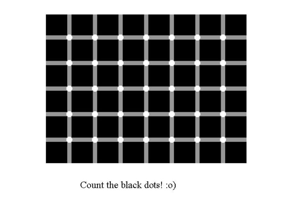
What do you see?

3
Do you see gray areas in between the squares? Now where did they come from?

4
Disappearing Pink Circles http://www.dougmoran.com/collections/optical-illusion-pink-dot-circle.html

5
Why is this happening? Are we seeing what we are supposed to be seeing?

6
Recall Images are formed on the light-sensitive layer of the eye. What is it? Retina

7
How did the Retina do it?

8
In the Retina… ContainsPhotoreceptors

9
Consists of Photoreceptors (light sensitive cells) RodsConesCones
 RodsConesCones")
10
Contain a pigment called visual purple. Allows them to be sensitive to dim light but only able to see black and white.

11
Rods Formation of visual purple requires: The lack of Vitamin A may cause night-blindness.

12
ConesCones 3 Types of Cones: Red Green Blue Each cone contains a different pigment which absorbs light of different wavelengths. All cones work together to allow us to see a variety of colour. Cones do not work well in dim light (associated with bright light and colour vision.

13
Recall There are 2 specialised spots on the retina. What are they? Fovea (Yellow Spot) Blind Spot
 Blind Spot")
14
Fovea (Yellow Spot) Blind Spot
 Blind Spot")
15
Fovea (Yellow Spot) Yellow depression in the retina (most central part of the retina) where images are sharply focused. Contains the greatest concentration of cones but no rods. Enables one to have detailed colour vision in bright light.

16
Blind Spot Region where the optic nerve leaves the eye. Does not contain any rods or cones. (no receptor) Not sensitive to light.
 Not sensitive to light..")
17
How does the light get focused on the retina?

18
Recall PHYSICS!!!

19
Ray Diagram

20
Focus of Image in the Eye

21
Characteristic of the Image on the Retina Upside down (inverted) Laterally Inverted Smaller in size than actual object (diminished)
 Laterally Inverted Smaller in size than actual object (diminished)")
22
But why do we see the objects as it is?

23
Recall Where is the image sent after it forms on the retina?

24
The Role of the Brain in Vision The Brain interprets the image so that objects may be seen to be the right way up, front to back, and the right size.

25
Light Transmission Process http://www.kscience.co.uk/animations/eye.swf

26
How the eye produces a focused image Light rays from an ______enter the transparent cornea. The cornea ______ the light rays towards the pupil. The light rays pass through the ______and the ________ humour. The _____ refracts the rays to focus on the retina. The light-sensitive cells on the ______ are stimulated by the light of the image, and convert the _____energy into _________ energy. Electrical energy, in the form of nerve_________, travel along the __________ to the brain. The brain decodes the impulses to produce the sensation of_______. object refracts pupil aqueous lens retina light electrical impulses optic nerve sight

27
Does this mean that our brain will process an out-of- focus image?

28
What we learn today. Photoreceptors (rods and cones) Functions of Rods and Cones The Fovea (Yellow Spot) and Blind Spot Image formation in the eye The role of the brain in image interpretation
 Functions of Rods and Cones The Fovea (Yellow Spot) and Blind Spot Image formation in the eye The role of the brain in image interpretation.")
Similar presentations


 17 April 2017 Biology Matters textbook page 281 Concept Map.>")


 McGraw Hill Ryerson 2007 6.1 Human Vision The pupil is the dark transparent region in the centre of the eye where light enters. The iris is the coloured.>")