Download presentation
Presentation is loading. Please wait.

2
DNA and Cell Division

3
Mitosis in Animals

4
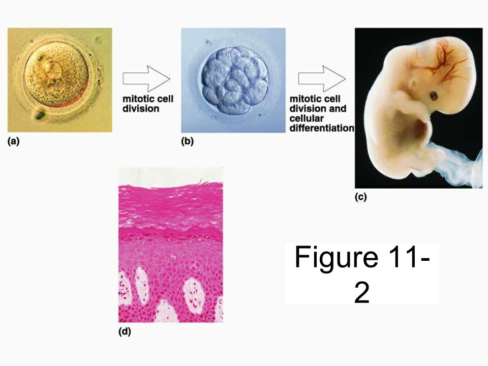
Background Information Once an egg becomes fertilized, cellular divisions begins, eventually producing a whole organism An Integrated Organism All cells derived from the zygote contain the same genetic material

5
Organization of DNA All cells have DNA (chromosomes). Almost all cells divide for reproduction, growth or repair. Each new cell needs the exact same DNA as the original cell. The original cell is called the mother cell and the two new cells are called daughter cells. The DNA in the nucleus must replicate before the cell divides.

6
Common NameGenus and Species Diploid Chromosome Number BuffaloBison bison60 CatFelis catus38 CattleBos taurus, B. indicus60 DogCanis familiaris78 DonkeyE. asinus62 GoatCapra hircus60 HorseEquus caballus64 HumanHomo sapiens46 PigSus scrofa38 SheepOvis aries54 Chromosome Number in Different Species

7
Number of genes in sequenced genomes E. coli4300 Yeast6000 Roundworm18,600 Fruit fly13-14,000 Mosquito13-14,000 Mouse30-35,000 Human30-35,000

9
A non-dividing cell: 90% of a cell’s life is spent growing, not dividing This phase is called interphase The DNA in this phase is not condensed; thus is chromatin At some point during this phase the DNA is doubled or replicated Two copies are made, one for each of the new cells

10
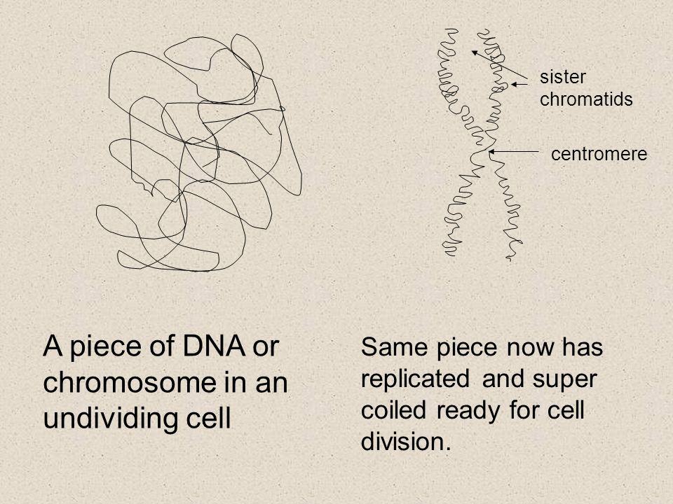
Once replication occurs, the chromatin folds up to form chromosomes This only occurs when the cell is about to divide The duplicated chromosomes attach to each other at the centromere Each individual copy of one chromosome is known as a chromatid When chromatids are joined at the centromere, they are known as a single chromosome.

12
A piece of DNA or chromosome in an undividing cell Same piece now has replicated and super coiled ready for cell division. sister chromatids centromere

14
Also during interphase… Additional organelles are produced Cell membrane enlarged to allow cell growth When the cell becomes too big to function it must divide –What would the SA/V ratio of this cell be…large or small?

15
Interphase The cell grows New organelles are formed Duplicate chromosomes are produced The chromosomes are uncoiled and invisible This uncoiled chromosomes are known as a chromatin

16
Cell division/reproduction Interphase is not considered to be part of cell reproduction. It is simply the growth of the cell and the duplication of the chromosomes. Cell reproduction consists of two separate stages known as mitosis and cytokinesis. Cell division = mitosis + cytokinesis A parent cell will produce 2 daughter cells.

17
In cell division each chromosome is replicated and then the cell (and nucleus) divides
 divides")
18
Cell Reproduction Original cell divides into two genetically identical daughter cells Complete set of genetic information passed onto each daughter cell –DNA must be accurately duplicated before cell division Mitosis: paired chromatids separate and move to opposite ends of the cell Cytokinesis: cytoplasm + organelles divide into roughly equal halves

19
http://www.stolaf.edu/people/gia nnini/flashanimat/celldivision/cro me3.swf Mitosis Animation

20
MITOSIS/ CELL DIVISION Stages include: Prophase Metaphase Anaphase Telophase

22
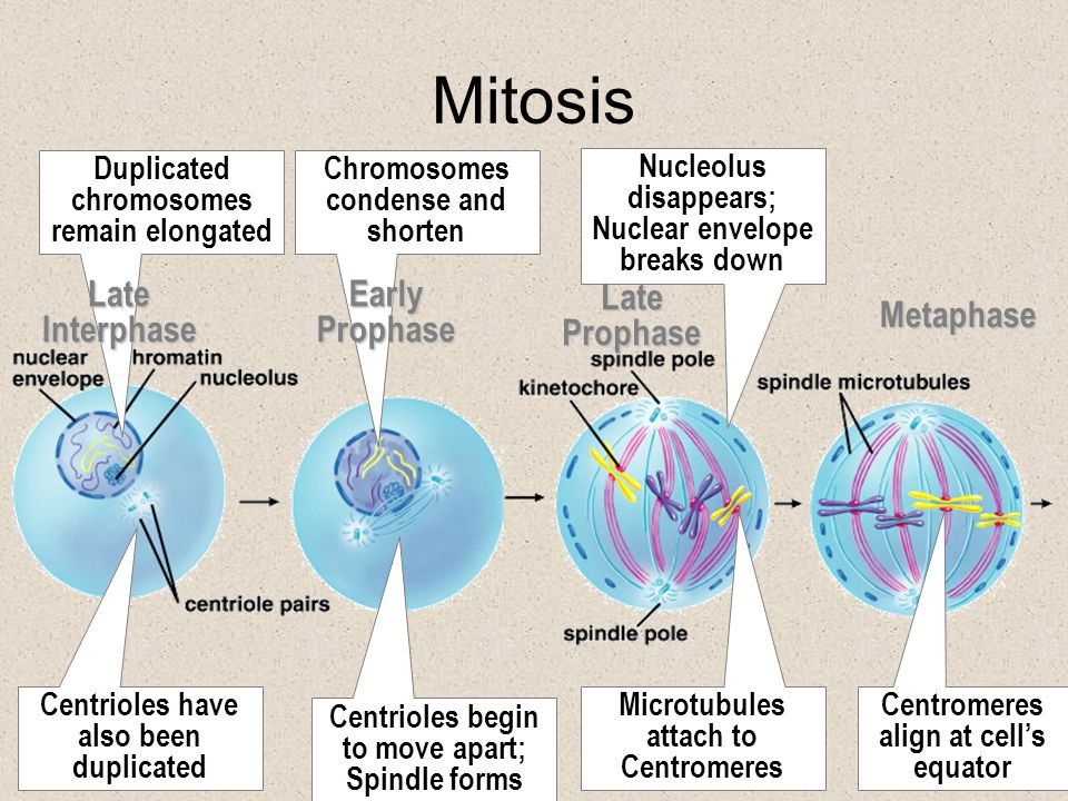
Mitosis Centromeres align at cell’s equator Nucleolus disappears; Nuclear envelope breaks down Microtubules attach to Centromeres Chromosomes condense and shorten Centrioles begin to move apart; Spindle forms Duplicated chromosomes remain elongated Centrioles have also been duplicated Late Interphase Early Prophase Late Prophase Metaphase

23
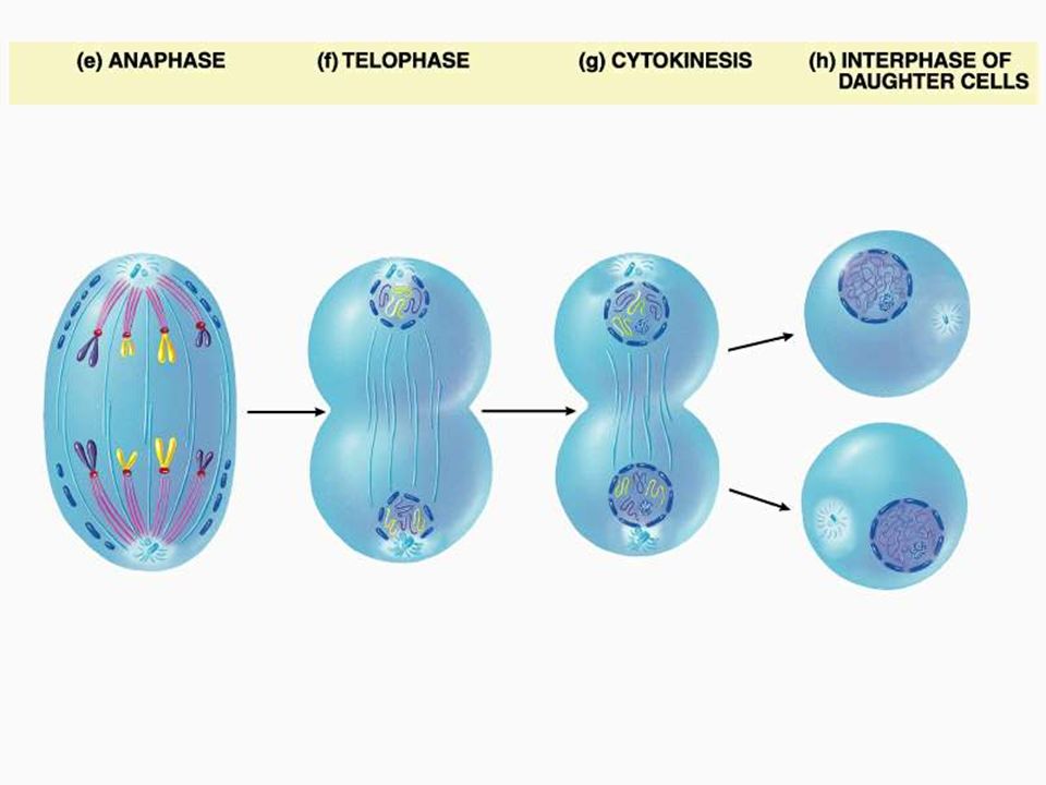
Mitosis: Spindle fibers pull chromatids to opposite poles Chromatids become independent chromosomes Chromosomes begin unwinding Nuclear envelope re- forms, spindle fibres disappear Cytoplasm divided along equator Each daughter gets 1 nucleus & half of cytoplasm AnaphaseTelophase Cytokinesis Next Interphase

26
Prophase Chromatin condenses (into chromosomes) Chromosomes appear as “X”s. Nuclear envelope dissolves (DNA free in cell) Animals cells only: Centrioles move to opposite ends of cell + form spindle fibers Centromere attaches to spindle fibre
 Animals cells only: Centrioles move to opposite ends of cell + form spindle fibers Centromere attaches to spindle fibre.")
27
Metaphase Each chromosome lines up in the middle of the cell. Highly organized so that both cells get exactly the same DNA. Spindle fibers attached to centromeres of chromosomes

28
Anaphase Each pair of chromatids splits at the centromere Each chromatid is now an individual chromosome Paired chromosomes are pulled to opposite ends by spindle fibres

29
Telophase Chromosomes end up at separate poles, spindle fibers begin to dissolve. New nuclear envelope begins to form around chromosomes chromosomes begin to uncoil Cell starts to pinch off through cytokinesis

30
Cytokinesis Division of all the rest of cell parts but not equally (organelles) Animals: cell membrane pinches to form two cells Plants: new cell plate created between the two cells (becomes cell wall)
 Animals: cell membrane pinches to form two cells Plants: new cell plate created between the two cells (becomes cell wall)")
31
The Cell Cycle: An Overview 1)Interphase 2)Mitosis a)Prophase b)Metaphase c)Anaphase d)Telophase 3)Cytokinesis Cell Division (Cell Reproduction) = mitosis + cytokinesis
Interphase 2)Mitosis a)Prophase b)Metaphase c)Anaphase d)Telophase 3)Cytokinesis Cell Division (Cell Reproduction) = mitosis + cytokinesis")
32
Use an Acronym For Mitosis: Prophase = P Metaphase = M Anaphase = A Telophase = T Make a sentence: Please Meet At Ten Phil, Mary, And Tom

35
Figure 11- 2

38
Links http://en.wikipedia.org/wiki/Trisomy#Triso myhttp://en.wikipedia.org/wiki/Trisomy#Triso my http://www.medgen.ubc.ca/wrobinson/mos aic/mos_how.htm

39
Mitosis Drawing Fold a large sheet of paper in 3 –You should have 3 columns on the front and 3 on the back You will draw a cell in each stage of mitosis and include a written description of what is occurring at each stage Stages: Interphase, Prophase, Metaphase, Anaphase, Telophase, Cytokinesis Notice that there are 6 stages and 6 coulmns on your sheet...1 stage per column!

40
Some facts 500,000 deaths per year (more males) Older age group stricken more often More than 100 types of cancer, many due to mutations triggered by environmental factors Highest cancer incidence: male - prostate female - breast Highest cancer deaths: lung
 Older age group stricken more often More than 100 types of cancer, many due to mutations triggered by environmental factors Highest cancer incidence: male - prostate female - breast Highest cancer deaths: lung")
41
Normal cells in culture Organized structure Limited cell growth No overlapping

42
Cancer cells in culture Disorganized Overlapping structure Uncontrolled cell growth

43
Some images to make this real: look first at normal skin

44
Cancerous Skin

45
What causes a normal cell to become a cancer cell?

Similar presentations


 A.)>")


