Download presentation
Presentation is loading. Please wait.

1
Trauma Spring 2011 FINAL

2
Some Trauma Stats 1.Most common cause of death for those 1.1-44 years of age 2.Medical costs for trauma 1.200 billion annually 3. Mostly results from MVA, unintentional accidents, gunshot wounds, stabbing, fights, domestic violence

3
Trimodal Distribution Immediate Early Late

4
Immediate Deaths Lacerations of the _________________ Lacerations of the _________________

5
Early Deaths 1.Within first __ hours 2._______hemorrhage 3.Lacerations of _____or _________ 4.Significant ____ loss Liver laceration with extravasation

6
Late Deaths 1.________after injury 2.____________ and ______ ____ failure

7
Level I, II & III Trauma Centers 1.Level 1 1.Usually in _____ metro areas and serve as both primary and tertiary care institutions 2.Must be avail _____ 3.Must treat ______admissions or ______major trauma patients per year 1.Level II 1.__________to level I when necessary 2.Serve ________cites and towns 3.Must be avail ___ hrs 2.Level III 1.__________&______ 2.______________ on nights and weekends

8
Skeletal Trauma

9
Fracture Classifications

10
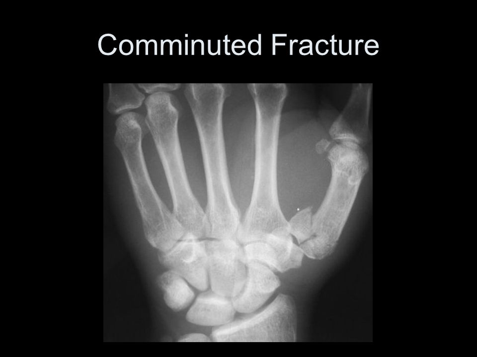
FRACTURE TYPES

11
_____________ reduction

12
__________ Reduction

13
_________ FRACTURES

14
Open Fracture 1.Bone has _____________ skin 2.May lead to infection 3.Precautions must be taken to _______ ___________from setting into the bone

15
Closed Fracture 1.__________ is not penetrated 2.Fractures can be classified by the _______ of the stress that caused the break 1.________ 2.________ 3.________

16
16 Closed Fracture- Clavicle

17
Forearm Closed fracture

18
____________Fracture- Wrist When the fractured bone is ________into the cancellous tissue of another fragment

19
Impacted Fracture- Hip

20
Fibular Impacted Fracture

21
Comminuted Fracture 1.Do not represent the full thickness of the bone. 2.Usually extensively ________________ 3.Particularly apt to be open fractures

22
Comminuted Fracture

24
Non-Comminuted Fracture

25
1._________ fracture in which the bone is separated into to fragments 2.Can be classified according to the direction of its fracture line 1.______________ 2.______________

26
________________ Fracture 1.Fragment of the bone is __________ from the shaft 2.Occur around the joints because of ligaments, tendons, muscles, associated with sprain or dislocation

27
Avulsion Fracture

29
Incomplete Fracture 1.Part of bony structure gives way with ________or no ________________ 1.Common example is a _________ fracture 2.Torus fracture

30
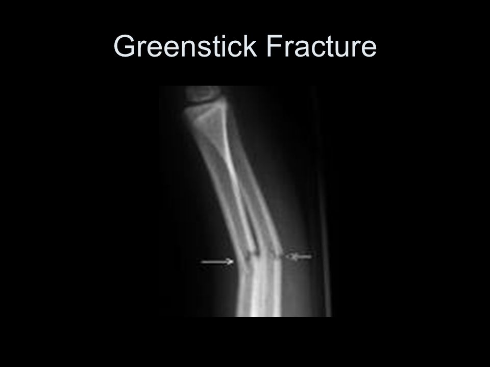
Greenstick :Incomplete Fracture 1.Cortex breaks on one side without separation or breaking of the opposite cortex 2.Found almost exclusively in children under the age of 10

31
Incomplete Fracture

32
Greenstick Fracture

35
________: Incomplete Fracture 1.AKA _____ Fracture 2.It is a greenstick fracture 3.Cortex bulges _______producing a slight irregularity

36
Torus Fracture

37
Growth Plate Fracture 1.Involve the end of the long bone 2.Not visible unless displacement occurs 3.Classified according to severity 1.____________________ 1.I-IV 2.Based on degree of epiphysis involvement

38
Growth Plate Fracture

40
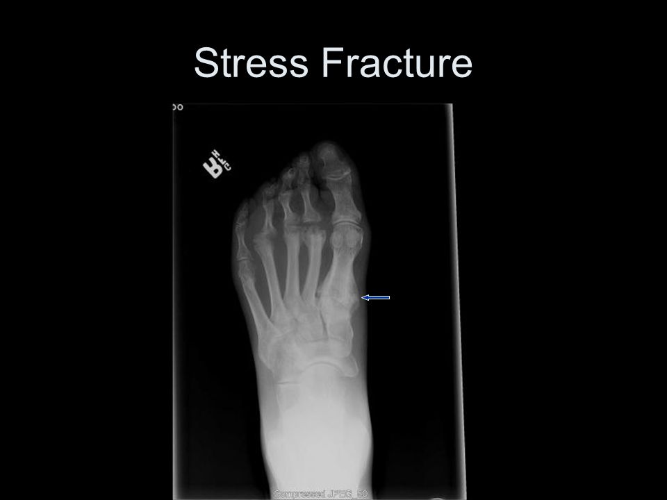
_____________ Fracture 1.Results from an _________degree of repetition 2.Generally found where __________ attachments are 1.EX: runners at tib/fib 3.Not always seen on plain x-ray

41
Stress Fracture

43
Occult Fracture 1.Gives ______________ without radiologic evidence 2.____ days later may show repairing itself or displacement

44
Occult Fracture

46
Colles Fracture 1.Fracture through distal inch of the __________ 2.Distal fragment angled ________on the shaft 3.Impaction along dorsal aspect 4.Avulsion fx of the______________ process

47
Colles Fracture

48
Boxer’s Fracture

49
Monteggia’s Fracture __________________________

50
Galeazzi Fracture ___________________________

51
____________ Fracture 1.Both ____________ 2.____________of the ankle joint 3.______________fx 1.Medial and post. malleoli of the tibia and lat. Malleolus of the fibula

52
Pott’s Fracture

53
____________ Fracture Severe ankle ______ Disruption of the _________________ between the distal tibia & fibula Fracture at prox third of the fibula, often missed

54
Maisonneuve Fracture

55
______________ No definitive fx is seen but the fat pads indicate an underlying fracture

56
Dislocations

58
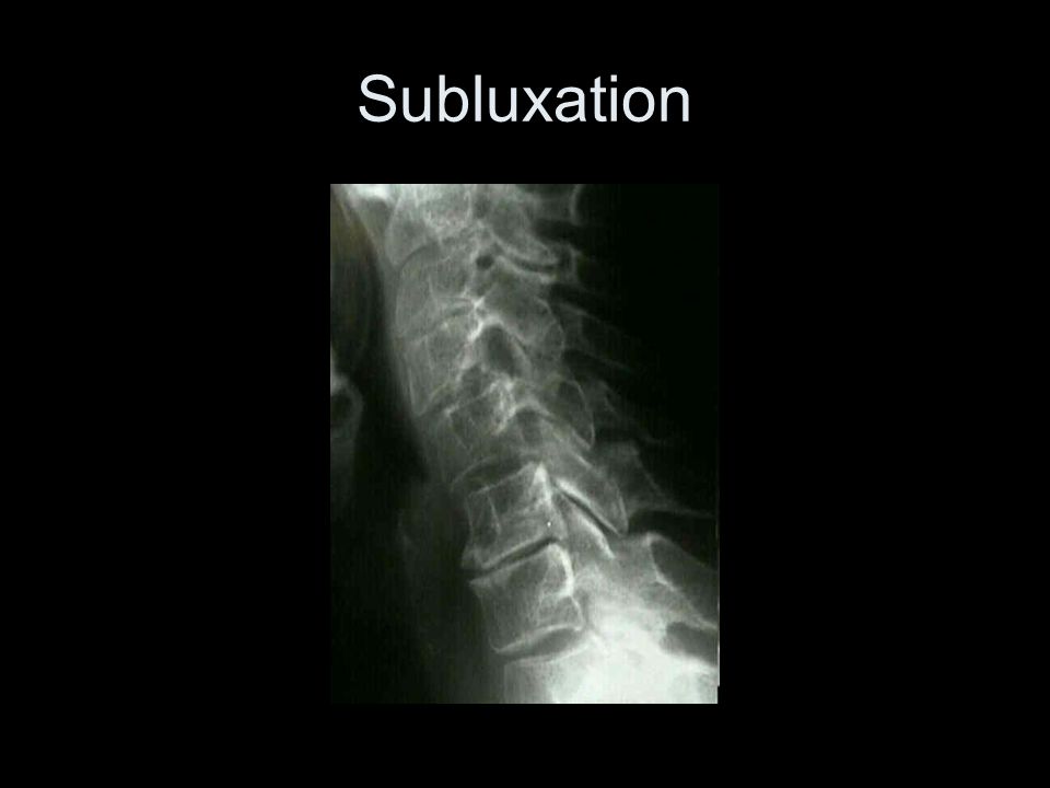
Subluxation

60
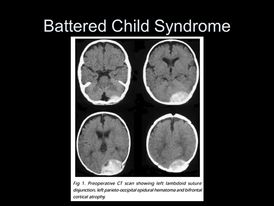
Skeletal Trauma Suspicious for Child Abuse Distal femur, wrist, ankle –Metaphyseal corner fractures Multiple –Fx’s in different stages of healing Femur, humerus, tibia –Spiral fx’s <1 year old Multiple skull fx’s –Occipital bone Post ribs, avulsed spinous processes, metacarpal & metatarsal fx’s, sternal& scapular fx’s, vertebral body fx’x and subluxation –Unusually naturally occurring fx’s <5 years old Fx’s with abundant callous formations –Implies repeated trauma with no immobilization

61
Battered Child Syndrome

65
Trauma of Chest and Thorax

66
PNEUMOTHORAX Common causes include a penetrating would such as: gun shot stabbing fractured ribs, thoracentesis

67
Atelectasis Refers to a condition with diminished air within lungs associated with reduced air volume Incomplete expansion of the lung caused by a partial or total collapse Often occurs from a penetrating wound in the chest

68
Abdominal Trauma

69
1.Can include GI tract, liver, spleen, kidneys, pancreas, aorta and pelvic organs. 2.Initially may show minimal symptoms 3.LLD is best for demonstrating small amounts of air fluid levels 1.Lay on side 10 minutes 4.CT very valuable to catch subtle abnormalities not detected with x-ray

70
Pneumoperitoneum 1.Presence of air in the peritoneum 2.LG amounts indicate a colon perforation 3.SM amounts indicate a duodenal perforation 4.Can be from trauma rupture or nontraumatic bowel perforation 5.Has a football sign

71
Pneumoperitoneum

72
Imaging Considerations 1.Radiography 1.First imaging modality for trauma 2.Portables often used 3.Primary means of evaluating skeletal trauma 2.MRI 1.For muscle, tendons, ligaments and soft tissue

73
Imaging Considerations 1.CT 1.Is excellent form imaging acute cerebral hemorrhage & fx's of the skull & facial bones 1.Quickly replacing x-ray as the standard for evaluating C-spine trauma 2.Better to visualize transverse processes of L- spine 2.Blunt trauma to abdomen can use CT or US 1.CT preferred for urinary trauma 2.Sometimes angio is used

Similar presentations


.>")


- Gastrocnemius, Soleus,>")
