Download presentation
Presentation is loading. Please wait.

1
Lecture 5 OCULAR INJURY Lecture 5 OCULAR INJURY Lecture is delivered by Ph. D., assistant of professor Tabalyuk Tetyana Anatolyivna

2
Classification of ocular injury By conditions: agricultural, industrial, military, criminal etc. By traumatic factor: By traumatic factor: mechanical, termal, chemical, radial, biological etc. By mechanism:blunt injuries (contusions) – lid injuries, orbital injuries, blunt injuries of the eyeball; By mechanism: A. blunt injuries (contusions) – lid injuries, orbital injuries, blunt injuries of the eyeball; B. penetrating trauma – corneal, limbal, scleral, corneo- scleral; C. burns.
 – lid injuries, orbital injuries, blunt injuries of the eyeball; By mechanism: A. blunt injuries (contusions) – lid injuries, orbital injuries, blunt injuries of the eyeball; B. penetrating trauma – corneal, limbal, scleral, corneo- scleral; C. burns..")
3
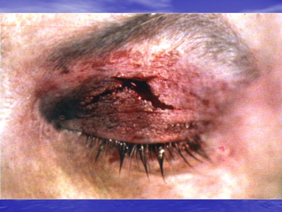
BLUNT INJURIES (contusions): lid injuries: haematoma, laceration; orbital injuries (fractures); blunt injuries of the eyeball: corneal abrasion, scleral rupture, iridodyalisis, hyphaema, traumatic mydriasis, lens dislocation, haemophthalmos, commotio retinae, choroidal rupture, choroidoretinal haemorrhage, retinal detachment
: lid injuries: haematoma, laceration; orbital injuries (fractures); blunt injuries of the eyeball: corneal abrasion, scleral rupture, iridodyalisis, hyphaema, traumatic mydriasis, lens dislocation, haemophthalmos, commotio retinae, choroidal rupture, choroidoretinal haemorrhage, retinal detachment")
10
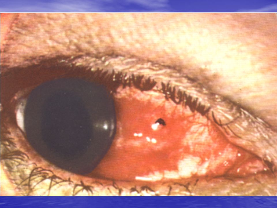
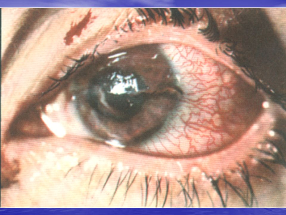
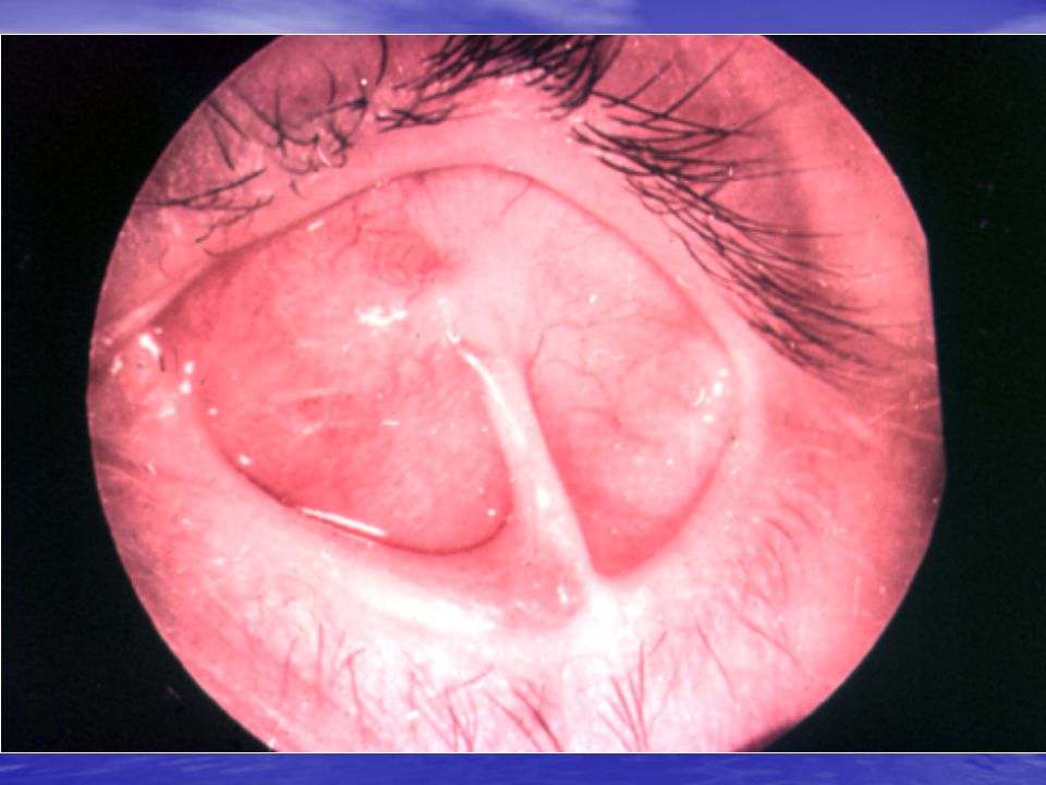
A small piece of iron has lodged near the margin of the cornea

11
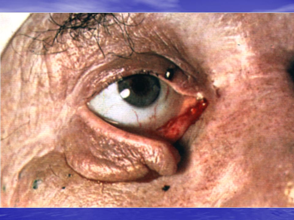
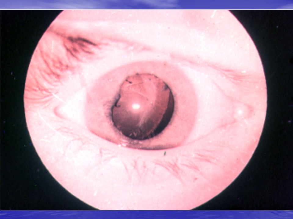
Iridodyalisis

12
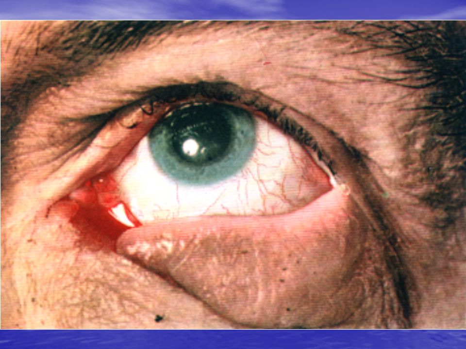
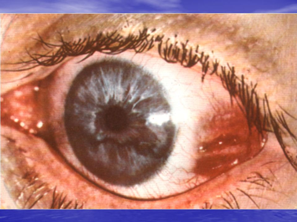
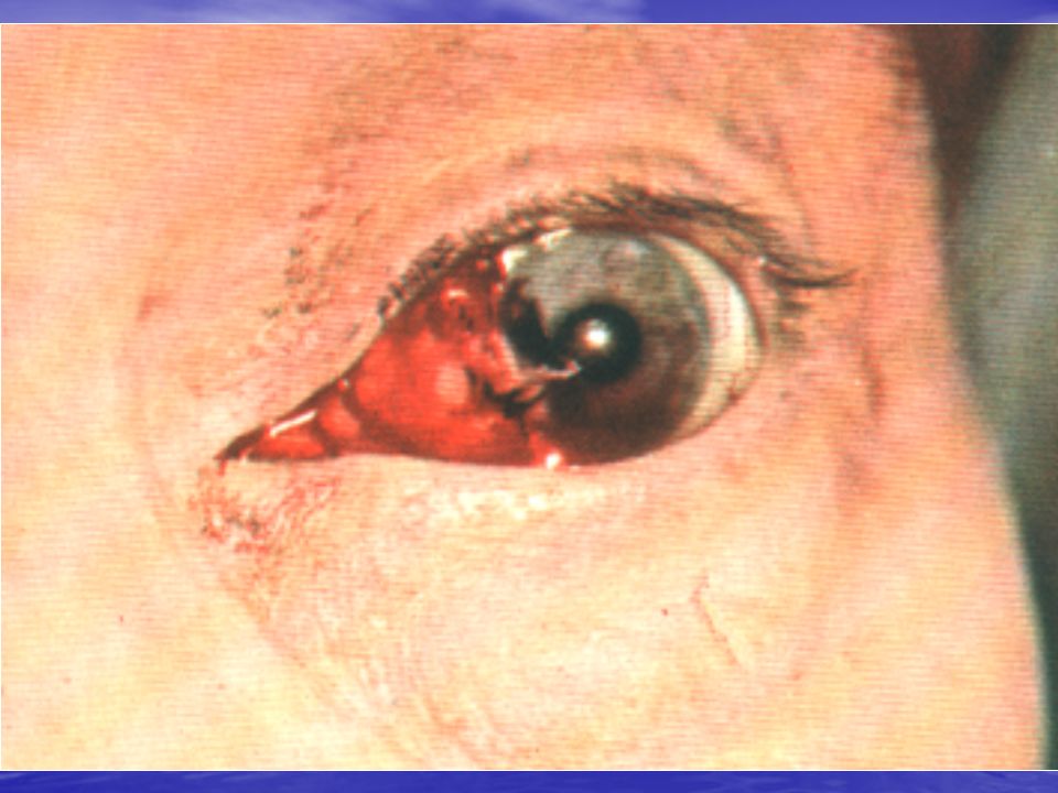
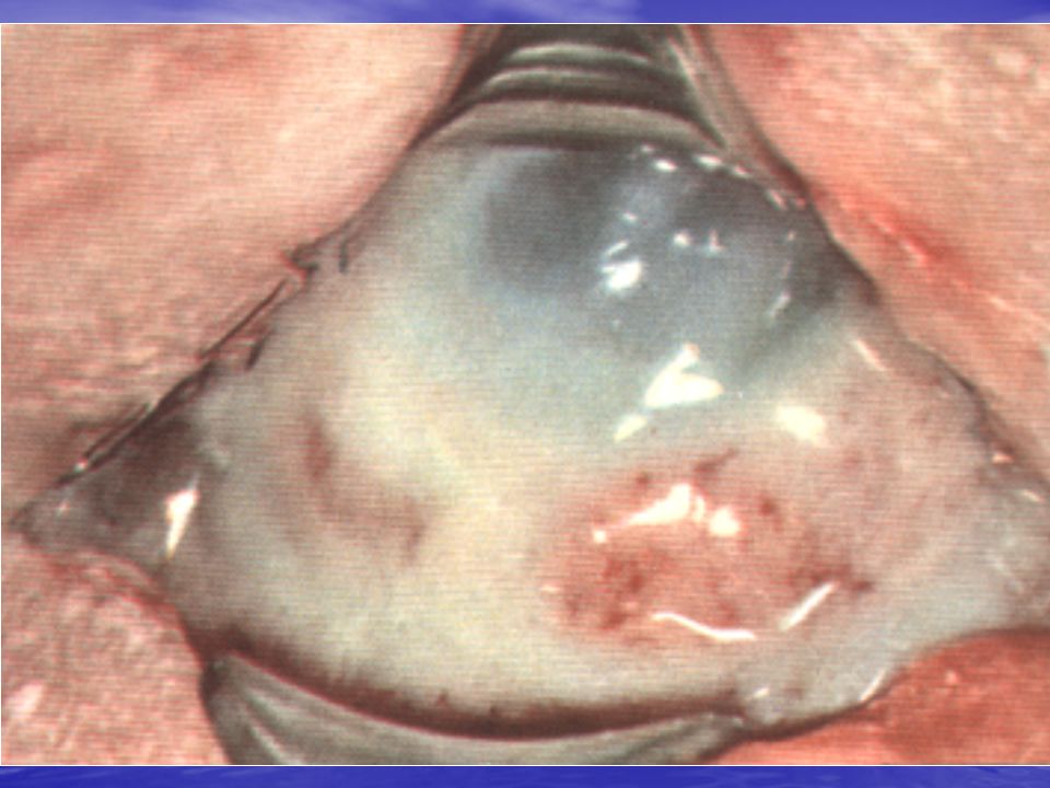
left - subconjunctival haemorrhage; right - blood behind the cornea, inside the eye (hyphaema), the iris has been bleeding
, the iris has been bleeding")
13
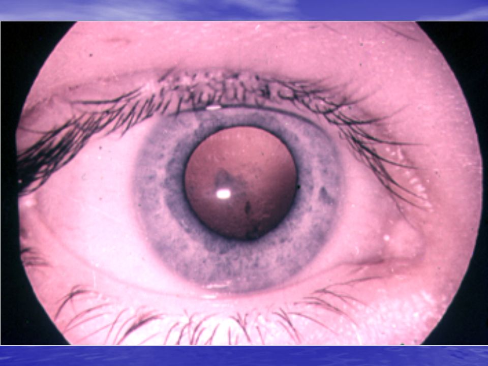
Hyphaema – blood in the anterior chamber

19
Emergency in blunt injuries: In corneal abrasion – antibacterial drops and ointments and medicines, which rapid corneal regeneration, for example, Tobramycini, Chinini hydrochloride 1% (2 drops into affected eye 4-6 times a day), Ung.”Floxali” and Corneregel (under lower eyelid 2-3 times a day). In hyphaema & haemophthalmos – haemostatic therapy, for example S. Dicinoni 12,5 % 2,0 i/m 2 times a day, Tab. ”Ascorutini” per os 3 times a day, ”Vikasoli” 0,015 per os 3 times a day, Sol. Ca chloridi 3 % in drops 4 times a day. Foreign body of conjunctiva is removed by wet cotton, pincet or injection needle. After removing, antiseptics are instillated and prescribed, for example S.Sulfacili Na or S. Oftadec 4 times a day during 5-7 days. Corneal foreign body is removed after anaesthesia (S. Alcaini) by injection needle. Antibacterial drops and ointments and medicines, which rapid corneal regeneration are dropped and prescribed, for example, S. Gentamycini 0,3 %, S. Taufoni 4 % (2 drops into affected eye 4-6 times a day), Ung.Tetracyclini and Actovegin gel (under lower eyelid 2-3 times a day).
 by injection needle. Antibacterial drops and ointments and medicines, which rapid corneal regeneration are dropped and prescribed, for example, S. Gentamycini 0,3 %, S. Taufoni 4 % (2 drops into affected eye 4-6 times a day), Ung.Tetracyclini and Actovegin gel (under lower eyelid 2-3 times a day)..")
20
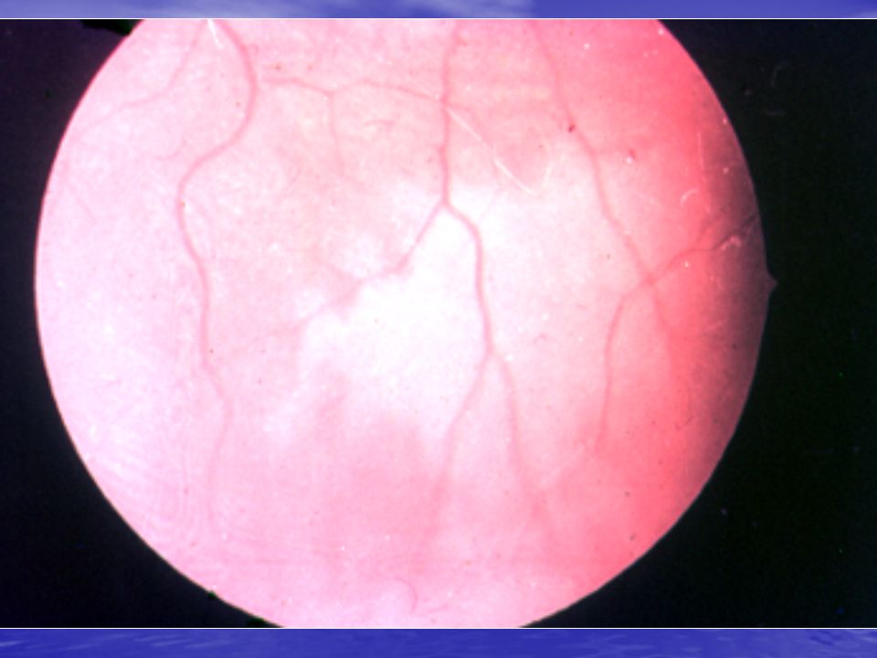
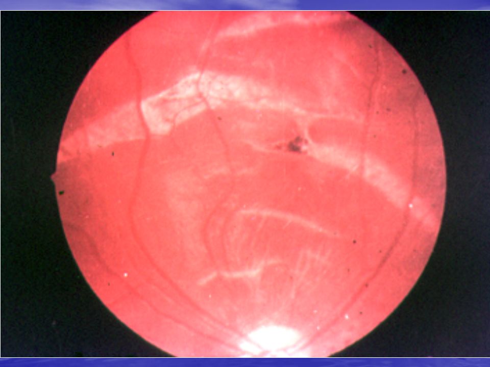
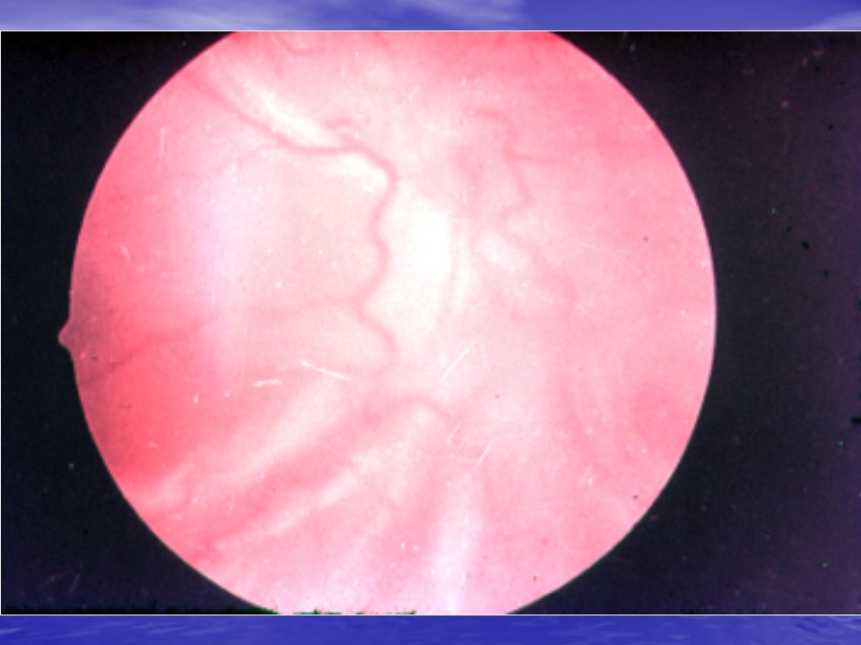
RETINAL DETACHMENT 1.Rhegmatogenous 2. Exudative 3. Tractional Signs of detachment – photopsia, metamorphopsia, “shadow” before eye, peripheral visual field loss controposite the localization of detached retina Surgical management: Transscleral photocoagulation or criopexy Scleral buckling procedures Vitrectomy & intraocular silicon oil or gas tamponade

21
Absolute signs of penetrating injury: corneal or scleral wound; intraocular foreign body; extrusion of intraocular tissues (iris, choroid, vitreous, lens, etc) through the wound Relative signs of penetrating injury: hypotonia; pupil deformation; changing of anterior chamber depth (flat or deep).
 through the wound Relative signs of penetrating injury: hypotonia; pupil deformation; changing of anterior chamber depth (flat or deep).")
25
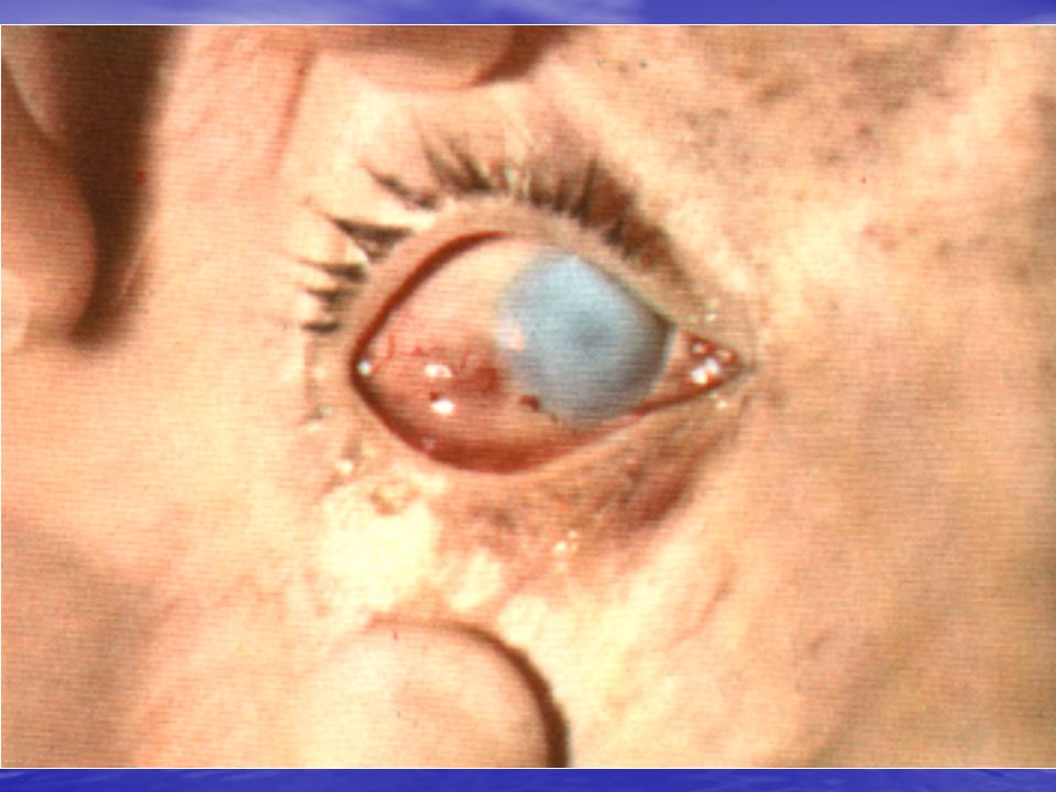
Eye injury by impact of small plastic body

26
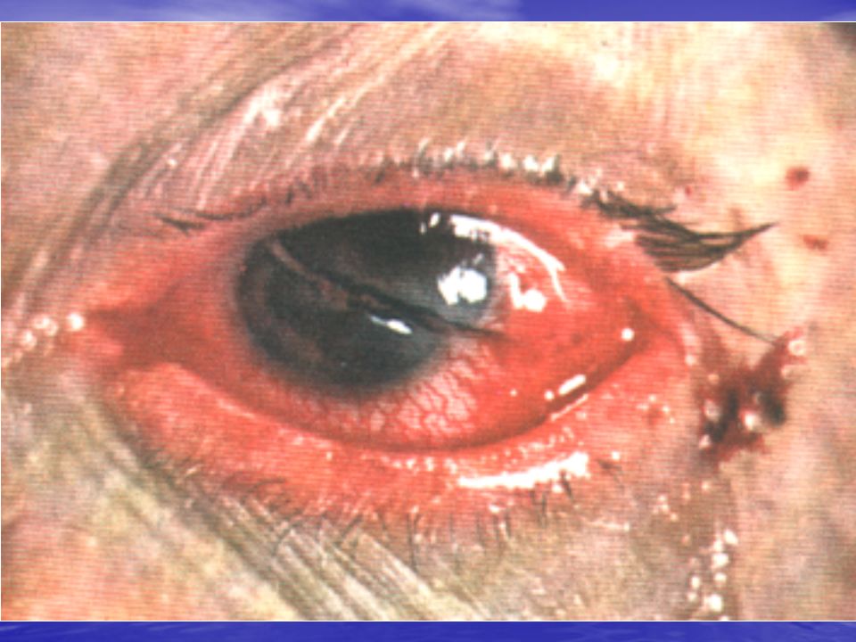
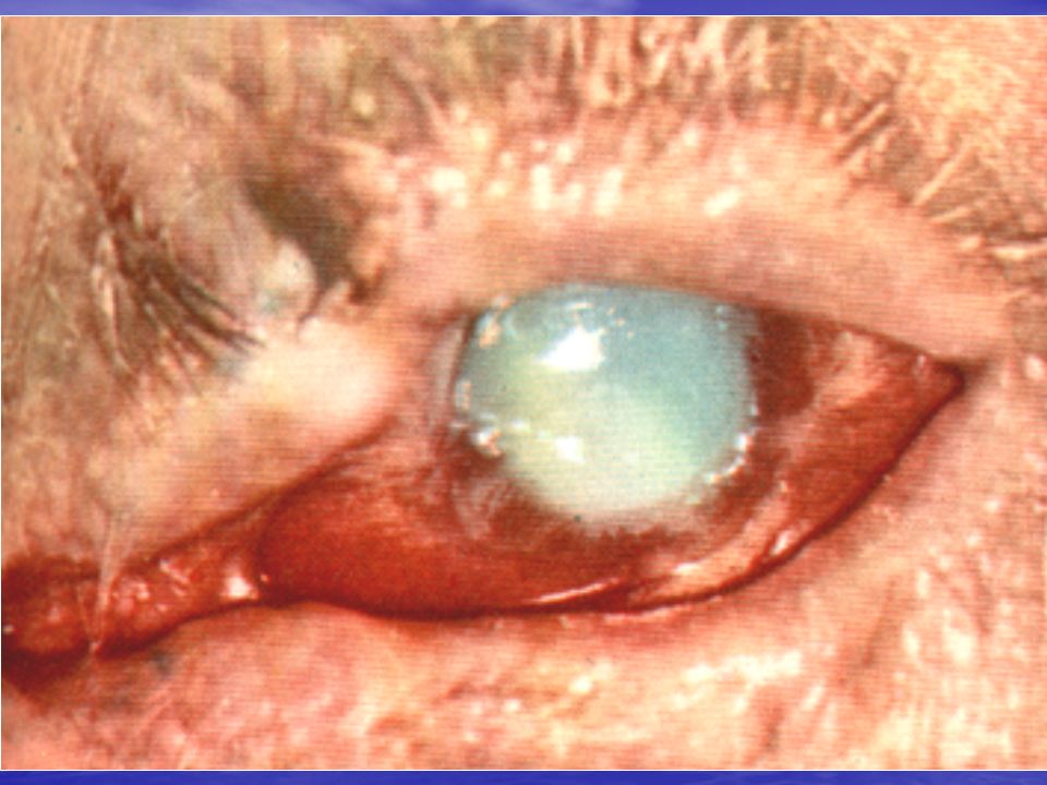
Open eye injury, with iris prolapsed through the cornea laceration. This type of injury needs immediate eye care attention

27
. Methods of localization of intraocular foreign body: X-ray examination (metal foreign body - with special protesis of Komberg-Baltin; nonmetal (glass) –X-ray examination by Fogt) Ultrasound examination; Complications of penetrating injury: traumatic cataract; traumatic iridocyclitis; endophthalmitis; panophthalmitis; sympathetic ophthalmia (chronic fibro-plastic autoimmune iridocyclitis of the unaffected eye) Emergency in eye penetrating injury cleaning the wound, using antiseptics locally (i.e. S. Furacilini 1: 5000 or S. Laevomycetini 0,25 %), analgetics (S. Analgini 50 % 2,0 i/m), antibiotics systemically (i.e.Tab. Ofloxacini 0,2 per os or S. Gentamycini sulfatis 4 % 1,0 i/m), binocular dressing, transportation the patient in horizontal position into the special department
, analgetics (S. Analgini 50 % 2,0 i/m), antibiotics systemically (i.e.Tab. Ofloxacini 0,2 per os or S. Gentamycini sulfatis 4 % 1,0 i/m), binocular dressing, transportation the patient in horizontal position into the special department.")
28
CLASSIFICATION of BURNS I degree – hyperemia of conjunctiva; superficial opacity of cornea or corneal abrasion which disappears without any changes II degree – superficial necrosis of conjunctiva gray & cloudy cornea (defect of epithelium & superficial layers of stroma) III degree – necrosis of hole conjunctiva defect of all corneal layers – “mat” cornea IV degree – necrosis not only of conjunctiva, but also sclera “porcelain” cornea Emergency in eye burns removing of foreign pieces especially in case of lime’s burn, watering of the eye by water, Sol. Na isotonici or S. Furacilini during 15-30 minutes, using of antiseptics or antibiotics in drops (S. Dimexidi 10 %, ”Ciloxan” or ” Tobrex ” )
.")
Similar presentations


 2-Perforating Injury 3-Perforating Injury & retained foreign body 4-Chemicals ( acid – alkaline ) & burns 5-Sonar.>")


 M echanism of injury 1) M echanism of injury 2)Size of the IOFB.>")


