Download presentation
Presentation is loading. Please wait.

1
Apoptosis Programmed cell death

2
OBJECTIVES DEFINITION, PHYSIOLOGIC AND PATHOLOGIC CONDITIONS. DESCRIBE THE MORPHOLOGY AND DISCUSS THE POSSIBLE MECHANISMS. COMPARE BETWEEN APOPTOSIS AND NECROSIS.

3
Apoptosis = “Falling away from” Apoptosis is responsible for the programmed cell death in several important physiologic and pathologic processes. Apoptosis: Programmed cell death that involves one cell or small group of cells

4
APOPTOSIS, PHYSIOLOGIC CONDITIONS 1- Destruction of unwanted cells during embryogenesis. 2- Mature tissue homeostasis, in proliferating cells. 3- Involution of hormone-dependent tissue as shedding of the endometrium during the menstrual cycle and regression of the breast following weaning. 4- Elimination of harmful self-reactive lymphocytes. *** Failure of cells to undergo physiologic apoptosis may result in developmental defects, autoimmune diseases and others.

5
APOPTOSIS, PATHOLOGIC CONDITIONS Apoptosis eliminates injured cells that are not able to repair 1- DNA damage, as in radiations; cytotoxic drugs; and hypoxia, to prevent malignant transformations. 2- Cell death in certain infections as viral infections by response from cytotoxic T-lymphocytes. 3- In tumors and transplant rejection. 4- Pathological atrophy in parynchemal organs after duct obstruction: pancreas, parotid, and kidney.

6
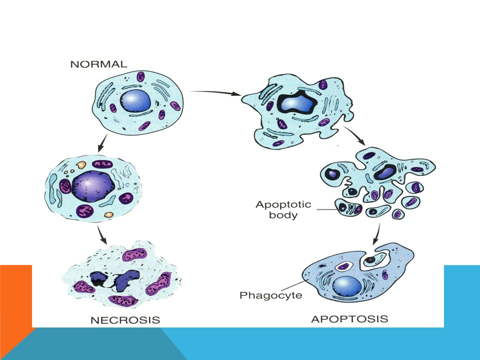
Morphology of apoptosis Usually involves single or small clusters of cells. Microscopically, apoptotic cells appear as round or oval masses with intense eosinophilic cytoplasm. 1.The nuclear chromatin undergoes condensation and fragmentation. 2.The cells rapidly shrink, forming cytoplasmic buds and then fragment into apoptotic bodies composed of membrane- bound vesicles of cytoplasm and organelles. 3.Digestion of apoptotic bodies by macrophages. 4.Lack of inflammation in the surrounding.

7
Apoptosis of a keratinocyte in skin ( arrow )
")
9
Apoptosis may be histologically inapparent, possible reasons include: Apoptosis may be histologically inapparent, possible reasons include: 1- Apoptotic fragments are rapidly phagocytozed and degraded. 2- Apoptosis does not induce inflammatory reaction, thus it becomes difficult to be recognised microscopically.

10
Mechanisms of apoptosis 1- Signalling 2- Control and integration 3- Execution 4- Removal of dead cells The basic processes include four separable but overlapping components:

11
1- Signalling Variety of signals (intrinsic & extrinsic) induce apoptosis: 1. Intrinsic (as in development) 2. Lack of growth factor 3. Toxin from cytotoxic T cells 4. Radiation, heat, chemicals 5. Receptor-ligand interactions: Tumor necrosis factor (TNF) family of plasma membrane receptors (e.g., FAS receptor) is a major initiator of death signals.
 2. Lack of growth factor 3. Toxin from cytotoxic T cells 4. Radiation, heat, chemicals 5. Receptor-ligand interactions: Tumor necrosis factor (TNF) family of plasma membrane receptors (e.g., FAS receptor) is a major initiator of death signals..")
12
Specific proteins connect the original death signals to the final execution program. Two pathways are involved in this process: 1- Direct transmission of death signals by specific adapter proteins to the executive mechanism. 2- Regulation of mitochondrial permeability by members of BCL-2 family of proteins. 2- Control and integration

13
Mitochondrial BCL-2 family members (BCL-X ) inhibit and others (BAX & BAD or BAK) promote pore formation in mitochondria: Promotion leads to less ATP production with mitochondrial swelling. Outer mito. membrane permeability increases leading to a release of apoptotic trigger (cytochrome c) into cytosol. Cytochrome c binds some cytosolic proteins, activating them and then eventually induces caspase activation. Caspases, when activated, begin proteolytic events that kill the cell.
 into cytosol. Cytochrome c binds some cytosolic proteins, activating them and then eventually induces caspase activation. Caspases, when activated, begin proteolytic events that kill the cell..")
14
3- Execution Distinctive biochemical events resulting from activity of catabolic enzymes in the cytosol. 1- Protein cleavage by caspases: Caspases: Specific proteases with an active site cysteine.

15
Activation of one or more caspase enzyme leads to cascade of activation of other proteases. E.g., activation of endonuclease result in DNA fragmentation 2- Proteins are cross-linked by transglutaminase Forming condensed shell that readily fragments into apoptotic bodies.

16
3- DNA breakdown giving 180 – 200 base pair fragments, by action of endonucleases. This may be visualized as a distinctive “Ladders” of DNA fragments on electrophoresis. This laddering pattern is not specific for apoptosis. Necrosis usually gives a more random pattern of fragmentation, forming a smear.

17
Gel electrophoresis of DNA extracted from cultured cells: 1- Lane a, Viable control cells 2- Lane b, Extensive apoptosis, with laddered DNA demonstrating fragmentation 3- Lane c, necrosis However, DNA laddering is not diagnostic for apoptosis as it may be seen in early stages of necrosis as well!

18
Apoptotic bodies have plasma membrane surface markers that facilitate uptake and degradation by adjacent cells or by phagocytes. A “flip” of inner plasma membrane phosphatidylserine to the outer surface is a sufficient signal to attract other cells for phagocytosis without the harmful secondary effects of inflammation. 4- Removal of dead cells

19
COMPARISON BETWEEN NECROSIS & APOPTOSIS FeatureNecrosisApoptosis Cell sizeswellingshrinkage NucleusPyknosis, karyorrhexis, karyolysis Fragmentation to apoptotic bodies Plasma membranedisruptedintact Cell contentsLeak out of cellintact Adjacent inflammation frequentno Physiologic or pathologic role pathologicPhysiologic and can be pathologic

20
THANK YOU DR/MONA KAMAL

Similar presentations


, death domain, cytochrome.>")
 Apoptosis is a cell mechanism used to eliminate cells that are unnecessary to or that contain mutations that.>")


 In adult tissues cell death exactly balances cell division In apoptosis the cell destroys itself from within.>")


