Download presentation
Presentation is loading. Please wait.

1
Neoplasia Lecture 3 Dr. Maha Arafah Dr. Abdulmalik Alsheikh, MD, FRCPC CARCINOGENESIS Foundation block 2012 Pathology

2
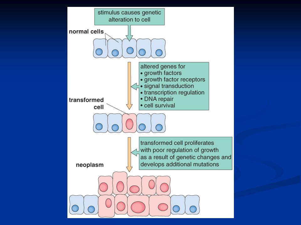
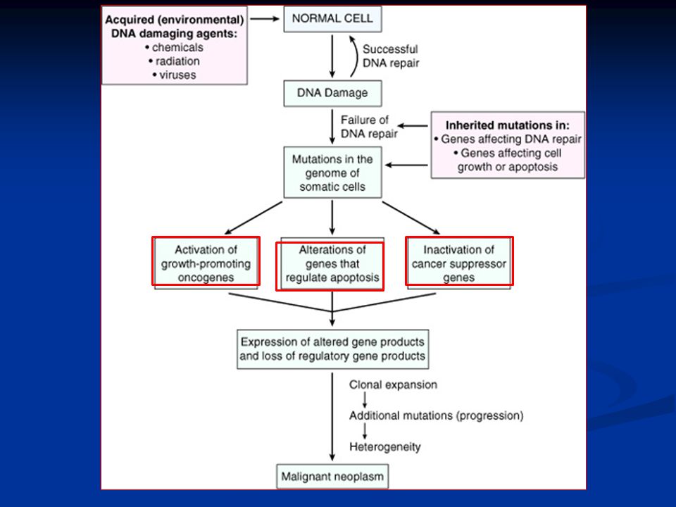
CARCINOGENESIS Carcinogenesis is a multistep process at both the phenotypic and the genetic levels. Carcinogenesis is a multistep process at both the phenotypic and the genetic levels. It starts with a genetic damage: It starts with a genetic damage: Environmental Environmental Chemical Chemical Radiation Radiation Viral Viral Inhereted Inhereted

3
Carcinogenesis Genetic damage lead to “ mutation” Genetic damage lead to “ mutation” single cell which has the genetic damage undergoes neoplastic proliferation ( clonal expansion) forming the tumor mass single cell which has the genetic damage undergoes neoplastic proliferation ( clonal expansion) forming the tumor mass
 forming the tumor mass single cell which has the genetic damage undergoes neoplastic proliferation ( clonal expansion) forming the tumor mass")
5
Carcinogenesis Where are the targets of the genetic damage?? Where are the targets of the genetic damage?? Four regulatory genes are the main targets: Four regulatory genes are the main targets: Growth promoting protooncogenes Growth promoting protooncogenes Protooncogene > mutation > oncogene Protooncogene > mutation > oncogene Growth inhibiting (supressors) genes Growth inhibiting (supressors) genes Genes regulating apoptosis Genes regulating apoptosis DNA repair genes DNA repair genes
 genes Growth inhibiting (supressors) genes Genes regulating apoptosis Genes regulating apoptosis DNA repair genes DNA repair genes.")
7
Carcinogenesis Main changes in the cell physiology that lead to formation of the malignant phenotype: Main changes in the cell physiology that lead to formation of the malignant phenotype: Self-sufficiency in growth signals Self-sufficiency in growth signals Insensitivity to growth-inhibitory signals Insensitivity to growth-inhibitory signals Evasion of apoptosis Evasion of apoptosis Limitless replicative potential Limitless replicative potential Sustained angiogenesis Sustained angiogenesis Ability to invade and metastsize Ability to invade and metastsize

8
Carcinogenesis A - Self-sufficiency in Growth signals: Oncogene: Gene that promote autonomous cell growth in cancer cells Oncogene: Gene that promote autonomous cell growth in cancer cells They are derived by mutations in protooncogenes They are derived by mutations in protooncogenes They are characterized by the ability to promote cell growth in the absence of normal growth- promoting signals They are characterized by the ability to promote cell growth in the absence of normal growth- promoting signals Oncoproteins : are the products Oncoproteins : are the products

9
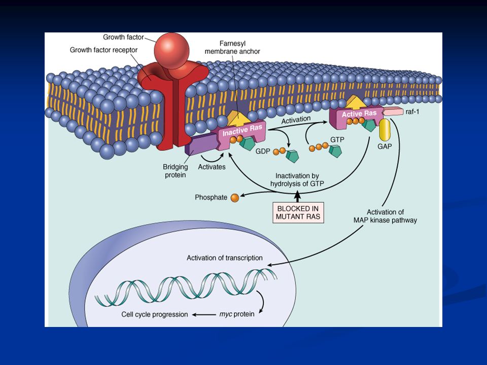
Carcinogenesis Remember the cell cycle !! Remember the cell cycle !! Binding of a growth factor to its receptor on the cell membrane Binding of a growth factor to its receptor on the cell membrane Activation of the growth factor receptor leading to activation of signal-transducing proteins Activation of the growth factor receptor leading to activation of signal-transducing proteins Transmission of the signal to the nucleus Transmission of the signal to the nucleus Induction of the DNA transcription Induction of the DNA transcription Entry in the cell cycle and cell division Entry in the cell cycle and cell division

10
Carcinogenesis HOW CANCER CELLS ACQUIRE SELF- SUFFICIENCY IN GROWTH SIGNALS?? HOW CANCER CELLS ACQUIRE SELF- SUFFICIENCY IN GROWTH SIGNALS??

11
Carcinogenesis 1- Growth factors: Cancer cells are capable to synthesize the same growth factors to which they are responsive Cancer cells are capable to synthesize the same growth factors to which they are responsive E.g. Sarcomas ---- > TGF- E.g. Sarcomas ---- > TGF- Glioblastoma-----> PDGF Glioblastoma-----> PDGF

13
Carcinogenesis 2-Growth factors receptors: Receptors --- mutation ----continous signals to cells and uncontroled growth Receptors --- mutation ----continous signals to cells and uncontroled growth Receptors --- overexpression ---cells become very sensitive ----hyperresponsive to normal levels of growth factors Receptors --- overexpression ---cells become very sensitive ----hyperresponsive to normal levels of growth factors

15
Carcinogenesis Example : Example : Epidermal Growth Factor ( EGF ) Receptor family Epidermal Growth Factor ( EGF ) Receptor family HER2 HER2 Amplified in breast cancers and other tumors Amplified in breast cancers and other tumors High levels of HER2 in breast cancer indicate poor prognosis High levels of HER2 in breast cancer indicate poor prognosis Anti- HER2 antibodies are used in treatment Anti- HER2 antibodies are used in treatment
 Receptor family Epidermal Growth Factor ( EGF ) Receptor family HER2 HER2 Amplified in breast cancers and other tumors Amplified in breast cancers and other tumors High levels of HER2 in breast cancer indicate poor prognosis High levels of HER2 in breast cancer indicate poor prognosis Anti- HER2 antibodies are used in treatment Anti- HER2 antibodies are used in treatment")
16
Carcinogenesis 3- Signal-transducing proteins : They receive signals from activated growth factors receptors and transmitte them to the nucleus. Examples : They receive signals from activated growth factors receptors and transmitte them to the nucleus. Examples : RAS RAS ABL ABL

17
Carcinogenesis RAS : RAS : 30% of all human tumors contain mutated RAS gene. E.g : colon. Pancreas cancers 30% of all human tumors contain mutated RAS gene. E.g : colon. Pancreas cancers Mutations of the RAS gene is the most common oncogene abnormality in human tumors Mutations of the RAS gene is the most common oncogene abnormality in human tumors Mutations in RAS --- cells continue to proliferate Mutations in RAS --- cells continue to proliferate

19
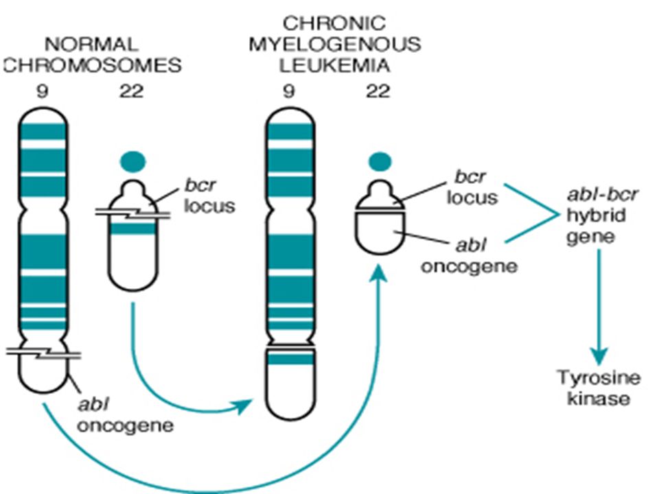
Carcinogenesis ABL gene ABL gene ABL protooncogene has a tyrosine kinase activity ABL protooncogene has a tyrosine kinase activity Its activity is controlled by negative regulatory mechanism Its activity is controlled by negative regulatory mechanism E.g. : chronic myeloid leukemia ( CML ) : E.g. : chronic myeloid leukemia ( CML ) : t( 9,22) ---ABL gene transferred from ch. 9 to ch. 22 t( 9,22) ---ABL gene transferred from ch. 9 to ch. 22 Fusion with BCR ---> BCR-ABL Fusion with BCR ---> BCR-ABL BCR-ABL has tyrosine kinase acttivity ---( oncogenec) BCR-ABL has tyrosine kinase acttivity ---( oncogenec)
 : E.g. : chronic myeloid leukemia ( CML ) : t( 9,22) ---ABL gene transferred from ch. 9 to ch. 22 t( 9,22) ---ABL gene transferred from ch. 9 to ch. 22 Fusion with BCR ---> BCR-ABL Fusion with BCR ---> BCR-ABL BCR-ABL has tyrosine kinase acttivity ---( oncogenec) BCR-ABL has tyrosine kinase acttivity ---( oncogenec).")
21
Carcinogenesis CML patients are treated with ( Gleevec) which is inhibitor of ABL kinase CML patients are treated with ( Gleevec) which is inhibitor of ABL kinase
 which is inhibitor of ABL kinase CML patients are treated with ( Gleevec) which is inhibitor of ABL kinase")
22
Carcinogenesis 4- Nuclear transcription factors : Mutations may affect genes that regulate transcription of DNA growth autonomy Mutations may affect genes that regulate transcription of DNA growth autonomy E.g. MYC E.g. MYC MYC protooncogene produce MYC protein when cell receives growth signals MYC protooncogene produce MYC protein when cell receives growth signals MYC protein binds to DNA leading to activation of growth-related genes MYC protein binds to DNA leading to activation of growth-related genes

23
Carcinogenesis Normally … MYC decrease when cell cycle begins …but..in tumors there is sustained expression of MYC continuous proliferation Normally … MYC decrease when cell cycle begins …but..in tumors there is sustained expression of MYC continuous proliferation E.g. Burkitt Lymphoma ; MYC is dysregulated due to t( 8,14) E.g. Burkitt Lymphoma ; MYC is dysregulated due to t( 8,14)
 E.g. Burkitt Lymphoma ; MYC is dysregulated due to t( 8,14).")
24
Carcinogenesis 5- Cyclins and cyclins- dependent kinases (CDKs) Progression of cells through cell cycles is regulated by CDKs after they are activated by binding with cyclins Progression of cells through cell cycles is regulated by CDKs after they are activated by binding with cyclins Mutations that dysregulate cyclins and CDKs will lead to cell proliferation …e.g. Mutations that dysregulate cyclins and CDKs will lead to cell proliferation …e.g. Cyclin D genes are overexpressed in breast, esophagus and liver cancers. Cyclin D genes are overexpressed in breast, esophagus and liver cancers. CDK4 is amplified in melanoma and sarcomas CDK4 is amplified in melanoma and sarcomas

26
Carcinogenesis Main changes in the cell physiology that lead to formation of the malignant phenotype: Main changes in the cell physiology that lead to formation of the malignant phenotype: A- Self-sufficiency in growth signals B- Insensitivity to growth-inhibitory signals C- Evasion of apoptosis D- Limitless replicative potential E- Sustained angiogenesis F- Ability to invade and metastsize

27
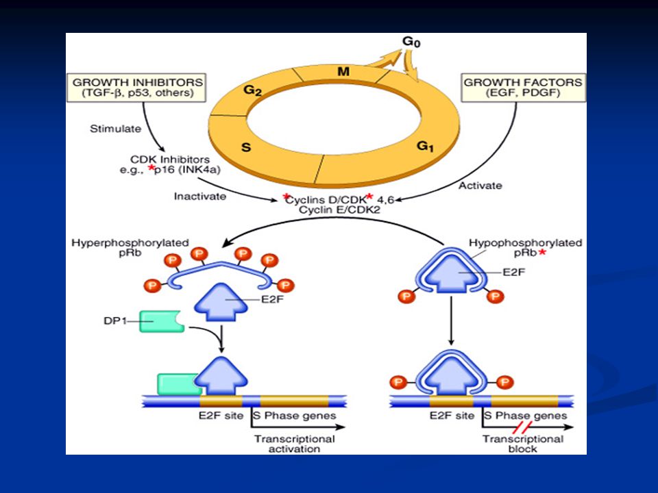
Carcinogenesis 2. Insensitivity to growth-inhibitory signals Tumor suppressor genes control ( apply brakes) cells proliferation Tumor suppressor genes control ( apply brakes) cells proliferation If mutation caused disruption to them cell becomes insensitive to growth inhibition uncontrolled proliferation If mutation caused disruption to them cell becomes insensitive to growth inhibition uncontrolled proliferation Examples: RB, TGF- , APC, P53 Examples: RB, TGF- , APC, P53
 cells proliferation Tumor suppressor genes control ( apply brakes) cells proliferation If mutation caused disruption to them cell becomes insensitive to growth inhibition uncontrolled proliferation If mutation caused disruption to them cell becomes insensitive to growth inhibition uncontrolled proliferation Examples: RB, TGF- , APC, P53 Examples: RB, TGF- , APC, P53.")
29
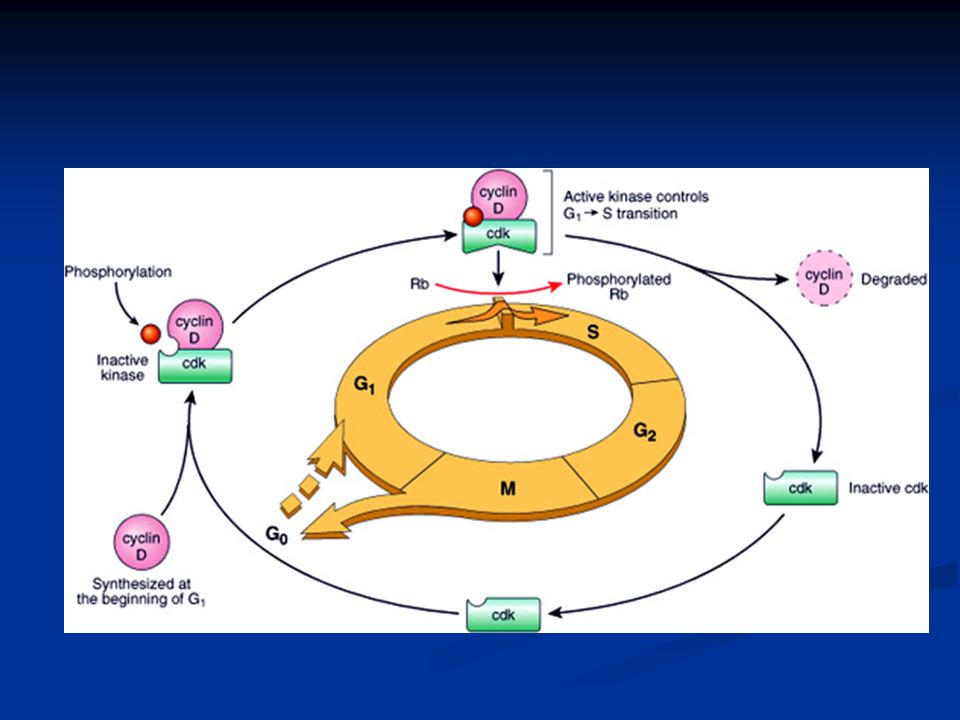
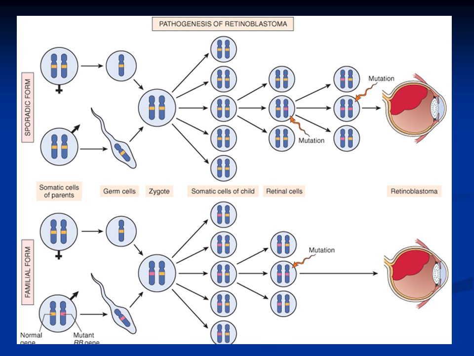
Carcinogenesis RB ( retinoblastoma ) gene : RB ( retinoblastoma ) gene : First tumor supressor gene discovered First tumor supressor gene discovered It was discovered initially in retinoblastomas It was discovered initially in retinoblastomas Found in other tumors, e.g. breast ca Found in other tumors, e.g. breast ca RB gene is a DNA-binding protein RB gene is a DNA-binding protein RB is located on chromosome 13 RB is located on chromosome 13

30
Carcinogenesis RB gene exists in “ active “ and “ inactive” forms RB gene exists in “ active “ and “ inactive” forms If active will stop the advancing from G1 to S phase in cell cycle If active will stop the advancing from G1 to S phase in cell cycle If cell is stimulated by growth factors inactivation of RB gene brake is released cells start cell cycle …G1 S M …then RB gene is activated again If cell is stimulated by growth factors inactivation of RB gene brake is released cells start cell cycle …G1 S M …then RB gene is activated again

32
Carcinogenesis Retinoblastoma is an uncommon childhood tumor Retinoblastoma is an uncommon childhood tumor Retinoblastoma is either sporadic (60%) or familial ( 40% ) Retinoblastoma is either sporadic (60%) or familial ( 40% ) Two mutations required to produce retinoblastoma Two mutations required to produce retinoblastoma Both normal copies of the gene should be lost to produce retinoblastoma Both normal copies of the gene should be lost to produce retinoblastoma
 or familial ( 40% ) Retinoblastoma is either sporadic (60%) or familial ( 40% ) Two mutations required to produce retinoblastoma Two mutations required to produce retinoblastoma Both normal copies of the gene should be lost to produce retinoblastoma Both normal copies of the gene should be lost to produce retinoblastoma")
34
Carcinogenesis Transforming Growth Factor- pathway: Transforming Growth Factor- pathway: TGF- is an inhibitor of proliferation TGF- is an inhibitor of proliferation It regulate RB pathway It regulate RB pathway Inactivation of TGF- lead to cell proliferation Inactivation of TGF- lead to cell proliferation Mutations in TGF- pathway are present in : of pancreatic cancers of colon cancers

35
Carcinogenesis Adenomatous Polyposis Coli – Catenin pathway: Adenomatous Polyposis Coli – Catenin pathway: APC is tumor supressor gene APC is tumor supressor gene APC gene loss is very common in colon cancers APC gene loss is very common in colon cancers It has anti-proliferative action through inhibition of Catenin which activate cell proliferation It has anti-proliferative action through inhibition of Catenin which activate cell proliferation Individuals with mutant APC develop thousands of colonic polyps Individuals with mutant APC develop thousands of colonic polyps

36
Adenomatous Polyposis Coli

37
Carcinogenesis One or more of the polyps will progress to colonic carcinoma One or more of the polyps will progress to colonic carcinoma APC mutations are seen in 70% to 80% of sporadic colon cancers APC mutations are seen in 70% to 80% of sporadic colon cancers

38
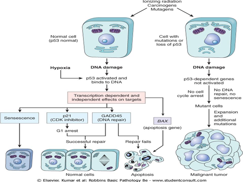
Carcinogenesis P53 P53 It has multiple functions It has multiple functions Mainly : Mainly : Tumor suppressor gene ( anti-proliferative ) Tumor suppressor gene ( anti-proliferative ) Regulates apoptosis Regulates apoptosis
 Tumor suppressor gene ( anti-proliferative ) Regulates apoptosis Regulates apoptosis")
39
Carcinogenesis P53 senses DNA damage P53 senses DNA damage Causes G1 arrest to give chance for DNA repair Causes G1 arrest to give chance for DNA repair Induce DNA repair genes Induce DNA repair genes If a cell with damaged DNA cannot be repaired, it will be directed by P53 to undergo apoptosis If a cell with damaged DNA cannot be repaired, it will be directed by P53 to undergo apoptosis

40
Carcinogenesis With loss of P53, DNA damage goes unrepaired With loss of P53, DNA damage goes unrepaired Mutations will be fixed in the dividing cells, leading to malignant transformation Mutations will be fixed in the dividing cells, leading to malignant transformation

42
P53 is called the “ guardian of the genome” P53 is called the “ guardian of the genome” 70% of human cancers have a defect in P53 70% of human cancers have a defect in P53 It has been reported with almost all types of cancers : e.g. lung, colon, breast It has been reported with almost all types of cancers : e.g. lung, colon, breast In most cases, mutations are acquired, but can be inhereted, e.g : Li-Fraumeni syndrome In most cases, mutations are acquired, but can be inhereted, e.g : Li-Fraumeni syndrome Carcinogenesis

43
Carcinogenesis Main changes in the cell physiology that lead to formation of the malignant phenotype: Main changes in the cell physiology that lead to formation of the malignant phenotype: A- Self-sufficiency in growth signals B- Insensitivity to growth-inhibitory signals C- Evasion of apoptosis D- Limitless replicative potential E- Sustained angiogenesis F- Ability to invade and metastsize

44
Carcinogenesis Evasion of apoptosis: Evasion of apoptosis: Mutations in the genes regulating apoptosis are factors in malignant transformation Mutations in the genes regulating apoptosis are factors in malignant transformation Cell survival is controlled by genes that promote and inhibit apoptosis Cell survival is controlled by genes that promote and inhibit apoptosis

45
Evasion of apoptosis Evasion of apoptosis Reduced CD95 level inactivate death – induced signaling cascade that cleaves DNA to cause death tumor cells are less susceptible to apoptosis Reduced CD95 level inactivate death – induced signaling cascade that cleaves DNA to cause death tumor cells are less susceptible to apoptosis DNA damage induced apoptosis (with the action of P53 ) can be blocked in tumors DNA damage induced apoptosis (with the action of P53 ) can be blocked in tumors loss of P53 and up- regulation of BCL2 prevent apoptosis e.g. follicular lymphoma loss of P53 and up- regulation of BCL2 prevent apoptosis e.g. follicular lymphoma

46
Carcinogenesis Main changes in the cell physiology that lead to formation of the malignant phenotype: Main changes in the cell physiology that lead to formation of the malignant phenotype: A- Self-sufficiency in growth signals B- Insensitivity to growth-inhibitory signals C- Evasion of apoptosis D- Limitless replicative potential E- Sustained angiogenesis F- Ability to invade and metastsize

47
Limitless replicative potential: Limitless replicative potential: Normally there is progressive shortening of telomeres at the ends of chromosomes Normally there is progressive shortening of telomeres at the ends of chromosomes Telomerase is active in normal stem cells but absent in somatic cells Telomerase is active in normal stem cells but absent in somatic cells In tumor cells : activation of the enzyme telomerase, which can maintain normal telomere length In tumor cells : activation of the enzyme telomerase, which can maintain normal telomere length

48
Carcinogenesis Main changes in the cell physiology that lead to formation of the malignant phenotype: Main changes in the cell physiology that lead to formation of the malignant phenotype: A- Self-sufficiency in growth signals B- Insensitivity to growth-inhibitory signals C- Evasion of apoptosis D- Limitless replicative potential E- Sustained angiogenesis F- Ability to invade and metastsize

49
Carcinogenesis Sustained angiogenesis Sustained angiogenesis Neovascularization has two main effects: Neovascularization has two main effects: Perfusion supplies oxygen and nutrients Perfusion supplies oxygen and nutrients Newly formed endothelial cells stimulate the growth of adjacent tumor cells by secreting growth factors, e.g : PDGF, IL-1 Newly formed endothelial cells stimulate the growth of adjacent tumor cells by secreting growth factors, e.g : PDGF, IL-1 Angiogenesis is required for metastasis Angiogenesis is required for metastasis

50
How do tumors develop a blood supply? How do tumors develop a blood supply? Tumor-associated angiogenic factors Tumor-associated angiogenic factors These factors may be produced by tumor cells or by inflammatory cells infiltrating the tumor e.g. macrophages These factors may be produced by tumor cells or by inflammatory cells infiltrating the tumor e.g. macrophages Important factors : Important factors : Vascular endothelial growth factor( VEGF ) Vascular endothelial growth factor( VEGF ) Fibroblast growth factor Fibroblast growth factor
 Vascular endothelial growth factor( VEGF ) Fibroblast growth factor Fibroblast growth factor.")
51
Carcinogenesis Main changes in the cell physiology that lead to formation of the malignant phenotype: Main changes in the cell physiology that lead to formation of the malignant phenotype: A- Self-sufficiency in growth signals B- Insensitivity to growth-inhibitory signals C- Evasion of apoptosis D- Limitless replicative potential E- Sustained angiogenesis F- Ability to invade and metastsize

53
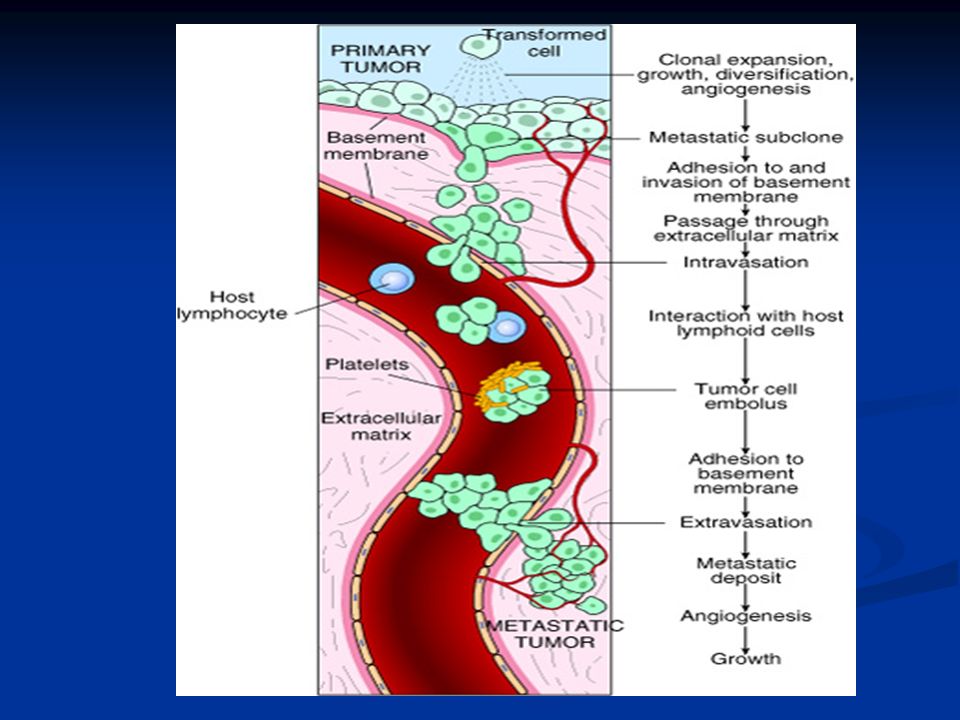
Carcinogenesis Ability to invade and metastsize: Ability to invade and metastsize: Two phases : Two phases : Invasion of extracellular matrix Invasion of extracellular matrix Vascular dissimenation and homing of tumor cells Vascular dissimenation and homing of tumor cells

54
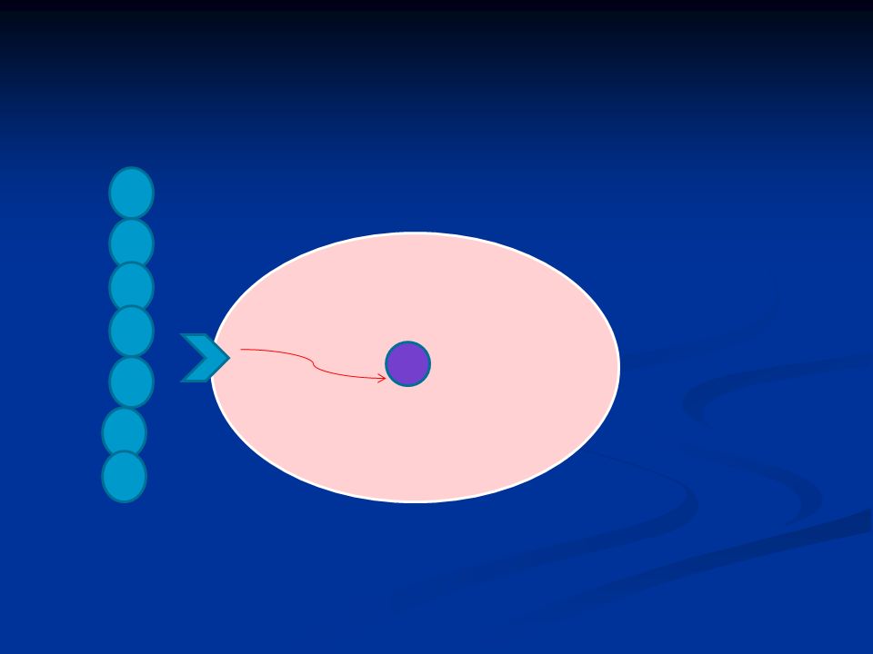
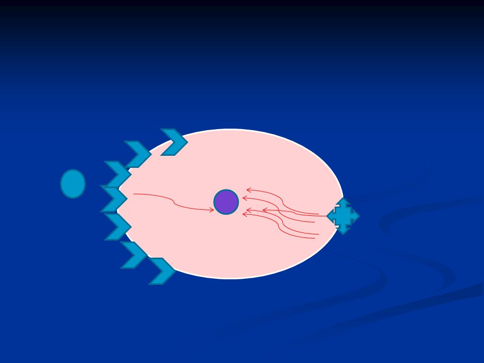
Carcinogenesis Invasion of ECM: Invasion of ECM: Malignant cells first breach the underlying basement membrane Malignant cells first breach the underlying basement membrane Traverse the interstitial tissue Traverse the interstitial tissue Penetrate the vascular basement membrane Penetrate the vascular basement membrane Gain access to the circulation Gain access to the circulation Invasion of the ECM has four steps:

55
1. Detachment of tumor cells from each other

56
2. Attachments of tumor cells to matrix components

57
3. Degradation of ECM by collagenase enzyme

58
4. Migration of tumor cells

59
Carcinogenesis Vascular dissemination and homing of tumor cells: Vascular dissemination and homing of tumor cells: May form emboli May form emboli Most travel as single cells Most travel as single cells Adhesion to vascular endothelium Adhesion to vascular endothelium extravasation extravasation

60
Carcinogenesis Main changes in the cell physiology that lead to formation of the malignant phenotype: Main changes in the cell physiology that lead to formation of the malignant phenotype: A- Self-sufficiency in growth signals B- Insensitivity to growth-inhibitory signals C- Evasion of apoptosis D- Limitless replicative potential E- Sustained angiogenesis F- Ability to invade and metastsize

61
Genomic Instability Enabler of malignancy Enabler of malignancy Due to defect in DNA repair genes Due to defect in DNA repair genes Examples: Examples: Hereditary Nonpolyposis colon carcinoma(HNPCC) Hereditary Nonpolyposis colon carcinoma(HNPCC) Xeroderma pigmentosum Xeroderma pigmentosum Familial breast cancer Familial breast cancer
 Hereditary Nonpolyposis colon carcinoma(HNPCC) Xeroderma pigmentosum Xeroderma pigmentosum Familial breast cancer Familial breast cancer")
62
Genomic Instability Familial breast cancer: Familial breast cancer: Due to mutations in BRCA1 and BRCA2 genes Due to mutations in BRCA1 and BRCA2 genes These genes regulate DNA repair These genes regulate DNA repair Account for 80% of familial breast cancer Account for 80% of familial breast cancer They are also involved in other malignancies They are also involved in other malignancies

63
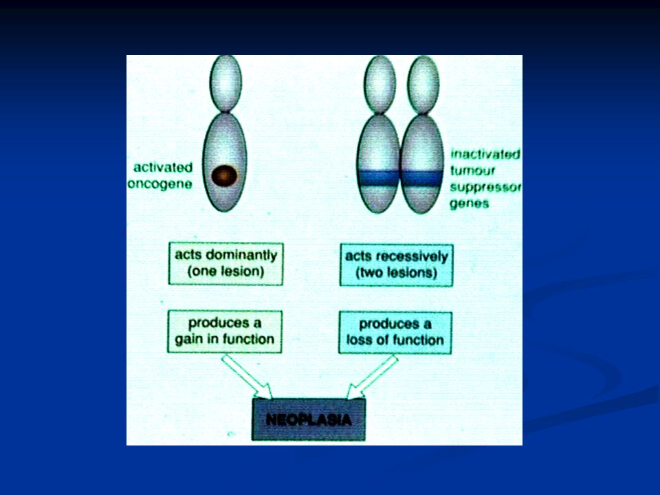
Molecular Basis of multistep Carcinogenesis Cancer results from accumulation of multiple mutations Cancer results from accumulation of multiple mutations All cancers have multiple genetic alterations, involving activation of several oncogenes and loss of two or more tumor suppressor genes All cancers have multiple genetic alterations, involving activation of several oncogenes and loss of two or more tumor suppressor genes

64
Molecular Basis of multistep Carcinogenesis

65
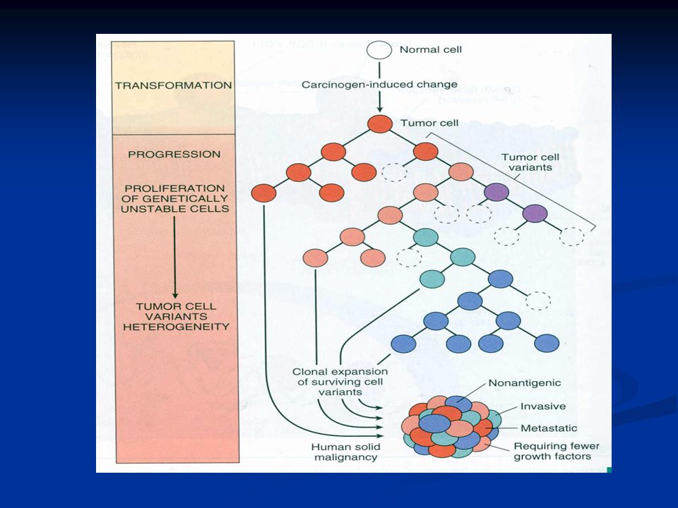
Tumor progression Many tumors become more aggressive and acquire greater malignant potential…this is called “ tumor progression” …not increase in size!! Many tumors become more aggressive and acquire greater malignant potential…this is called “ tumor progression” …not increase in size!! By the time, the tumor become clinically evident, their constituent cells are extremely heterogeneous By the time, the tumor become clinically evident, their constituent cells are extremely heterogeneous

67
Karyotypic Changes in Tumors Translocations: Translocations: In CML : t(9,22) …” Philadelphia chromosome” In CML : t(9,22) …” Philadelphia chromosome” In Burkitt Lymphoma : t(8,14) In Burkitt Lymphoma : t(8,14) In Follicular Lymphoma : t(14,18) In Follicular Lymphoma : t(14,18) Deletions Deletions Gene amplification: Gene amplification: Breast cancer : HER-2 Breast cancer : HER-2
 … Philadelphia chromosome In CML : t(9,22) … Philadelphia chromosome In Burkitt Lymphoma : t(8,14) In Burkitt Lymphoma : t(8,14) In Follicular Lymphoma : t(14,18) In Follicular Lymphoma : t(14,18) Deletions Deletions Gene amplification: Gene amplification: Breast cancer : HER-2 Breast cancer : HER-2")
68
TranslocationsGene amplification

70
NEOPLASIA Lecture 4 Maha Arafah, MD, KSFP Abdulmalik Alsheikh, M.D, FRCPC ETIOLOGY OF CANCER: CARCINOGENIC AGENTS Foundation block 2012 Pathology Foundation block 2012 Pathology

71
Objectives List the various causes of neoplasms List the various causes of neoplasms

72
Carcinogenic Agents Chemicals Chemicals Radiation Radiation Microbial agents Microbial agents

73
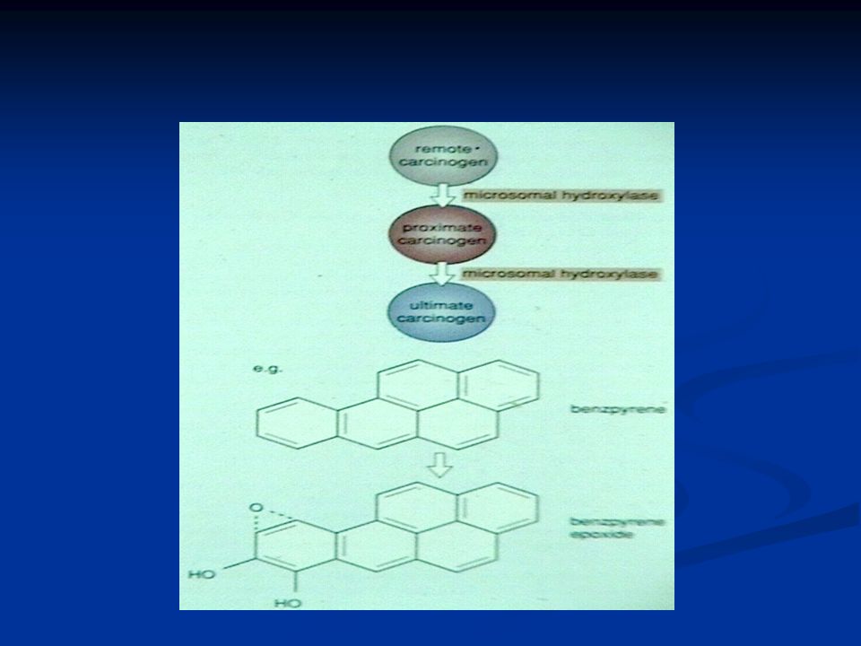
Carcinogenic Agents Chemical Carcinogens Natural or synthetic Natural or synthetic Direct reacting or indirect Direct reacting or indirect Indirect need metabolic conversion to be active and carcinogenic Indirect need metabolic conversion to be active and carcinogenic Indirect chemicals are called “ procarcinogens “ and their active end products are called “ ultimate carcinogens” Indirect chemicals are called “ procarcinogens “ and their active end products are called “ ultimate carcinogens”

75
Carcinogenic Agents Chemical Carcinogens All direct reacting and ultimate chemical carcinogens are highly reactive as they have electron-deficient atoms All direct reacting and ultimate chemical carcinogens are highly reactive as they have electron-deficient atoms They react with the electron rich atoms in RNA, DNA and other cellular proteins They react with the electron rich atoms in RNA, DNA and other cellular proteins

76
Carcinogenic Agents Chemical Carcinogens Examples: Examples: Alkylating agents Alkylating agents Polycyclic hydrocarbons: Polycyclic hydrocarbons: Cigarette smoking Cigarette smoking Animal fats during broiling meats Animal fats during broiling meats Smoked meats and fish Smoked meats and fish

77
Carcinogenic Agents Chemical Carcinogens Aromatic amines and azo dyes: Aromatic amines and azo dyes: -naphthylamine cause bladder cancer in rubber industries and aniline dye -naphthylamine cause bladder cancer in rubber industries and aniline dye Some azo dyes are used to color food also can cause bladder cancer Some azo dyes are used to color food also can cause bladder cancer

78
Carcinogenic Agents Chemical Carcinogens Other substances: Other substances: Nitrosamines and nitrosamides are used as preservatives. They cause gastric cancer. Nitrosamines and nitrosamides are used as preservatives. They cause gastric cancer. Aflatoxin B: produced by aspirigillus growing on improperly stored grains. It cause hepatocellular carcinoma Aflatoxin B: produced by aspirigillus growing on improperly stored grains. It cause hepatocellular carcinoma

79
Carcinogenic Agents Chemical Carcinogens Mechanism of action of chemical carcinogens: Mechanism of action of chemical carcinogens: Most of them are mutagenic. i.e. cause mutations Most of them are mutagenic. i.e. cause mutations RAS and P53 are common targets RAS and P53 are common targets

80
Carcinogenic Agents Radiation Carcinogenesis UV rays of sunlight UV rays of sunlight X-rays X-rays Nuclear radiation Nuclear radiation Therapeutic irradiations Therapeutic irradiations Radiation has mutagenic effects: chromosomes breakage, translocations, and point mutations Radiation has mutagenic effects: chromosomes breakage, translocations, and point mutations

81
Carcinogenic Agents Radiation Carcinogenesis UV rays of sunlight : UV rays of sunlight : Can cause skin cancers: melanoma, squamous cell carcinoma, and basal cell carcinoma Can cause skin cancers: melanoma, squamous cell carcinoma, and basal cell carcinoma It is capable to damage DNA It is capable to damage DNA With extensive exposure to sunlight, the repair system is overwhelmed skin cancer With extensive exposure to sunlight, the repair system is overwhelmed skin cancer They cause mutations in P53 gene They cause mutations in P53 gene

82
Carcinogenic Agents Viral and Microbial oncogenesis Viral and Microbial oncogenesis DNA viruses DNA viruses RNA viruses RNA viruses other organisms other organisms

83
Carcinogenic Agents Viral Carcinogenesis carry genes that induce cell replication as part of the viral life cycle host cell has endogenous genes that maintain the normal cell-cycle Viral infection mimics or blocks these normal cellular signals necessary for growth regulation

84
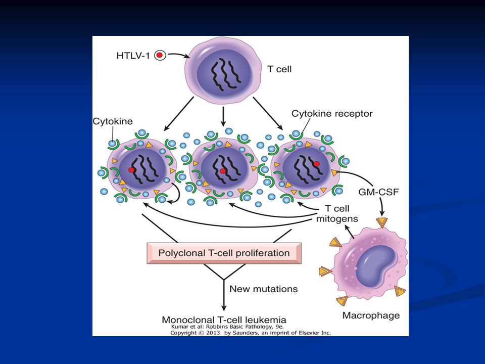
Carcinogenic Agents Viral Carcinogenesis RNA Oncogenic viruses Human T-Cell Leukemia Virus type 1 (HTLV-1) RNA retrovirus targets / transforms T-cells RNA retrovirus targets / transforms T-cells causes T-Cell leukemia/Lymphoma causes T-Cell leukemia/Lymphoma Endemic in Japan and Caribbean Endemic in Japan and Caribbean Transmitted like HIV but only 1% of infected develop T-Cell leukemia/Lymphoma Transmitted like HIV but only 1% of infected develop T-Cell leukemia/Lymphoma 20-30 year latent period 20-30 year latent period
 RNA retrovirus targets / transforms T-cells RNA retrovirus targets / transforms T-cells causes T-Cell leukemia/Lymphoma causes T-Cell leukemia/Lymphoma Endemic in Japan and Caribbean Endemic in Japan and Caribbean Transmitted like HIV but only 1% of infected develop T-Cell leukemia/Lymphoma Transmitted like HIV but only 1% of infected develop T-Cell leukemia/Lymphoma year latent period year latent period")
85
Carcinogenic Agents Viral Carcinogenesis RNA Oncogenic viruses Human T-Cell Leukemia Virus type 1 (HTLV-1) No cure or vaccine No cure or vaccine Treatment : chemotherapy with common relapse Treatment : chemotherapy with common relapse
 No cure or vaccine No cure or vaccine Treatment : chemotherapy with common relapse Treatment : chemotherapy with common relapse")
87
Carcinogenic Agents Viral Carcinogenesis DNA Oncogenic Viruses virus DNA forms stable association with host’s DNA virus DNA forms stable association with host’s DNA transcribed viral DNA transforms host cell transcribed viral DNA transforms host cell Examples: papilloma viruses Examples: papilloma viruses Epstein-Barr (EBV) Hepatitis B (HBV) Kaposi sarcoma herpes virus
 Hepatitis B (HBV) Kaposi sarcoma herpes virus")
88
Carcinogenic Agents Viral Carcinogenesis Human Papillomavirus (HPV) 70 types 70 types squamous cell carcinoma of squamous cell carcinoma of cervix cervix anogenital region anogenital region mouth mouth larynx larynx
 70 types 70 types squamous cell carcinoma of squamous cell carcinoma of cervix cervix anogenital region anogenital region mouth mouth larynx larynx")
89
Carcinogenic Agents Human Papillomavirus (HPV) sexually transmitted sexually transmitted Cervical cancer Cervical cancer 85% have types 16 and 18 85% have types 16 and 18 Genital warts Genital warts types 6 and 11 types 6 and 11
 sexually transmitted sexually transmitted Cervical cancer Cervical cancer 85% have types 16 and 18 85% have types 16 and 18 Genital warts Genital warts types 6 and 11 types 6 and 11")
90
Carcinogenic Agents Human Papillomavirus (HPV) HPV causing benign tumors: HPV causing benign tumors: types 6, 11 types 6, 11 HPV causing malignant tumors : HPV causing malignant tumors : types 16, 18, 31 types 16, 18, 31 vDNA integrates w/ host vDNA integrates w/ host
 HPV causing benign tumors: HPV causing benign tumors: types 6, 11 types 6, 11 HPV causing malignant tumors : HPV causing malignant tumors : types 16, 18, 31 types 16, 18, 31 vDNA integrates w/ host vDNA integrates w/ host")
91
Carcinogenic Agents Viral Carcinogenesis HPV (types 16 and 18) HPV (types 16 and 18) over-expression of Exon 6 and 7 over-expression of Exon 6 and 7 E6 protein binds to Rb tumor suppressor E6 protein binds to Rb tumor suppressor replaces normal transcription factors replaces normal transcription factors decreases Rb synthesis decreases Rb synthesis E7 protein binds to P53 E7 protein binds to P53 facilitates degradation of P53 facilitates degradation of P53
 HPV (types 16 and 18) over-expression of Exon 6 and 7 over-expression of Exon 6 and 7 E6 protein binds to Rb tumor suppressor E6 protein binds to Rb tumor suppressor replaces normal transcription factors replaces normal transcription factors decreases Rb synthesis decreases Rb synthesis E7 protein binds to P53 E7 protein binds to P53 facilitates degradation of P53 facilitates degradation of P53")
92
Carcinogenic Agents Viral Carcinogenesis HPV infection alone is not sufficient - HPV infection alone is not sufficient - other risk factors: other risk factors: cigarette smoking cigarette smoking coexisting infections coexisting infections hormonal changes hormonal changes

93
Carcinogenic Agents Viral Carcinogenesis Epstein-Barr Virus Epstein-Barr Virus common virus worldwide common virus worldwide Infects B lymphocytes and epithelial cells of oropharynx Infects B lymphocytes and epithelial cells of oropharynx causes infectious mononucleosis causes infectious mononucleosis EBV infection may cause malignancy EBV infection may cause malignancy Burkitt’s Lymphoma Burkitt’s Lymphoma B cell lymphoma in immunosuppressed B cell lymphoma in immunosuppressed Nasopharyngeal carcinoma Nasopharyngeal carcinoma

94
Carcinogenic Agents Viral Carcinogenesis Epstein-Barr Virus related Nasopharyngeal carcinoma Nasopharyngeal carcinoma Cancer of nasopharygeal epithelium Cancer of nasopharygeal epithelium Endemic in South China, parts of Africa Endemic in South China, parts of Africa 100% of tumors contain EBV genome in endemic areas 100% of tumors contain EBV genome in endemic areas

95
Carcinogenic Agents Viral Carcinogenesis Epstein-Barr Virus related Burkitt Lymphoma Burkitt Lymphoma highly malignant B cell tumor sporadic rare occurrence worldwide most common childhood tumor in Africa ) all cases have t(8:14)
 all cases have t(8:14)")
96
Carcinogenic Agents Viral Carcinogenesis Epstein-Barr Virus related causes B lymphocyte cell proliferation causes B lymphocyte cell proliferation loss of growth regulation loss of growth regulation predisposes to mutation, esp. t(8:14) predisposes to mutation, esp. t(8:14)
 predisposes to mutation, esp. t(8:14).")
97
Carcinogenic Agents Viral Carcinogenesis Hepatitis B virus (HBV) Strong association with Liver Cancer World-wide, but HBV infection is most common in Far East and Africa HBV infection incurs up to 200-fold risk
 Strong association with Liver Cancer World-wide, but HBV infection is most common in Far East and Africa HBV infection incurs up to 200-fold risk")
98
Carcinogenic Agents Helicobacter Pylori bacteria infecting stomach implicated in: peptic ulcers gastric lymphoma Mucosal Associated Lymphoid Tumor (MALT) gastric carcinoma
 gastric carcinoma")
Similar presentations


?>")


 –Lung tissue –Breast tissue (glands/ducts) –Prostate (gland) –White blood cells.>")


