Download presentation
Presentation is loading. Please wait.

1
Drosophila melanogaster Genetic studies Microsurgical manipulation One of the best understood developmental systems 13,600 genes Axis determination Signaling pathway Transcriptional regulation P48-52

2
4 stages: embryo, larva, pupa, adult

3
Rapid division 9 mins/division 9 divisions 13 divisions Single cell

4
Transgenic flies

5
One single epithelial layer –all tissues Mesoderm—muscle, connective tissues Endoderm---midgut (foregut and hidgut- Ectoderm) Ectoderm---nervous tissue and epidermis
 Ectoderm---nervous tissue and epidermis")
6
gastrulation

7
Larva hatch 24 hrs Acron: associated with head Telson:posterior terminal structure 3 thorcic and 8 abdominal segments—specialization in cuticle (denticle belts and cuticular structure)
")
8
Genitalia Sex comb Pigmentation Small wing p. 421-431

9
Sex determining signal--- Sex-lethal (X chromosome) Transformer-spliced + transformer 2
 Transformer-spliced + transformer 2")
10
2X higher numerator Repression by autosome

11
Dosage compensation Barr body Xist-non-coding RNA Male specific gene Repressed by Sxl

12
Primordial germ cell -special cytoplasm Germ plasm-polar granules, pole plasm

13
Oskar—organization and assembly of the pole plasm mRNA-posterior pole—3’ untranslated region

14
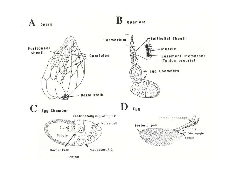
Polarization of the body axes during oogenesis

16
Cyst formation: 16 cell cyst enter a long S phase only one (Oocyte) continues meiosis Oocyte—4 ring canals 15 cells become nurse cells after germarium nurse cells left meiotic cycle, grow rapidly without division, and form polytene chromosomes
 continues meiosis Oocyte—4 ring canals 15 cells become nurse cells after germarium nurse cells left meiotic cycle, grow rapidly without division, and form polytene chromosomes")
17
A/P during oogenesis The oocyte move towards one end in contact with follicle cells Both the oocyte and the posterior follicle cells express high levels of the E-cadherin If E-cadherin is removed, the oocyte is randomly positioned. Then the oocyte induces surrounding follicle cell to adopt posterior fate.

18
Microtubule cytoskeleton reorganization is essential for localization of bicoid and oskar mRNA

19
Maternal effect mutations---reguired for pole plasm assembly Lack polar granules: grandchildless mutation (homozygote female— Progeny—sterile) Central role: Oskar, Vasa, and Tudor Pole cell number = amount of oskar RNA Ectopic pole cells: oskar RNA at the anterior pole MtlrRNA (mitochondrically encoded large ribosomal RNA) + gcl RNA for pole cell formation antisense reduce pole cells mtlrRNA rescue UV-irradation Vasa: DEAD-box RNA helicase— translational regulator
 Central role: Oskar, Vasa, and Tudor Pole cell number = amount of oskar RNA Ectopic pole cells: oskar RNA at the anterior pole MtlrRNA (mitochondrically encoded large ribosomal RNA) + gcl RNA for pole cell formation antisense reduce pole cells mtlrRNA rescue UV-irradation Vasa: DEAD-box RNA helicase— translational regulator")
20
Germ cell—extragonadal origin, migrate to reach the somatic gonad a. posterior end b. gastrulation c. migrate dorsally through the wall of the posterior midgut d. associate with the somatic gonadal precursors e. GC align with somatic gonadal mesoderm f. coalesce to form the embryonic gonad Germ cell migration

21
PGC migration----Genes and mechanisms Genetic screen—somatically expressed genes Guidance (cues): Wunen: repulsive signal (exclude migrating pole cells from wrong places) Misexpression wunen: transform a tissue permissive to PGC to repulsive one Phosphatidic acid phosphatase 2 (transmembrane protein) Columbus: factor (gonadal mesoderm) attracts pole cells Misexpression Columbus—attract PGCs to tissues other than gonadal mesoderm 3-hydroxy-3-methylglutaryl coenzymeA reductase (cholesterol biosynthesis in human, but fly does not make cholesterol) nanos, pumilio mutants stall at the outer gut surface differentiate prematurely---act as complete migration to the somatic gonads nanos target: RNA binding protein Sex lethal (Sxl)---splicing and translational regulation also depend on specific germ plasm components, e.g polar granule component (Pgc)
: Wunen: repulsive signal (exclude migrating pole cells from wrong places) Misexpression wunen: transform a tissue permissive to PGC to repulsive one Phosphatidic acid phosphatase 2 (transmembrane protein) Columbus: factor (gonadal mesoderm) attracts pole cells Misexpression Columbus—attract PGCs to tissues other than gonadal mesoderm 3-hydroxy-3-methylglutaryl coenzymeA reductase (cholesterol biosynthesis in human, but fly does not make cholesterol) nanos, pumilio mutants stall at the outer gut surface differentiate prematurely---act as complete migration to the somatic gonads nanos target: RNA binding protein Sex lethal (Sxl)---splicing and translational regulation also depend on specific germ plasm components, e.g polar granule component (Pgc)")
22
Patterning of the fly embryo

23
Localized mRNA and Proteins Translated after fertilization— Positional information to activate zygotic genes parasegment Pattern in the segment Segment identities Temporal sequence

24
Appendages:imaginal discs—pattern formation Ectoderm invagination-epithelium(20-40 cells—larva 1000X) Specification occurs –segment being patterned-according to it p.350-358
 Specification occurs –segment being patterned-according to it p")
25
A/P and D/V compartment

26
Wing blade Ventral fold under dorsal-double layers of epithelium

27
Signal region and the compartment Maintain compartment boundaries—communication between compartments Hh—10 cells, induces expression of Dpp through activation of Ci

28
The hedgehog signaling pathway Without signal—Ci is processed as a repressor into nucleus With signal---full length Ci acts as an activator in the nucleus

29
Intercellular signaling set up PS boundary Wg distributed asymmetrically—less in posterior (endocytosis and degradation)
")
30
TGFb, Activin: R-Smad 2,3 BMPs: R-Smad 1, 5, 8 Common Smad4 Inhibitory Smads: I-Smad6, 7—recruting Smurf (ubiquitin ligase to receptor) Cell, 95,737,1998
 Cell, 95,737,1998")
31
Smad= Sma + Mad Sma-C. elegans Mad-Fly

32
Dpp-secreted into both compartment Long range signal—expression of spalt

33
Patterning the A/P axis of the wing disc Dpp-morphogen Low level—omb High level—spalt 1.Clones can’t respond to Dpp— no spalt and omb 2. Ectopic hh-Dpp—localized activation of spalt and omb around the hh clones 3. Ts mutant of dpp—reduction in the region with expression of low Threshold genes—omb 4. Clones expression low or high Dpp—distinquish these two types of genes

34
Ectopic expression of Hh and Dpp L4—compartment boundary L3– Hh L2 ---adjacent to cells expressing spalt Ectopic Hh in posterior—no effects In anterior—mirrow-symmetric repeated pattern Hh--Dpp

35
Expression of Wingless (green) and vestigial Homeotic selector gene— apterous (Lmx-1) induces fringe and Serrate, then Notch receptor activation – Leading to Wingless expression Wg—achaete, distal-less, vestigial Wingless (green) Vestigial (red) D/V boundary Dpp, Wg morphogen GFP-dpp active transportation— Endocytosis Regulate their receptors Dpp inhibits receptor—thick veins Dpp high--receptor low, and dpp low Receptor high—1, prevent spreading 2,cells reach threshold at low Dpp
 and vestigial Homeotic selector gene— apterous (Lmx-1) induces fringe and Serrate, then Notch receptor activation – Leading to Wingless expression Wg—achaete, distal-less, vestigial Wingless (green) Vestigial (red) D/V boundary Dpp, Wg morphogen GFP-dpp active transportation— Endocytosis Regulate their receptors Dpp inhibits receptor—thick veins Dpp high--receptor low, and dpp low Receptor high—1, prevent spreading 2,cells reach threshold at low Dpp")
36
Notch –transmembrane protein DSL family—Delta, Serrate, Lag-2 Kuzbanian—cleave Notch ECD Presenilin—cleave Notch ICD nervous system: selection of a single neuroblast (lateral inhibition)
")
37
Leg disc extension Jointed tubes of epidermis—secrete the hard cuticle (exoskeleton), inside: Muscles, nerves
, inside: Muscles, nerves")
38
Fate map of the leg imaginal disc Proximo-distal segment Center—distal end

39
Signaling centers in A/P compartment Dpp, wg meets—Dll (distal end) homothorax (proximal)
 homothorax (proximal)")
40
Regional subdivision Dpp, and Wg induce Dll and inhibit homothorax Activates dachshund between Dll and hth

41
Butterfly wing pattern Eyespot center—distal-less

42
Segmental identity of imaginal disc Homeotic selector genes Similar signal into different structures— Different interpretation— controlled by Hox genes Antennapedia—PS4 and 5– 2 pairs of legs If in head, antennae into legs (clones) – which part of the leg—depends on their position along the P/D axis (positional values are similar) Hth (proximal) and Dll (distal)—in antennae and leg In combination as selector to specify antenna No Hth, antenna into leg In leg: antennapedia prevents Hth and Dll acting together Dominant antennapedia mutant (gene on)— blocks Hth and Dll in antennae disc, so leg forms
 – which part of the leg—depends on their position along the P/D axis (positional values are similar) Hth (proximal) and Dll (distal)—in antennae and leg In combination as selector to specify antenna No Hth, antenna into leg In leg: antennapedia prevents Hth and Dll acting together Dominant antennapedia mutant (gene on)— blocks Hth and Dll in antennae disc, so leg forms")
43
Imaginal discs and adult thoracic appendages Bithorax mutation—Ubx misexpressed T3 into T2 –anterior haltere into Anterior wing

44
Postbithorax muation (pbx)— Regulatory region of the Ubx— Posterior of the haltere into wing If both mutations—effect is additive— Four wings
— Regulatory region of the Ubx— Posterior of the haltere into wing If both mutations—effect is additive— Four wings")
Similar presentations


 page 1 © copyright Bruce Blumberg 2000. All rights reserved Bio 108 - 3/13/2000 Molecular Genetics of Pattern Formation.>")

