Download presentation
Presentation is loading. Please wait.

1
Threats to fetal oxygenation during labor– what is the context of your epidural? Tom Archer, MD, MBA January 31, 2012

2
http://darksideofthecatalogue.wordpress.com/2011/11/22/light-at-the-end-of-the-tunnel-is-glowing-thing-23-12/ The fetus floats at the far end of a tunnel of oxygen delivery. If the tunnel is blocked, the fetus dies.

3
Systematic approach to thinking about the “risk context” of an epidural We can do the “usual things” directly related to hypotension more intelligently (fluids, pressors, LUD, O2. Think about “less usual things” (hyperstimulation, nuchal cord, pre-existing disease that make patient more precarious. Epidural may only be tangentially “to blame”– or not at all!

4
med.yale.edu http://www.spiralzoom.com/Science/SpiralsHu manBody/SpiralsHumanBody.html The fetal oxygen supply is precarious– both on the fetal and maternal sides of the placental interface

5
Fetal-side (umbilical cord) problems with fetal oxygen supply
 problems with fetal oxygen supply")
6
Nuchal umbilical cord http://library.med.utah.edu/WebPath/PLACHTML/PLAC002.html

7
http://library.med.utah.edu/WebPath/PLACHTML/PLAC010.html Knotted umbilical cord

8
http://midwifemuse.wordpress.com/2008/02/27/vasa-previa/ Vasa previa– fetal blood vessels between presenting part and cervix– will rupture as presenting part descends.

9
http://www.empowher.com/media/reference/umbilical-cord-prolapse Prolapsed umbilical cord

10
Maternal-side threats to fetal oxygen supply

11
Minimal collateral venous return to heart via lumbar and azygos system Open IVC Uncompressed aorta and iliac arteries Figure 1 Healthy, abundant uteroplacental perfusion Upper body Fetal O2 supply

12
Minimal collateral venous return to heart via lumbar and azygos system Open IVC Uncompressed aorta and iliac arteries Figure 2 Uterine contractions periodically deprive placenta of perfusion. Upper body Uterine contractions Fetal O2 supply

13
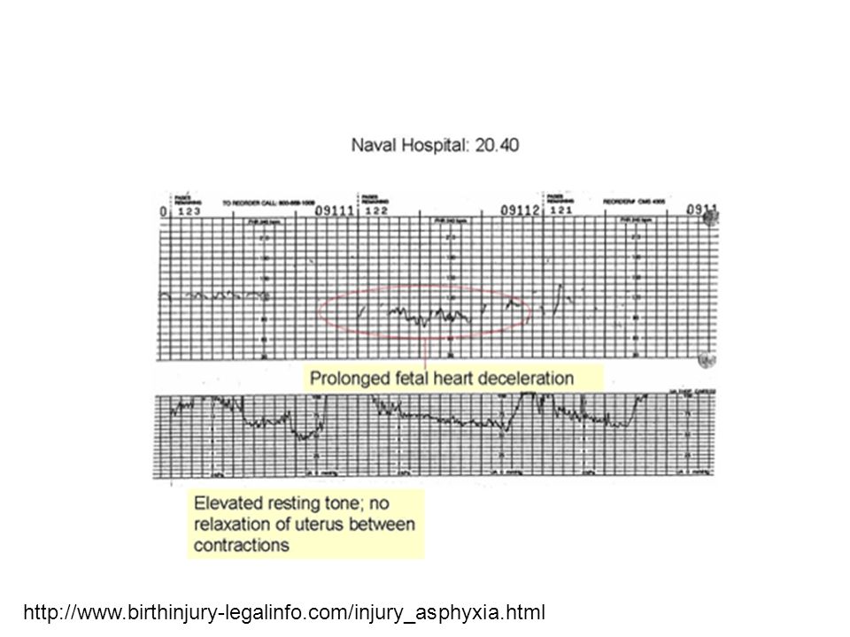
http://www.birthinjury-legalinfo.com/injury_asphyxia.html
14
Increased collateral venous return to heart via lumbar and azygos system Compressed IVC Compressed aorta and iliac arteries ACC Figure 3 Aortocaval compression reduces placental perfusion pressure. Upper body Uterine contractions Fetal O2 supply Uterine mass

15
Manbit images http://www.manbit.com/OA/f28-1.htm

17
Ballas, Mantell, Archer SOAP 2012

20
Michard Both positive pressure ventilation and uterine contractions in the presence of free venous return cause the heart to receive periodic increases in venous return. Could these periodic volume challenges shed light on the parturient’s “volume status?”

21
Increased collateral venous return to heart via lumbar and azygos system Compressed IVC Compressed aorta and iliac arteries ACC Figure 4 Placental vascular disease (e.g. preeclampsia) further reduces placental perfusion. Upper body Uterine contractions Uterine mass Fetal O2 supply Placental vascular disease
 further reduces placental perfusion. Upper body Uterine contractions Uterine mass Fetal O2 supply Placental vascular disease.")
22
www.siumed.edu/~dking2/erg/images/placenta.jpg Say “OUCH!” Pre-E mediators Poor placentation Pre-eclampsia: ischemic chorionic villi release pre-E mediators into maternal blood.

23
http://pharyngula.org/images/preeclampsia_model.jpg Poor-placentation theory of pre-E: Synciotrophoblast invades myometrium but does not denervate spiral arteries of mother properly. Hence, intervillous flow is sub- optimal. Chorionic villi are ischemic and release mediators (VEGF, etc) which damage maternal endothelium.
 which damage maternal endothelium..")
24
Increased collateral venous return to heart via lumbar and azygos system Compressed IVC Compressed aorta and iliac arteries ACC Figure 5 Placental abruption reduces placental volume available for gas exchange Upper body Uterine contractions Uterine mass Fetal O2 supply Placental spiral artery disease Placental abruption or thrombosis

25
http://emedicine.medscape.com/article/252810-overview Placental abruption decreases placental area available for gas exchange.

26
Increased collateral venous return to heart via lumbar and azygos system Compressed IVC Compressed aorta and iliac arteries ACC Figure 6 Epidural may be “straw that breaks camel’s back” and causes “fetal distress”. Upper body Uterine contractions Uterine mass Fetal O2 supply Placental vascular disease Epidural reduces arterial blood pressure Placental abruption or thrombosis

27
http://www.macllp.com/child_birth_anesthesia/epidural_ad.cfm “Routine” epidural.

28
Your next epidural Ask yourself, “What are the pre-existing threats to fetal oxygenation in my patient?” “What special precautions should I take to prevent fetal hypoxia in this patient?” Be attentive to hyperstimulation, preeclampsia, abruption, hypotension, etc.

29
The End

Similar presentations
RBC 2)WBC 3)Platelet.>")


