Download presentation
Presentation is loading. Please wait.

1
Pelvic Anatomy from a Laparoscopic Perspective Tommaso Falcone MD Professor & Chairman Cleveland Clinic Foundation

2
Anatomy & Advanced Laparoscopic Surgery Course

3
Anatomic Areas Anterior abdominal wall Pelvic sidewall Extra-peritoneal spaces –Retropubic space –Presacral space –Pararectal space

4
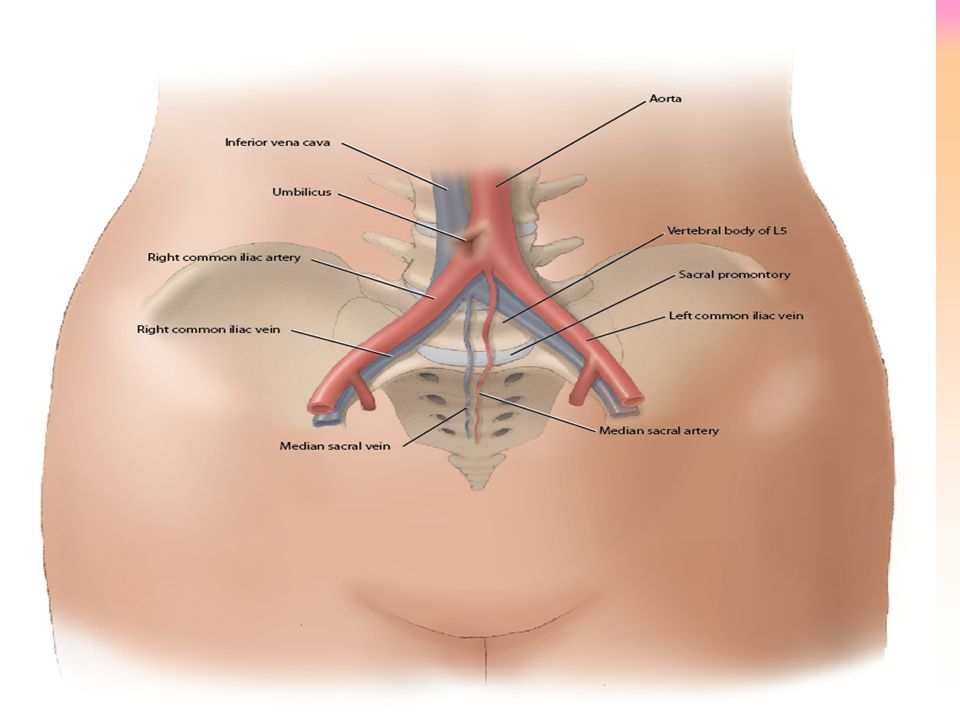
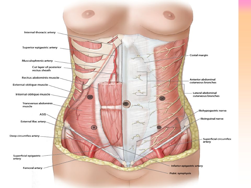
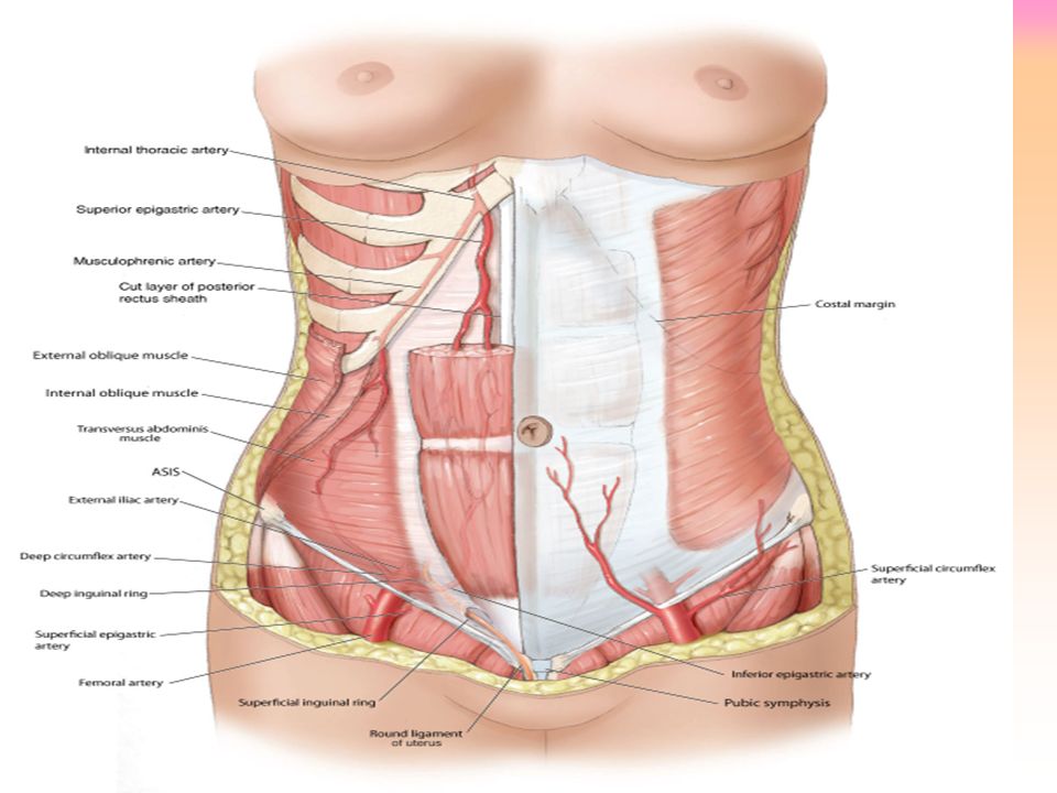
Anterior Abdominal Wall Relationship of the vessels & nerves to potential entry sites for trocars

5
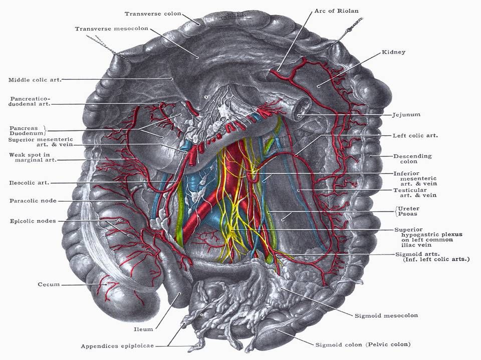
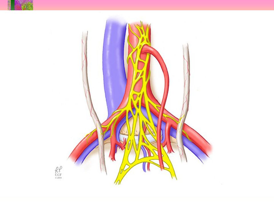
Retroperitoneal Vessels & Umbilicus Bifurcation of the aorta –thin patients at umbilicus –More caudad with increasing weight Left common iliac vein –inferior to the bifurcation of the aorta –crosses the sacrum

17
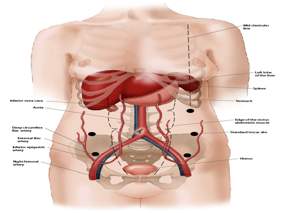
Left Upper Quadrant Insertion 2-cm below the subcostal margin mid- clavicular line Organs –Aorta-11 cm –Spleen-12cm –Stomach-4.4cm –Liver-4.0cm –Left kidney 13.2cm

18
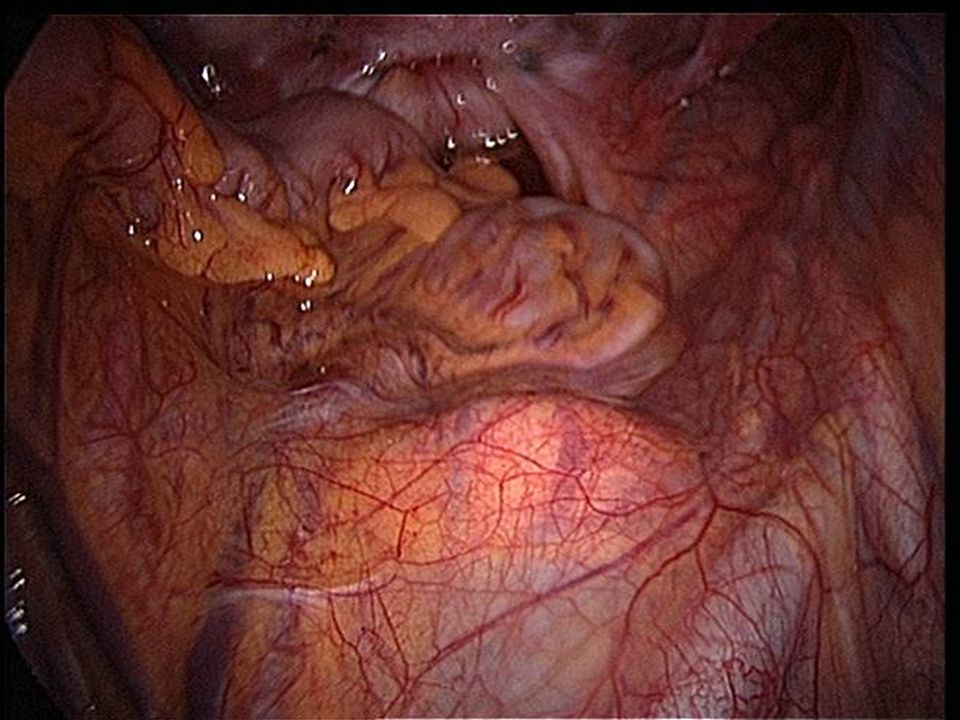
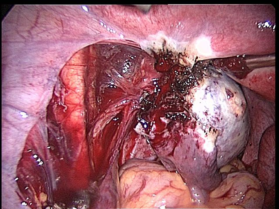
Laparoscopic view of the spleen Spleen is far from the LUQ, unless splenomegaly is present

21
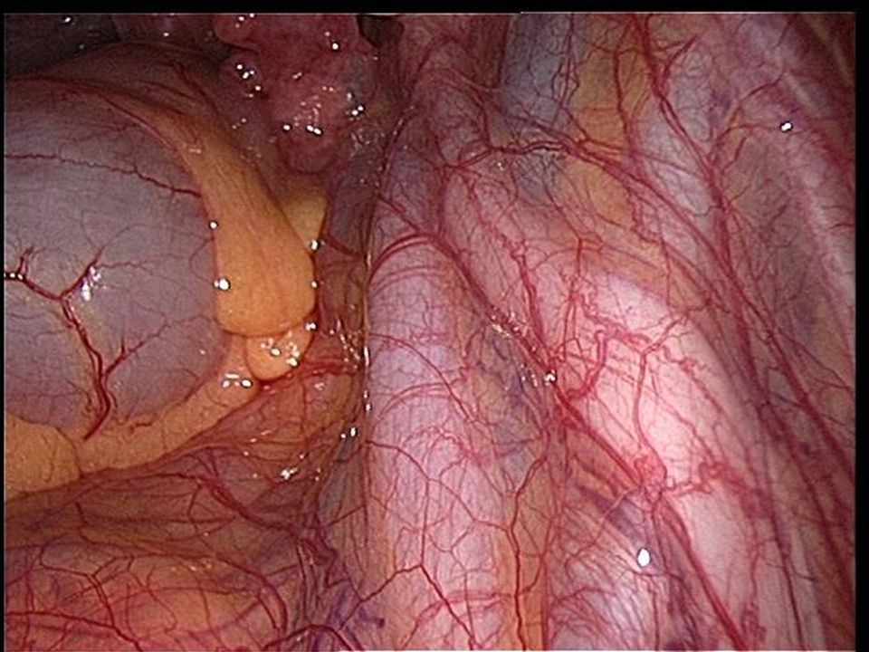
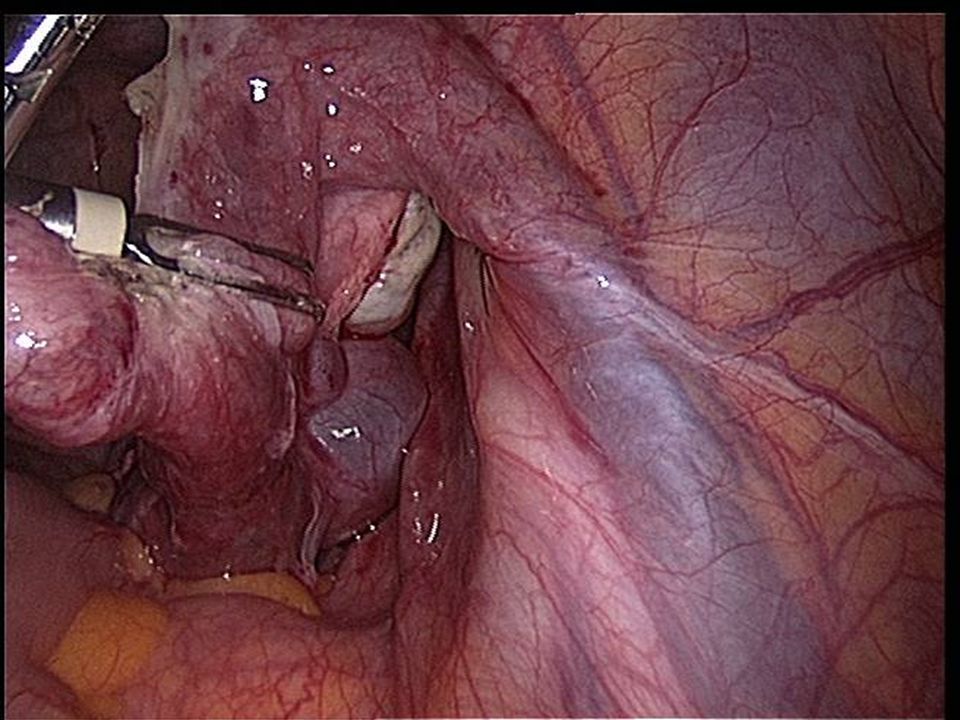
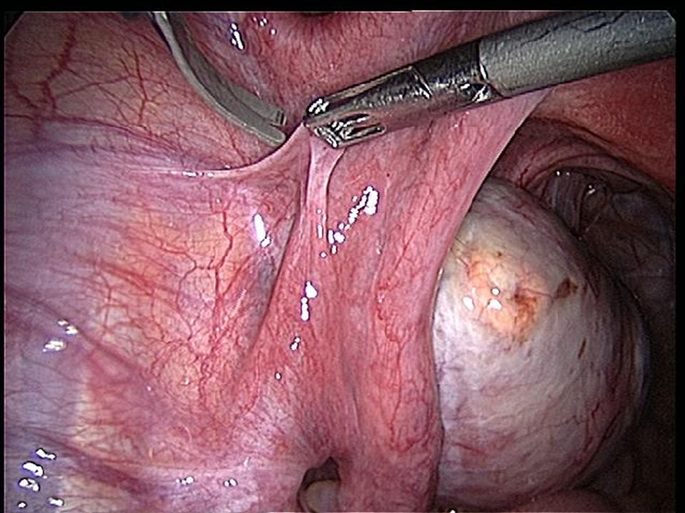
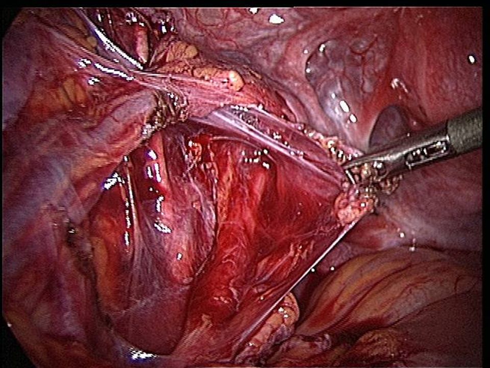
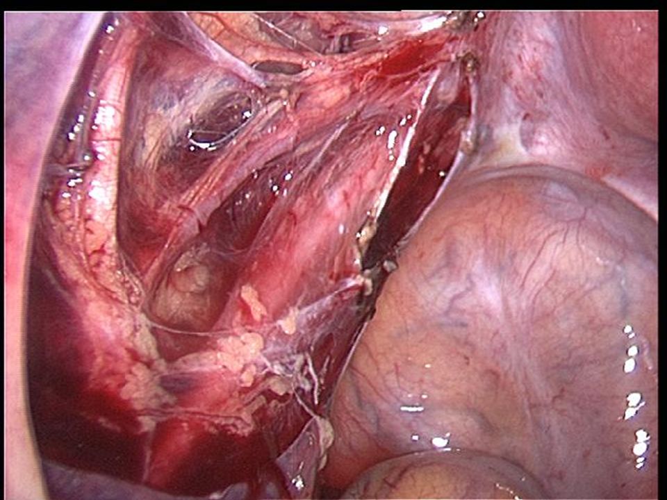
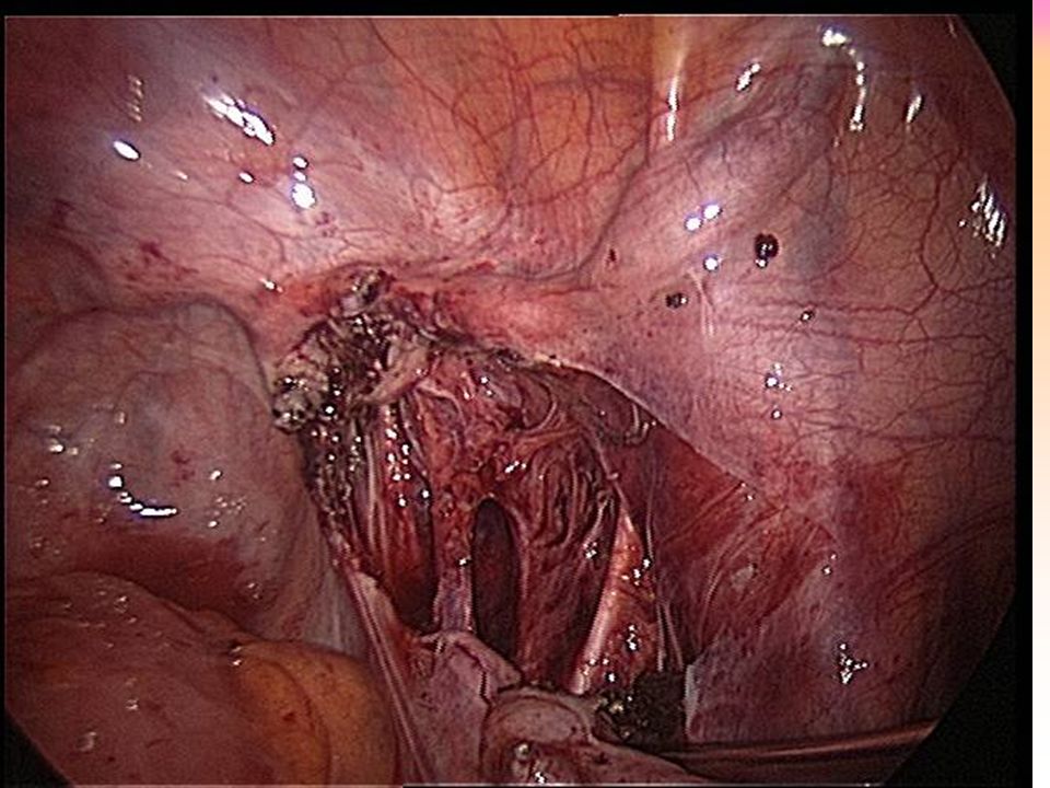
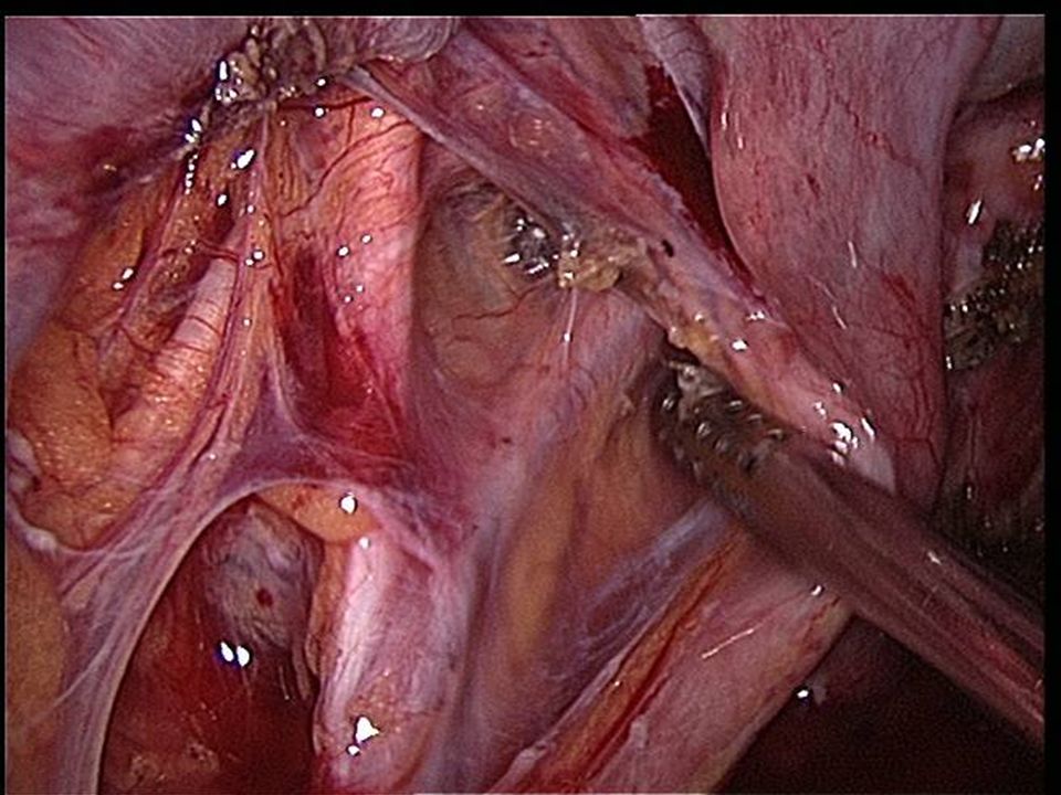
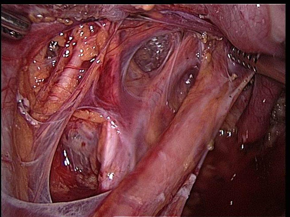
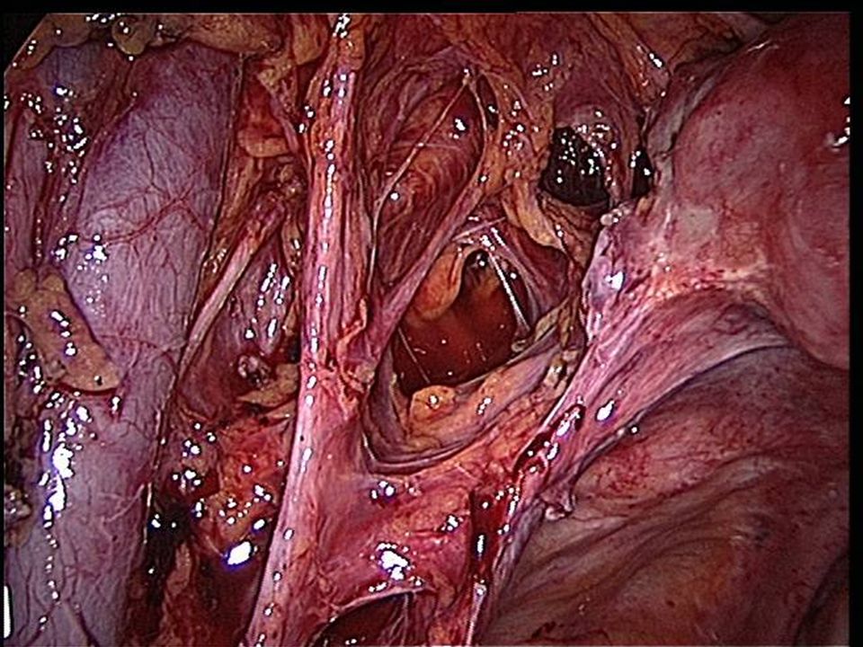
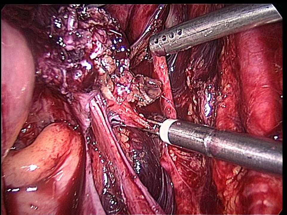
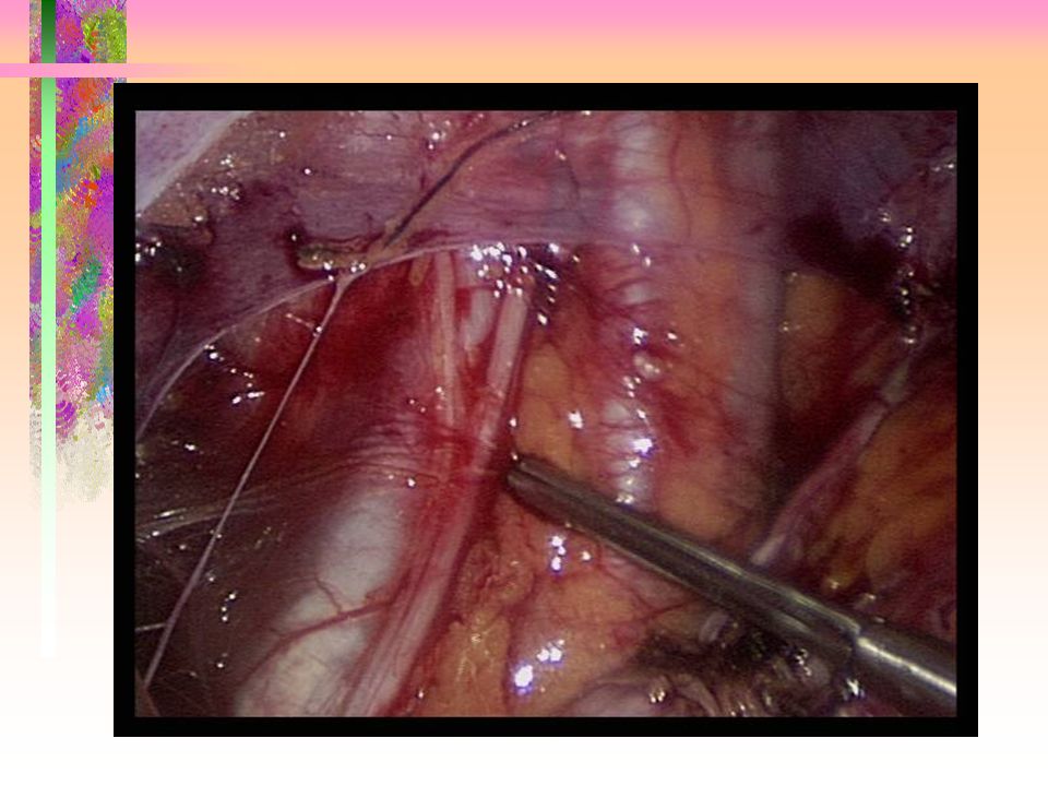
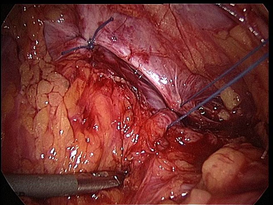
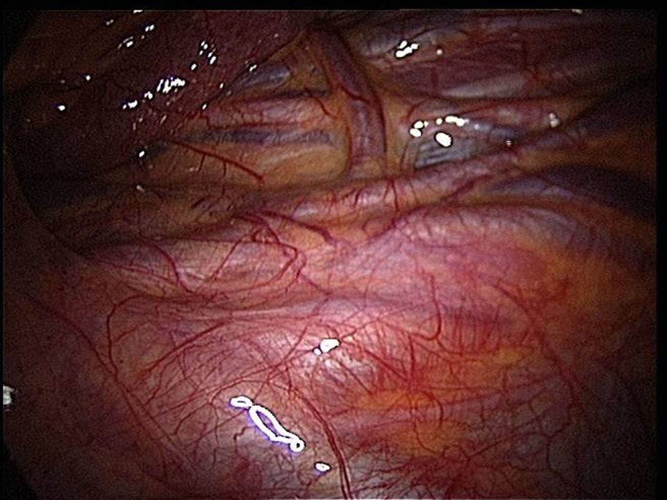
Pelvic Sidewall Anatomy 3 layers –Ureter –Branches of the int.iliac artery –Muscle & nerve

22
Pelvic Sidewall: ureter Pelvic brim –over the common or external iliac –under ovarian vessels Courses anterior to the internal iliac –UNDER THE OVARY –1.5 -2 CM LATERAL UTERO-SACRAL LIGAMENTS Cervix –WITHIN 2CM

32
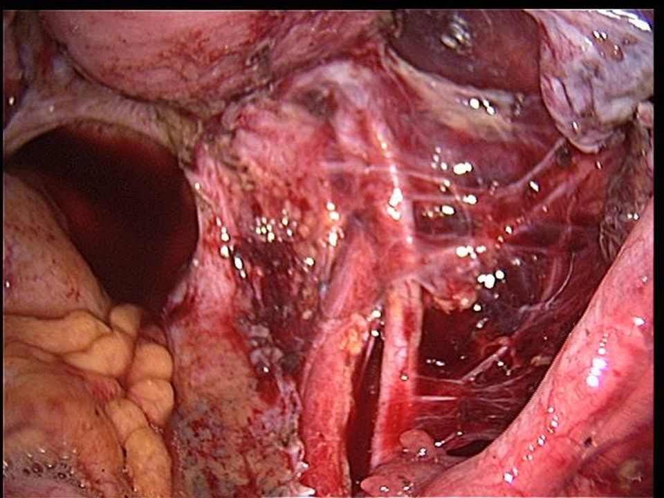
Pelvic Sidewall: Blood vessels Internal iliac artery –anterior & posterior division –Umbilical artery obliterated medial umbilical ligament relationship to the uterine artery

35
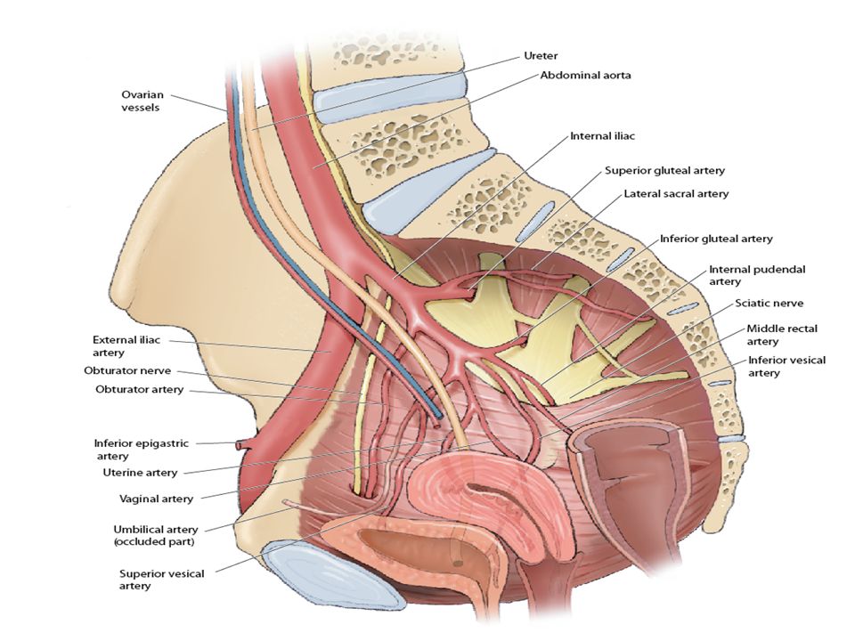
Pelvic Arteriogram

44
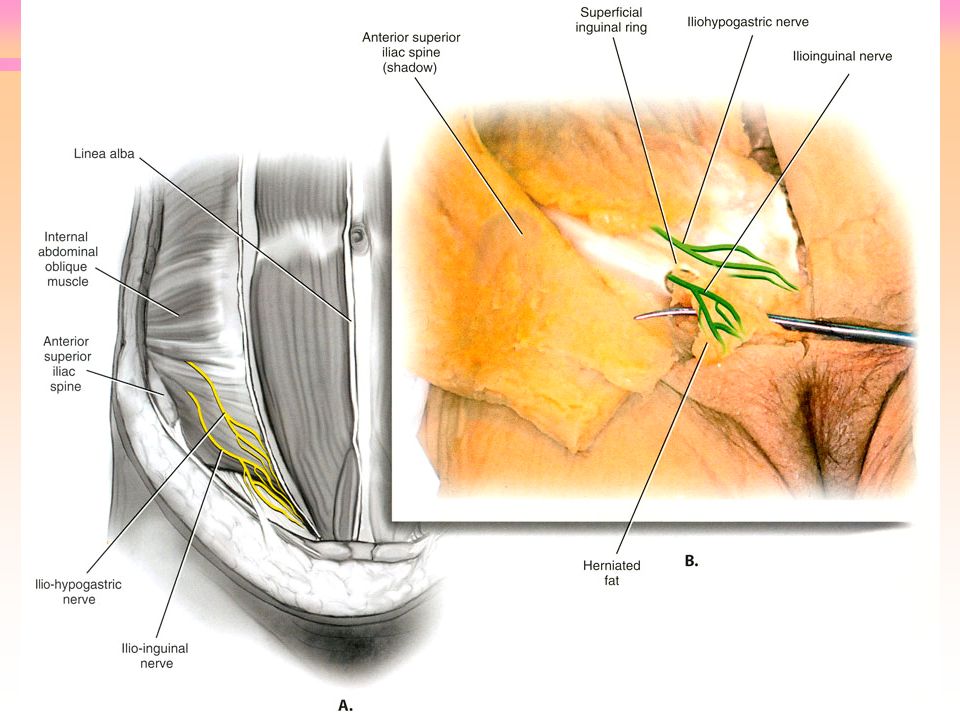
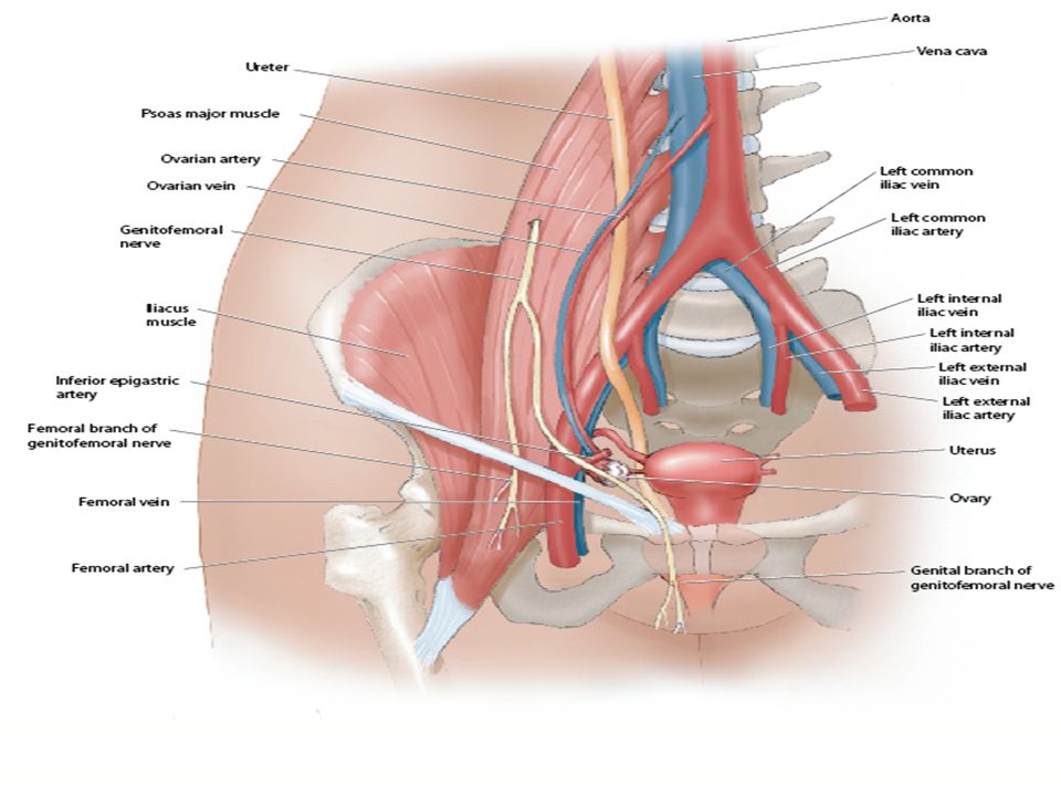
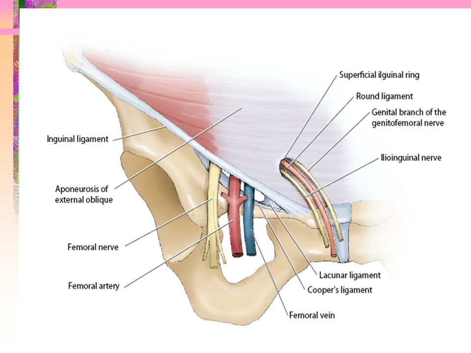
Pelvic & Inguinal Nerves Genito-femoral nerve Femoral nerve

49
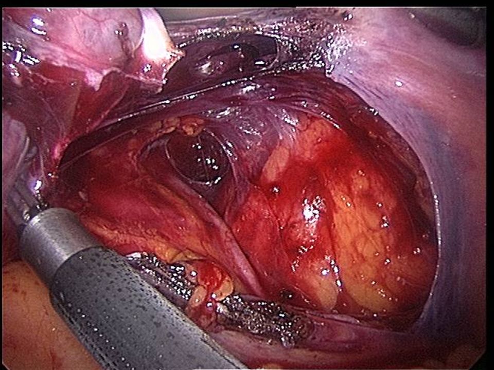
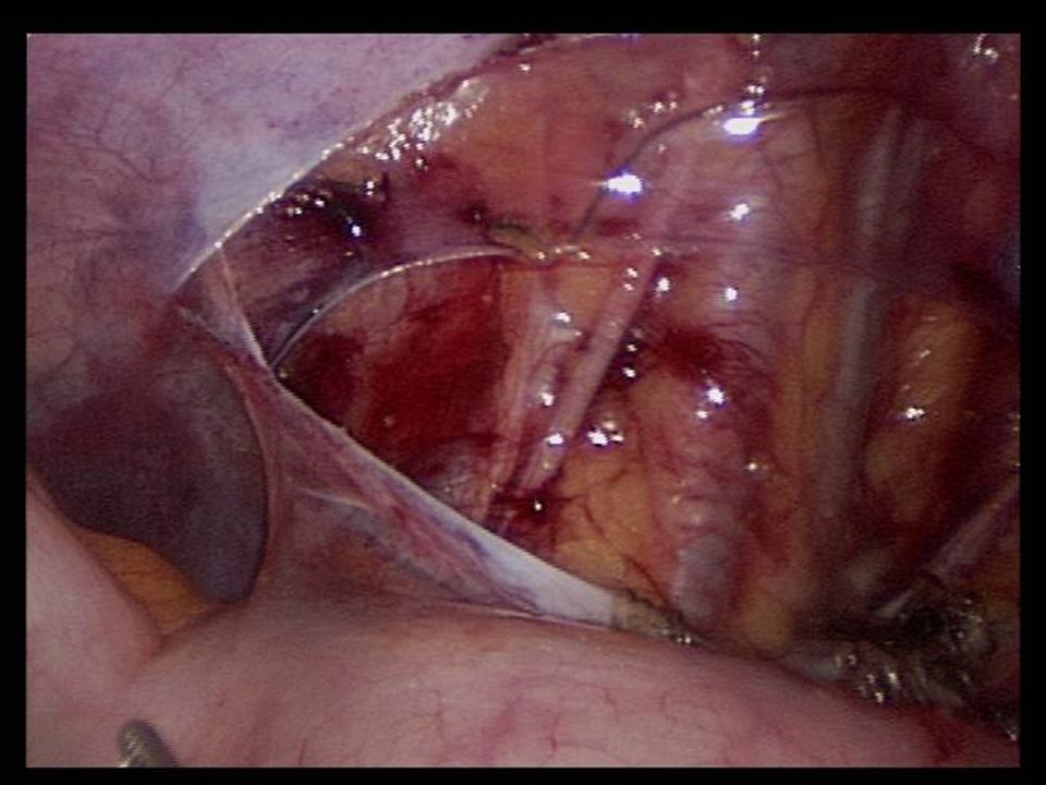
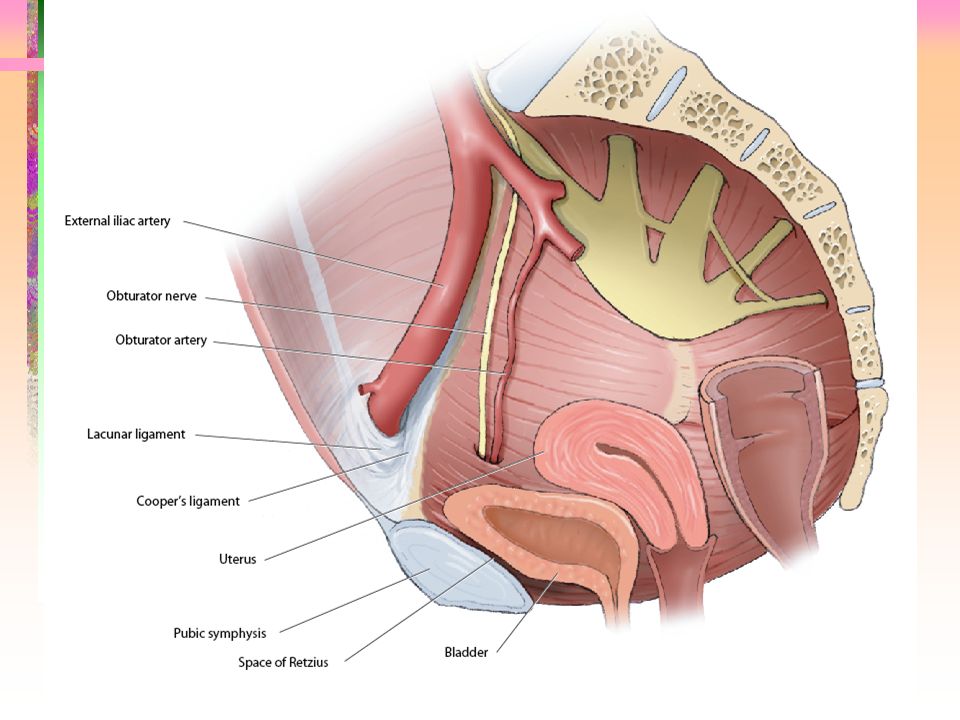
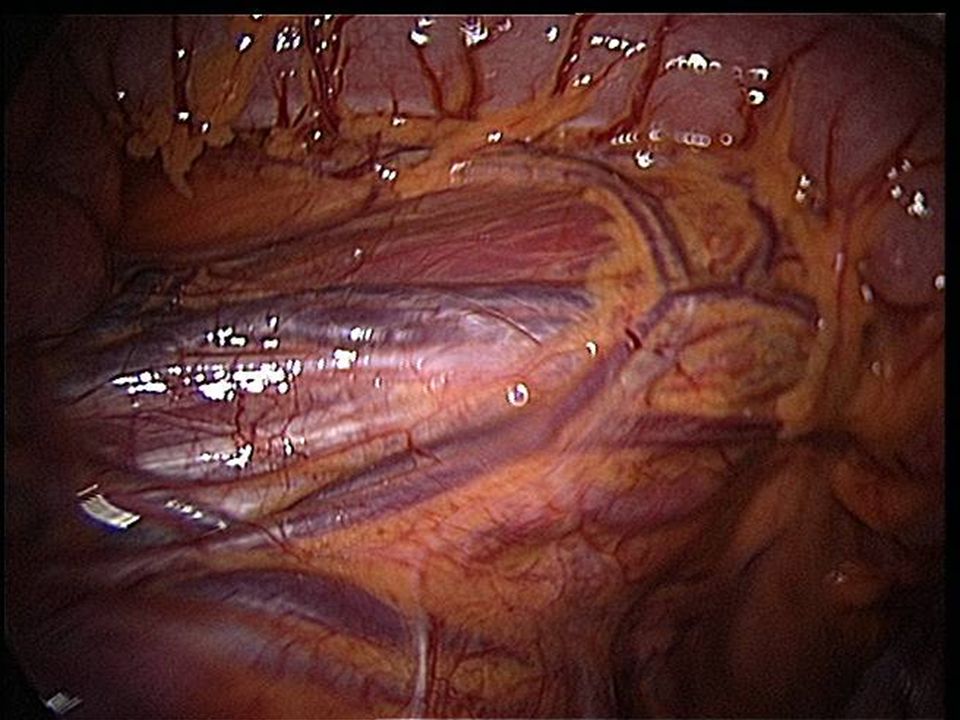
Retropubic Space Anterior –Pubic bone Lateral –Obturator internus muscle, fasciae, neurovascular bundle Posteriorly –bladder & pubocervical fasciae

61
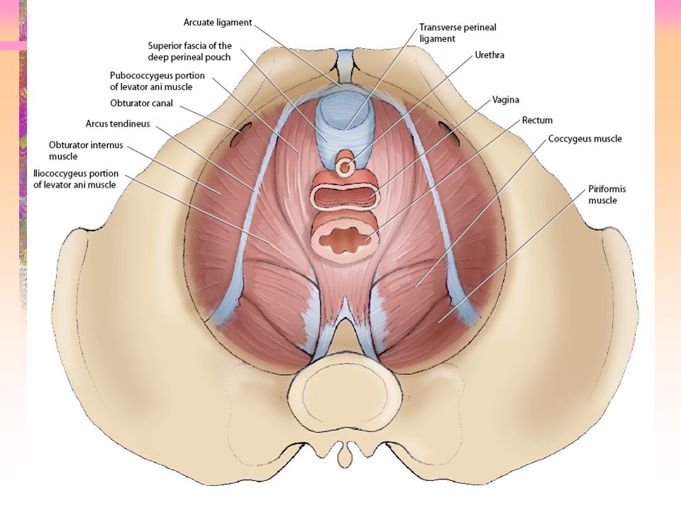
Pelvic Diaphragm Sheet of muscle (Levator ani & coccygeus) covered on both sides by fasciae From pubis to coccyx & is attached to the lateral pelvic wall by a thickened band of obturator fascia called arcus tendineus m. levator ani Anogenital hiatus

62
Pelvic Diaphragm:Muscle Levator Ani –Pubococcygeus (Puborectalis & pubovaginalis) –Iliococcygeus Iliococcygeus portion that arises from the obturator internus muscle (arcus tendineus m. levator ani) & ischial spine Arcus: spine of the ischium forward & upward.
 & ischial spine Arcus: spine of the ischium forward & upward..")
63
Pelvic Diaphragm: Fasciae Parietal fasciae on the muscles Endopelvic fasciae on the pelvic viscera –Attached to the parietal fasciae laterally –Connective tissue attachments stabilize the vagina –Attachment along a line of thickened parietal fasciae called Arcus tendineus fasciae pelvis or white line –Mid-vagina is supported by lateral connections to the white line

71
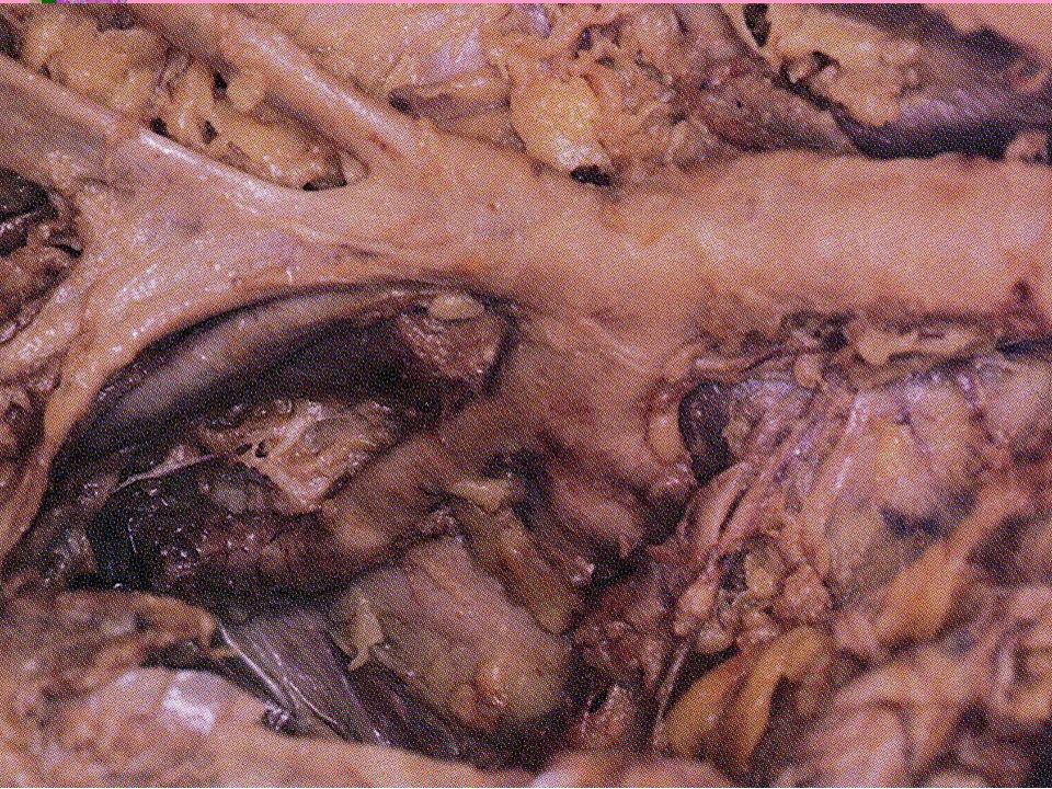
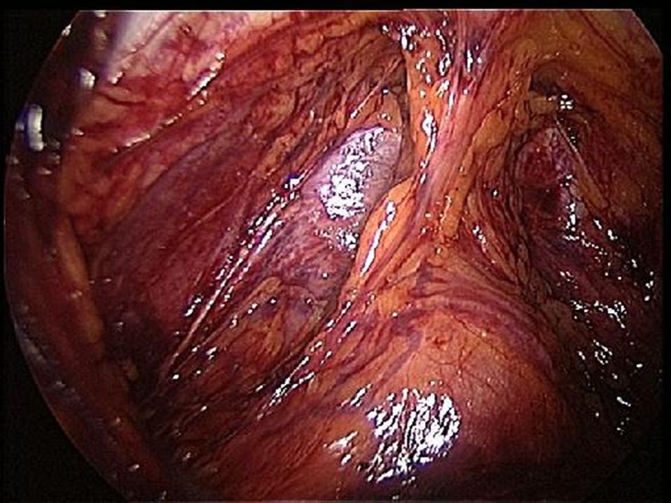
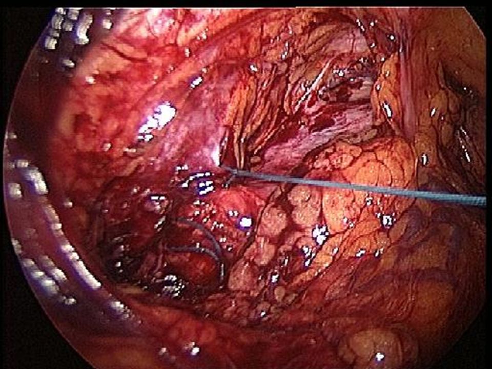
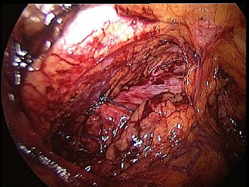
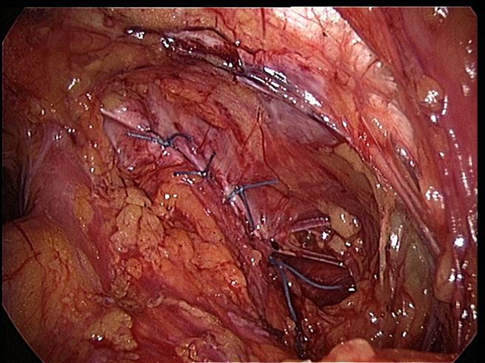
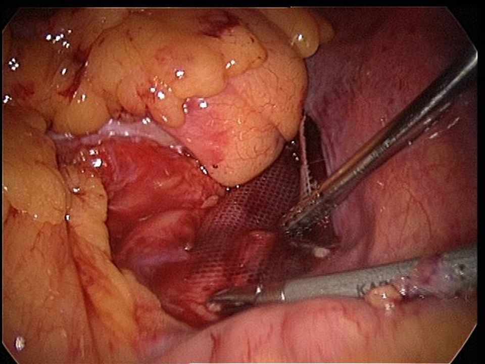
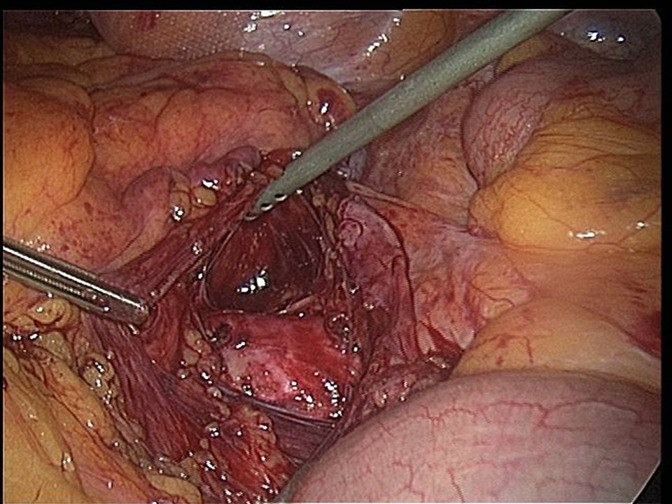
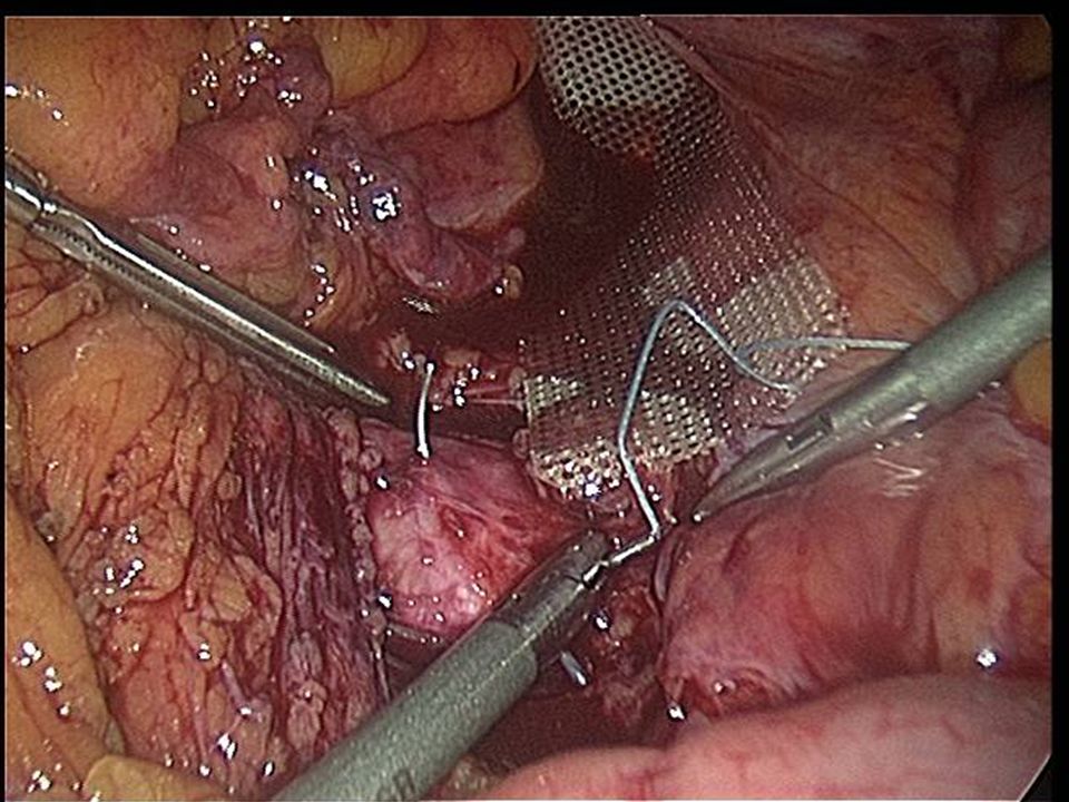
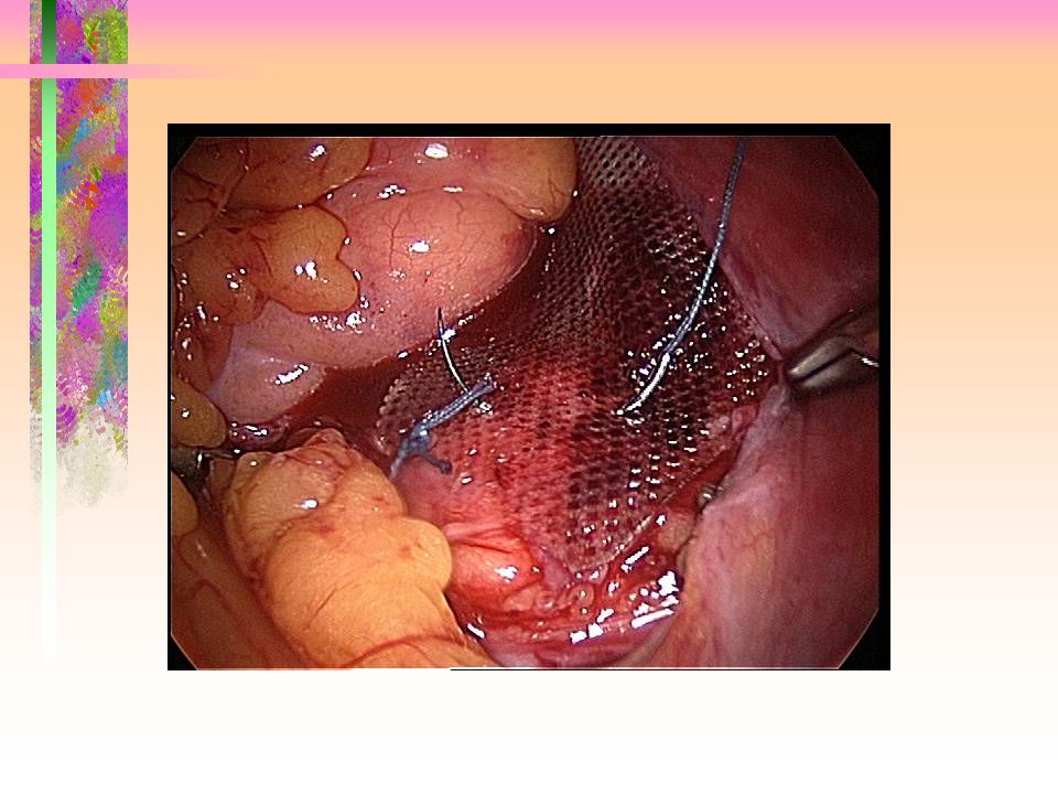
Pre sacral space

Similar presentations