Download presentation
Presentation is loading. Please wait.

1
DR. ABEER FAWZY EL SOBKY Master Degree In Radiodiagnosis INTERVENTIONAL RADIOLOGY AS MINIMALLY INVASIVE PROCEDURES

3
What is Interventional Radiology? What is Interventional Radiology? Interventional radiology procedures are minimally invasive, targeted treatments. Interventional radiologists use imaging equipments such as X-rays, ultrasound, CT and MRI to guide small instruments such as catheters or wires through the blood vessels or other pathways to treat as well as diagnose diseases percutaneously.

4
What are the Advantages of Interventional Radiology? Interventional radiology procedures are generally easier for the patient because: no general anesthesia no large incisions outpatient basis less risk less pain less blood loss less recovery times less expensive

5
What Procedures do Interventional Radiologist Perform? Balloon angioplasty Biliary drainage and stenting Biliary drainage and stenting Chemoembolization Chemoembolization Embolization Embolization Radiofrequency ablation (RFA) Radiofrequency ablation (RFA) Stenting Stenting Stent-graft Stent-graft Thrombolysis Thrombolysis TIPS (transjugular intrahepatic portosystemic shunt) TIPS (transjugular intrahepatic portosystemic shunt) Cryotherapy Cryotherapy Blood clot filters Blood clot filters
 Radiofrequency ablation (RFA) Stenting Stenting Stent-graft Stent-graft Thrombolysis Thrombolysis TIPS (transjugular intrahepatic portosystemic shunt) TIPS (transjugular intrahepatic portosystemic shunt) Cryotherapy Cryotherapy Blood clot filters Blood clot filters.")
6
CHEMOEMBOLIZATION Delivering cancer treatment directly to a tumor through its blood supply, then using clot-inducing substances to block the artery, ensuring that the delivered chemotherapy is not "washed out" by continued blood flow. Used mostly to treat primary liver cancers and metastases of the liver. In about two- third of cases the tumors are stopped or shrunk.

7
How Does Chemoembolization Work? Attacks the cancer in two ways: very high concentration of chemotherapy in the tumor is achieved by direct delivery through the hepatic artery, sparing most of the healthy liver tissue ischaemia of the tumor is caused by embolization of nourishing artery with embolic particles This treatment preserves liver function and relatively normal quality of life.

8
Superselective Chemoembolization of a Sole Liver Metastasis

9
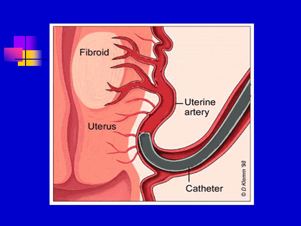
EMBOLIZATION Highly effective way of controlling bleeding from injury, esp. in abdomen or pelvis, or stomach ulcer, in an emergency situation. Worldwide succesful treatment of uterine fibroids Effective treatment of tumors that either cannot be removed surgically or would involve great risk of surgery.

10
Bleeding from a Stomach Ulcer Selective arteriography Stopped after of left gastric artery Gelfoam embolization

11
Bleeding from Jejunum Arteriography of superior Stopped after superselective mesenteric artery metal coils embolization

12
Uterine Fibroid Embolization Angiography of right uterine artery and embolization MRI of uterine fibroid visible before and invisible 6 months after embolization

13
Unilateral common femoral artery access sufficient to treat both uterine arteries

15
AORTIC STENTGRAFTS Endovascular repair of abdominal aorta aneurysm (AAA) with a fabric-wrapped flexible mesh tube. Prevention of enlargement or life- threatening rupture of AAA.

16
How Does Aortic Stentgrafts Work? An appropriate type of self-expanding stentgraft is inserted through the femoral artery and placed at the site of the aneurysm of abdominal aorta. Stentgraft bridges the aneurysm inside and eliminates the blood flow in aneurysmal sac. Reduction of the aneurysmal sac and decrease of the pressure on the aortic wall as an effective prevention of rupture.

17
Various Aortic Stentgrafts

18
Aortic Stentgrafts

19
Subrenal Abdominal Aortic Aneurysm Angiography before and after implantation of stentgraft

20
Angioscopy This is also an invasive method of studying intimal surface of the arterial wall An angioscope catheter containing optical fibers By flushing away the blood, which obscures the field of view, the inner surface of both arteries and grafts, atheroma and thrombus can be directly visualized.

21
Catheter-directed Thrombolysis Minimally invasive treatment that dissolves abnormal blood clots in blood vesselsblood clots

22
The blood clot will then be dissolved in one of two ways: By delivering medication directly to the blood clot. By positioning a mechanical device at the site to break up the clot.

23
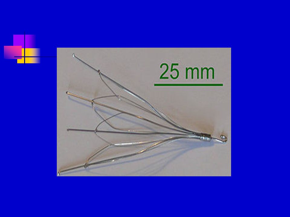
IVC filters Metallic filters placed in the inferior vena cavae to prevent propagation of deep venous thrombus, both temporary and permanent.

25
Common uses of the procedure: Patients who have a history of diagnosed deep vein thrombosis (DVT) or pulmonary embolism. IVC filters are used when patients cannot be successfully treated by other methods, including blood thinning agentsblood thinning agents

26
Complications IVC filter can lodge in the wrong place, change position or injure a nearby organ. The IVC filter may break loose and travel to the heart or lungs causing injury or death. Filled with clots Risk of infection.

27
Phlebotomy of Varicose Veins Phlebotomy is a minimally invasive procedure used to remove varicose veins on the surface of the leg. This is usually done in a physician’s office using local anesthesialocal anesthesia

28
Sclerotherapy of Varicose Veins Minimally invasive treatment used to treat varicose and spider veins. The procedure involves the injection of a solution directly into the affected veins, causing them to shrink and eventually disappear

29
Cholecystostomy Placement of a tube into the gallbladder to remove infected bile in patients with cholecystitis, an inflammation of the gallbladder, who are too sick to undergo surgery

30
Drain insertions Placement of tubes into different parts of the body to drain fluids (e.g., abscess drains to remove pus, pleural drains)
")
31
Radioembolization Embolization of liver with radioactive microspheres of glass or plastic, to kill tumors while minimizing exposure to healthy cells.

32
Nephrostomy Placing a catheter directly into the kidney to drain urine in situations where normal flow of urine is obstructed. NUS catheters are nephroureteral stents which are placed through the ureter and into the bladder

33
Radiologically Inserted Gastrostomy Placement of a feeding tube percutaneously into the stomach and/or jejunum

34
Vertebroplasty PercutaneousPercutaneous injection of biocompatible bone cement inside fractured vertebraevertebrae

35
Cryoablation localized destruction of tissue by freezing

36
Biopsy Taking of a tissue sample from the area of interest for pathological examination from a percutaneous or transjugular approach

37
TIPS Placement of a Transjugular Intrahepatic Porto-systemic Shunt (TIPS) for management of select patients with critical end-stage liver disease and portal hypertension
 for management of select patients with critical end-stage liver disease and portal hypertension")
38
Radiofrequency ablation (RF/RFA) localized destruction of tissue (e.g., tumors) by heating
 localized destruction of tissue (e.g., tumors) by heating")
39
Biliary intervention Placement of catheters in the biliary system to bypass biliary obstructions and decompress the biliary system. Also placement of permanent indwelling biliary stents

40
CONCLUSIONS Interventional radiology procedures are an advance in medicine that often replace open surgical procedures. These minimally invasive procedures involve less blood loss and thus play an important role in bloodless medicine.

Similar presentations


: Pericardium:>")


 LECT7 ALI B ALHAILIY.>")

Hypertension 2)Coronary Artery Disease - arteriosclerosis - atherosclerosis - angina - myocardial infarction.>")

