Download presentation
Presentation is loading. Please wait.

1
Jenelle Beadle, RDMS Inland Imaging December 15 th, 2014

2
Biceps Tendon Palm up and resting on the patient’s leg Transducer placed on the anterior shoulder find the best sonographic window with slight external/internal rotation dynamically evaluate (external/internal rotation) for subluxation or dislocation angle transducer as appropriate to avoid anisotropy Long Biceps Tendon Trans Biceps Tendon
 for subluxation or dislocation angle transducer as appropriate to avoid anisotropy Long Biceps Tendon Trans Biceps Tendon")
3
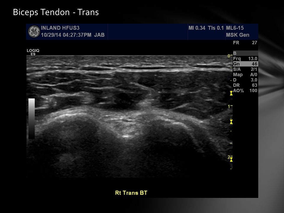
Biceps Tendon - Trans

7
Biceps Tendon - Long

9
Transverse Humeral Ligament over Synovial Tendon Sheath

10
Biceps Tendon - Long

11
Subscapularis Tendon Palm up and arm externally rotated Transducer placed on the anterior shoulder find the best sonographic window with slight external/internal rotation dynamically evaluate (external/internal rotation) for tears and impingement angle transducer as appropriate to avoid anisotropy Long SubscapularisTrans Subscapularis
 for tears and impingement angle transducer as appropriate to avoid anisotropy Long SubscapularisTrans Subscapularis")
12
Subscapularis Tendon - Long Musculotendonis junction coming from under the coracoid process

13
Subscapularis Tendon - Long Cartilage on the head of the humerus up to the point of insertion

14
Subscapularis Tendon - Long Tendon extends medially to the insertion on the lesser tuberosity

15
Subscapularis Tendon - Long Anisotropy where tendon fibers turn to insert

16
Subscapularis Tendon - Long

17
Tendon insert along this flattened area of the lesser tuberosity

18
Subscapularis Tendon - Long

20
Bicipital Groove

21
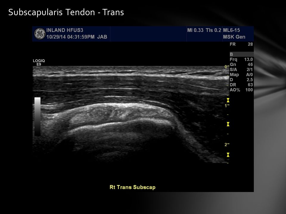
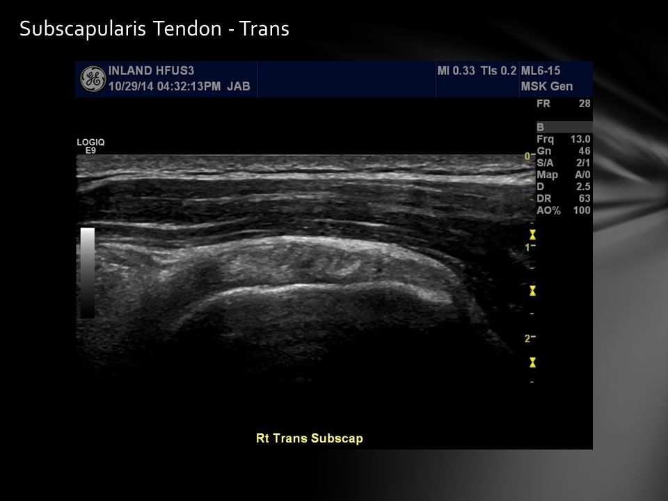
Subscapularis Tendon - Trans

25
Supraspinatus Tendon hand placed in back pocket with palm facing in transducer placed on the anterior shoulder scan anterior to posterior from biceps interval to infraspinatus tendon dynamically evaluate (external/internal rotation) for tears dynamically evaluate (abduction/adduction) for impingement angle transducer as appropriate to avoid anisotropy Long Supraspinatus
 for tears dynamically evaluate (abduction/adduction) for impingement angle transducer as appropriate to avoid anisotropy Long Supraspinatus")
26
Supraspinatus Tendon - Long Long Supraspinatus Neutral Position Long Supraspinatus Stressed Position Acromion

27
Supraspinatus Tendon - Long

28
Musculotendonis junction coming from under the acromion

29
Supraspinatus Tendon - Long

33
Supraspinatus Tendon - Trans Biceps Interval: biceps tendon should be seen adjacent to the supraspinatus tendon any separation here indicates an anterior full thickness supraspinatus tear BT Supra

34
Supraspinatus Tendon - Trans Biceps Interval: biceps tendon should be seen adjacent to the supraspinatus tendon any separation here indicates an anterior full thickness supraspinatus tear

35
Supraspinatus Tendon - Trans

39
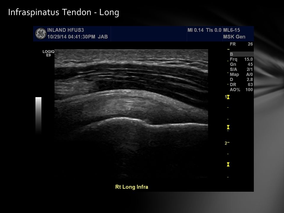
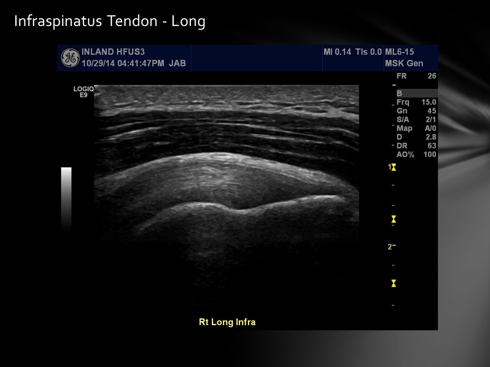
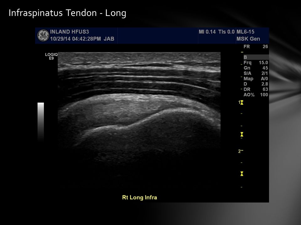
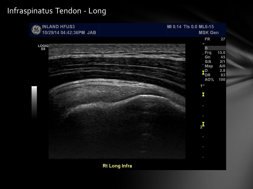
Infraspinatus Tendon atiene’s hand placed on contralateral shoulder transducer placed on the posterolateral shoulder scan anterior to posterior from the supraspinatus tendon to teres minor tendon dynamically evaluate (extreme external rotation and abduction) for impingement angle transducer as appropriate to avoid anisotropy Long Supraspinatus
 for impingement angle transducer as appropriate to avoid anisotropy Long Supraspinatus")
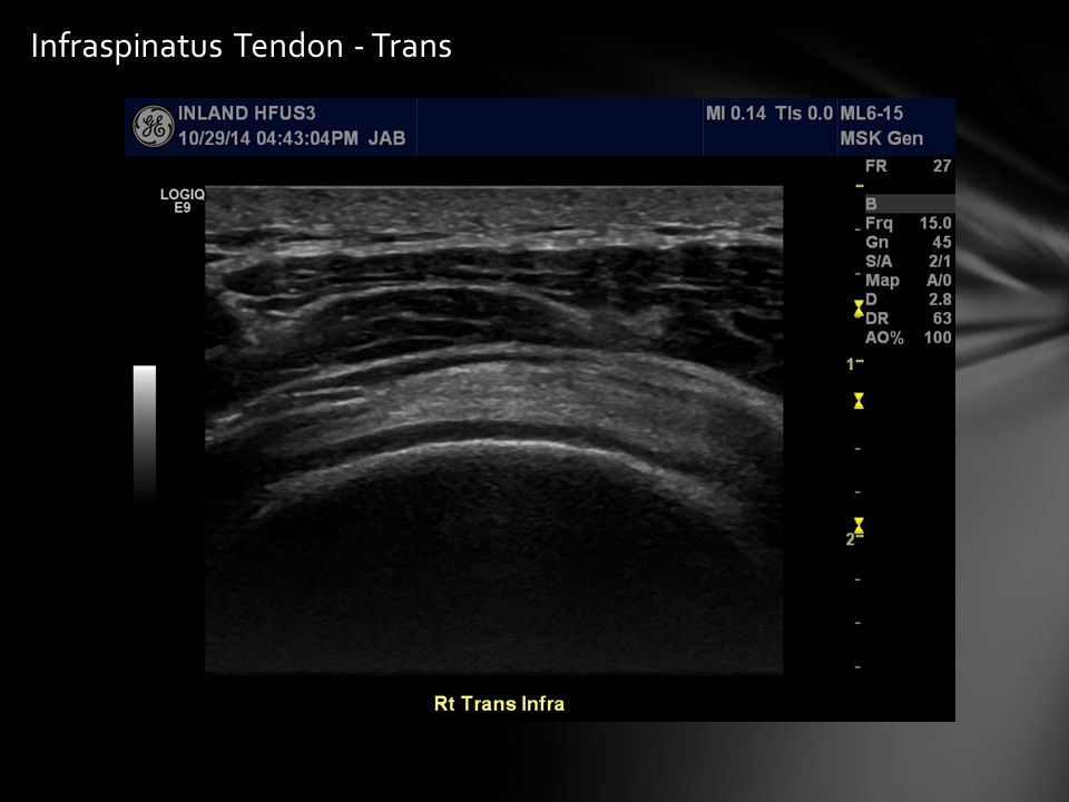
40
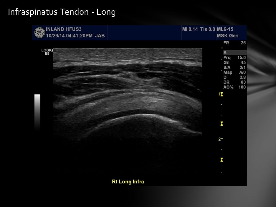
Infraspinatus Tendon - Long

47
Supraspinatus vs Infraspinatus Tendons - Long

48
Infraspinatus Tendon - Trans

52
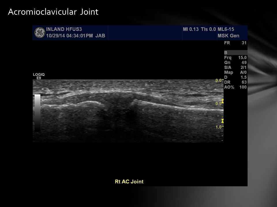
Acromioclavicular Joint Assessed for stability and fluid

53
Acromioclavicular Joint

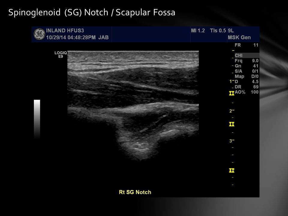
56
Posterior Glenohumeral (GH) Joint & Spinoglenoid Notch / Scapular Fossa
 Joint & Spinoglenoid Notch / Scapular Fossa")
57
Posterior Glenohumeral (GH) Joint
 Joint")
58
Labrum Humeral Head Glenoid Rim Infraspinatus Musculotendinous Junction

59
Posterior Glenohumeral (GH) Joint
 Joint")
60
Spinoglenoid (SG) Notch / Scapular Fossa
 Notch / Scapular Fossa")
63
“Skeletal Muscle Ultrasound” European Journal Translational Myology 2010; 1 (4): 145- 155 “Ultrasonographic Findings of Musculoskeletal Tissues” J Korean Orthop Assoc. 2013 Oct;48(5):334-341 “Sonography of Common Tendon Injuries” American Journal of Roentgenology. 2009;193: 607-618 “Tendon and Ligament Imaging” Br J Radiol. Aug 2012; 85(1016): 1157–1172 “Imaging of the Bursae” J Clin Imaging Sci 2011; 1:22 “Ultrasonography of tendon abnormalities” OA Musculoskeletal Medicine 2013 Jun 01;1(2):12 “Sonography of Lower Limb Muscle Injury” American Journal of Roentgenology. 2004;182: 341-351 “Full Thickness and Partial Thickness Supraspinatus Tendon Tears” Radiology 2004; 230:234–242 “Long Head of Biceps Brachii Tendon Evaluation...” AJR 2011; 197:942–948 “Ultrasound of the Shoulder” JBR–BTR, 2007, 90: 325-337 Gaitini D. “Shoulder Ultrasonography: Performance and Common Findings” J Clin Imaging Sci 2012; 2: 38-38 Read J, Perko M. “Ultrasound Diagnosis of Subacromial Impingement for Lesions of the Rotator Cuff” AJUM May 2010; 13 (2): 11-15 References
: Sonography of Common Tendon Injuries American Journal of Roentgenology. 2009;193: Tendon and Ligament Imaging Br J Radiol. Aug 2012; 85(1016): 1157–1172 Imaging of the Bursae J Clin Imaging Sci 2011; 1:22 Ultrasonography of tendon abnormalities OA Musculoskeletal Medicine 2013 Jun 01;1(2):12 Sonography of Lower Limb Muscle Injury American Journal of Roentgenology. 2004;182: Full Thickness and Partial Thickness Supraspinatus Tendon Tears Radiology 2004; 230:234–242 Long Head of Biceps Brachii Tendon Evaluation... AJR 2011; 197:942–948 Ultrasound of the Shoulder JBR–BTR, 2007, 90: Gaitini D. Shoulder Ultrasonography: Performance and Common Findings J Clin Imaging Sci 2012; 2: Read J, Perko M. Ultrasound Diagnosis of Subacromial Impingement for Lesions of the Rotator Cuff AJUM May 2010; 13 (2): References.")
64
http://www.sonoguide.com/soft_tissue.html http://www.dynamicultrasound.org/dugphysics.html http://www.ultrasoundcases.info/Slide-View.aspx?cat=405&case=1858 http://www.ultrasoundcases.info/Slide-View.aspx?cat=405&case=1858 http://www.shoulderdoc.co.uk/article.asp?section=904 http://www.radiologyassistant.nl/en/p50cf8392cbd97/us-guided- injection-of-joints.html http://www.radiologyassistant.nl/en/p50cf8392cbd97/us-guided- injection-of-joints.html http://radiopaedia.org/articles/posterosuperior-impingement-of-th- shoulder http://radiopaedia.org/articles/posterosuperior-impingement-of-th- shoulder References (continued…)
")
Similar presentations


 Joint>")


