Download presentation
Presentation is loading. Please wait.

1
Dr. Nelly M. Dabbour Pharmacist, MSc. Applied Medical Chemistry Medical Research Institute Alexandria University, Egypt

2
Liver cancer is the fifth most common cancer in men and ninth most common in women, with hepatocellular carcinoma (HCC) accounting for >90% of primary liver cancer cases. Hepatocellular carcinoma (HCC) is the second most common cause of cancer deaths worldwide (after lung cancer). The greatest burden of HCC is in the developing world, with cases in eastern and southeastern Asia, and central and western Africa accounting for more than 80% of the total; 50% of all cases occur in China alone.
 is the second most common cause of cancer deaths worldwide (after lung cancer). The greatest burden of HCC is in the developing world, with cases in eastern and southeastern Asia, and central and western Africa accounting for more than 80% of the total; 50% of all cases occur in China alone..")
3
In Middle Eastern countries, liver cancer is a major concern. In Egypt, liver cancer is the fourth most common cancer and is the second cause of cancer mortality in both sexes. Hospital-based studies have reported an overall increase in the relative frequency of all liver related cancers (>95% as HCC). The prevalence of HCC is high in the Nile Delta area.
. The prevalence of HCC is high in the Nile Delta area..")
4
Pathogenesis of HCC is a complex process, it is usually associated with liver damage, and subsequent development of cirrhosis, dysplastic lesions, and eventually invasive carcinoma. Tumors lesions are common in the liver. Liver is sensitive to chemical carcinogens such as 4-Dimethylaminoazobenzene (DAB, known as butter yellow) which acts as initiator and Phenobarbital (PB) which acts as promoter.
 which acts as initiator and Phenobarbital (PB) which acts as promoter..")
5
DAB produces liver damage followed by regeneration of parenchymal cells and tumors develop from parenchyma that end up with neoplastic transformation. PB enhances hepatocytes growth and suppresses apoptotic rate.

6
Liver cancer rapidly reduces quality of life and typically causes death 6 months to one year from diagnosis. Chemotherapy is a major therapeutic approach for the treatment of localized and metastasized cancers. 5-FU remains one of the most widely used anticancer agents as it shows activity in HCC, as metabolism eliminates 90% of 5-FU, half-life is short ranging from 10 to 20 minutes that necessitates frequent administration of the drug which may lead to severe side effects.

12
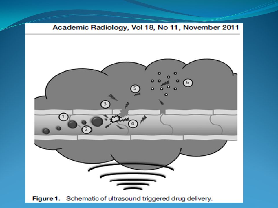
The technique is non-invasive Ultrasonic waves can penetrate deep into the interior of the body. They can be carefully controlled and focused on the tumor site. Since harmful drug is sequestered until the desired release place and time, the side effects of chemotherapy can be minimized. Ultrasonic cavitation is producing stress on the cell membrane to allow greater drug uptake than would occur without US.

13
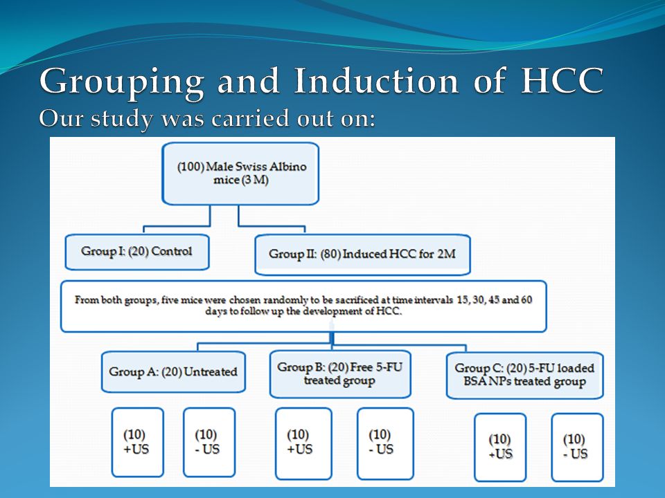
Preparation and characterization of NPs Induction of HCC by the administration of DAB and PB Studying the therapeutic effect of 5-FU loaded BSA NPs on the experimentally induced HCC in mice

15
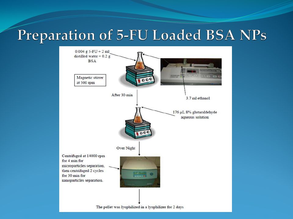
The pellet was redispersed to the original volume (3.5 ml) in phosphate buffered saline (PBS) at pH 7.4 using ultrasonication for 5 min. For complete liberation of entrapped drug, 50 μl of trypsin solution (1 mg/ml) was added and the resulted solution was incubated at 37°C for 8 h. Later, total drug concentration was determined spectrophotometrically at 266 nm.
 was added and the resulted solution was incubated at 37°C for 8 h. Later, total drug concentration was determined spectrophotometrically at 266 nm..")
16
Scanning electron microscope (SEM): Morphology of prepared particles was studied using SEM that indicates the formation of completely spherical particle with smooth surface and low level of agglomeration: (Mag. x10.000, 20 kV) (Mag. x50.000, 20 kV) (Mag. x10.000, 20 kV) (Mag. x50.000, 20 kV)
 (Mag. x50.000, 20 kV) (Mag. x10.000, 20 kV) (Mag. x50.000, 20 kV).")
17
(Mag. x7.500, 80 kV) (Mag. x15.000, 80 kV) (Mag. x7.500, 80 kV) (Mag. x15.000, 80 kV) Transmission Electron Microscope (TEM): TEM examination of blank BSA NPs revealed spherical particle with smooth surface and no agglomeration :
 (Mag. x15.000, 80 kV) Transmission Electron Microscope (TEM): TEM examination of blank BSA NPs revealed spherical particle with smooth surface and no agglomeration :.")
18
(Mag. x7.500, 80 kV) (Mag. x15.000, 80 kV) (Mag. x7.500, 80 kV) (Mag. x15.000, 80 kV) TEM examination of 5-FU loaded BSA NPs revealed spherical particle with smooth surface and low level of agglomeration :
 (Mag. x15.000, 80 kV) TEM examination of 5-FU loaded BSA NPs revealed spherical particle with smooth surface and low level of agglomeration :.")
19
Particle Size Analyzer (PSA): The particle size distribution of blank BSA NPs was carried out by PSA showed particle size with mean of 53 nm:
: The particle size distribution of blank BSA NPs was carried out by PSA showed particle size with mean of 53 nm:")
20
The particle size distribution of 5-FU loaded BSA NPs was carried out by PSA showed particle size with mean of 70 nm:

21
Determination of DLE and EE: Amount of entrapped 5-FU in BSA NPs was determined by drug loading efficiency (DLE) which was 19.23% and encapsulation efficacy (EE) which was 62.5%.
 which was 19.23% and encapsulation efficacy (EE) which was 62.5%.")
22
Comparing between release profiles of free drug and nanoparticle suspension confirms that this colloidal drug carrier is capable of releasing drug in a controlled manner with the ability of revealing burst release In vitro release profile:

23
Exposure to US accelerated the release of 5-FU from the NPs, the in vitro drug release of 5-FU under the effect of US reached 50% in the first 24 hrs compared with 33.3% in the absence of US. After 48 hrs the effect reached 76% compared with 53.8%, and then after 72 hrs the effect reached 87.5% compared with 67.4%:

24
The comparison showed a higher release percentage with the use of US. A maximum release of 5-FU under the effect of US reached 97% after 96 hrs, while maximum release of 88.9 % reached after 144 hrs from NPs in the absence of US irradiation:

26
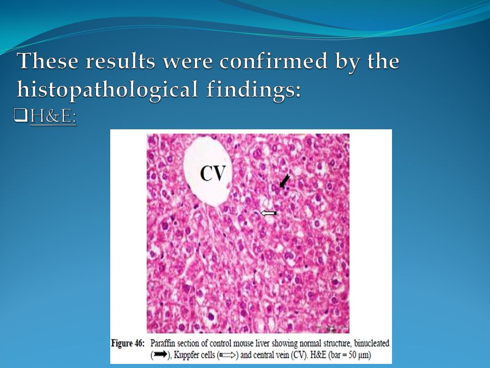
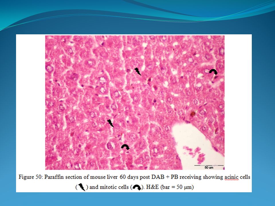
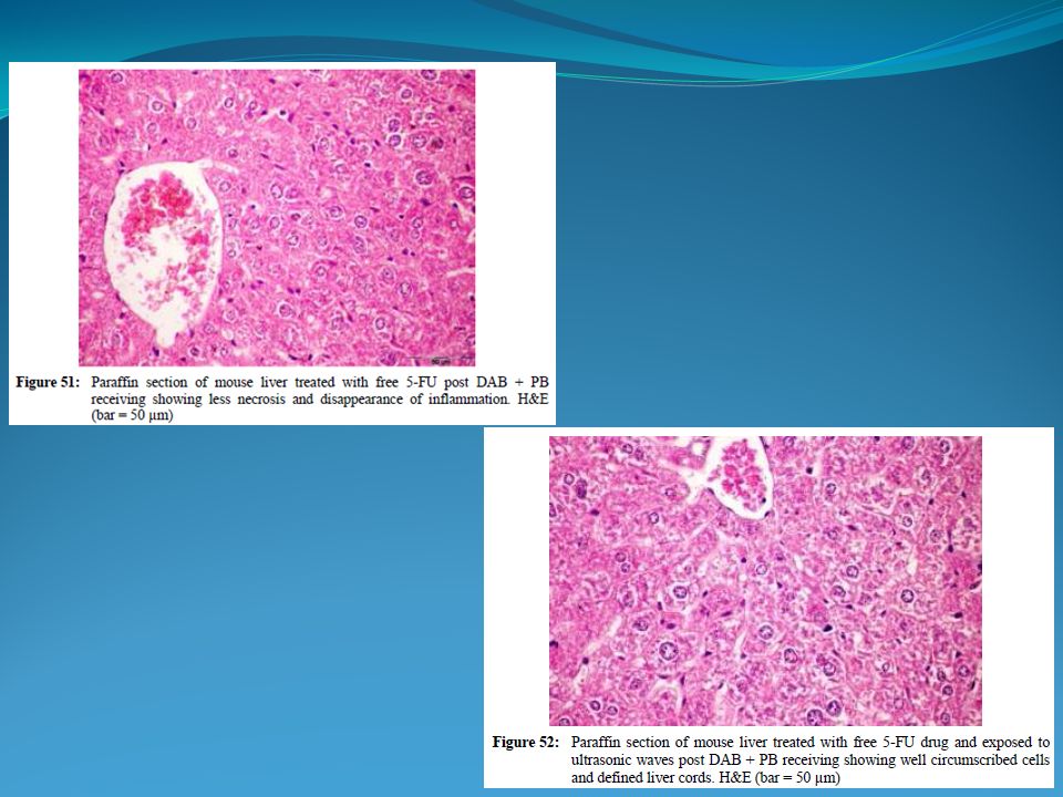
Biochemical: Hepatic malondialdehyde (MDA) level Serum and hepatic ALT activity. Histopathological: Histomorphological changes Histochemistry of alkaline phosphatase (ALP) Alpha-fetoprotein (AFP) immunohistochemicaly.
 Alpha-fetoprotein (AFP) immunohistochemicaly..")
27
MDA was significantly increased in DAB + PB feeding group of mice at all intervals when compared to the control group.

28
DAB + PB feeding caused a highly significant progressive increase in serum and hepatic ALT activity at all intervals when compared to the control group. Serum ALT activity: Serum ALT activity: Hepatic ALT activity: Hepatic ALT activity:

29
Treated mice showed improvement mostly after the use of 5- FU loaded BSA NPs and exposed to US. By measuring the biochemical parameters, hepatic MDA compared with mice bearing the liver nodules showed decreased levels by 9% and 58% after being treated with free 5-FU, and treated with 5-FU loaded BSA NPs respectively. While compared with mice bearing the liver nodules and exposed to US showed decrease levels by 27% and 75% after being treated with free 5-FU and exposed to US, and being treated with 5-FU loaded BSA NPs and exposed to US respectively.

30
Group B showed no significant decrease, while group C caused a highly significant decrease compared to group A. A significant decrease was obtained when compared group C with B. Exposing mice to US caused more significant decrease in group C compared to group A. Within the same group, a significant decline in groups B and C. Hepatic MDA level: The best correction in MDA level was obtained in the group received 5-FU loaded BSA and exposed to US.

31
Serum ALT compared with mice bearing the liver nodules showed decreased levels by 10% and 36% after being treated with free 5-FU, and treated with 5-FU loaded BSA NPs respectively. While compared with mice bearing the liver nodules and exposed to US, it showed decreased levels by 25% and 50% after being treated with free 5-FU and exposed to US, and being treated with 5-FU loaded BSA NPs and exposed to US respectively.

32
Group B showed no significant difference, while group C caused a highly significant decrease compared to group A. A non-significant decrease was obtained when compared group C with B. Exposing mice to US caused more significant decrease in group C compared to group A. Within the same group, a significant decline in group C only. Serum ALT activity: The best correction in ALT activity was obtained in the group received 5-FU loaded BSA and exposed to US.

33
Hepatic ALT compared with mice bearing the liver nodules showed decrease levels by 19% and 30% after being treated with free 5-FU, and treated with 5-FU loaded BSA NPs respectively. While compared with mice bearing the liver nodules and exposed to US showed decrease levels by 0% and 53% after being treated with free 5-FU and exposed to US, and being treated with 5-FU loaded BSA NPs and exposed to US respectively.

34
Group B showed a significant decrease, while group C caused a highly significant decrease compared to group A. A non-significant decrease was obtained when compared group C with B. Exposing mice to US caused more significant decrease in group C compared to group A. Within the same group, a significant decline in all groups. Hepatic ALT activity: The best correction in ALT activity was obtained in the group received 5-FU loaded BSA and exposed to US.

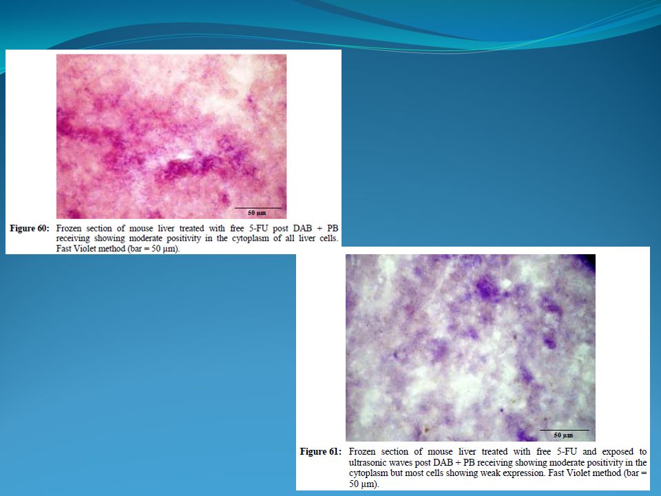
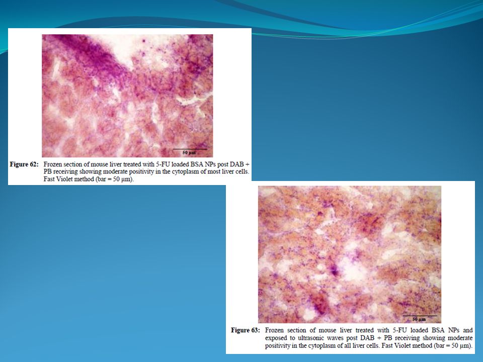
39
ALP:

43
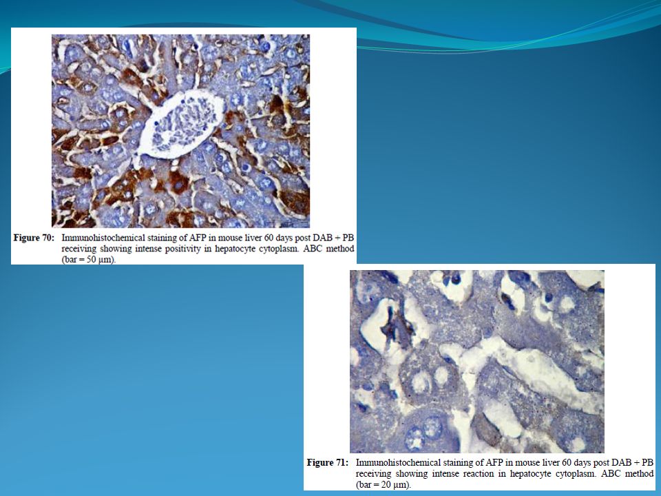
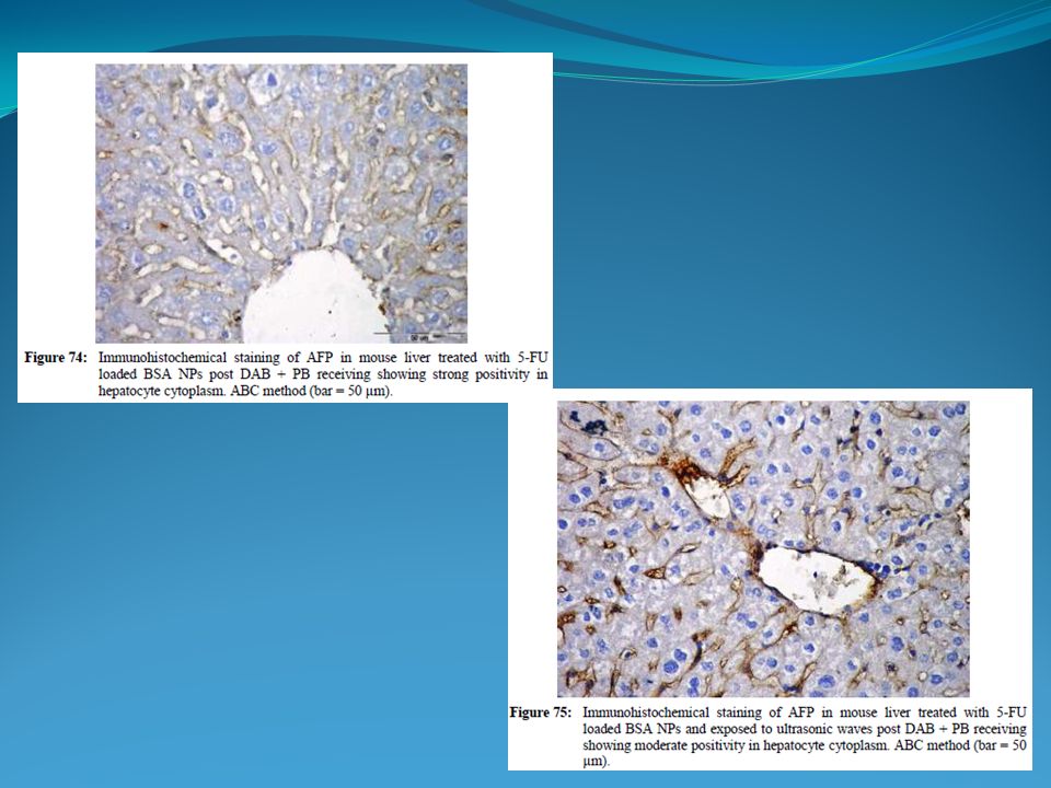
AFP:

47
This significant decline in the serum and hepatic biochemical parameters beside the histochemical findings proved that: The growth inhibitory effect was the best in the use of 5-FU loaded BSA NPs and exposed to US. There is a good response in liver tissues towards different modalities of treatment being better in the use of 5-FU loaded BSA NPs and exposed to US.

48
The investigation of BSA NPs containing 5-FU shows that they are promising carriers for delivery system through their enhanced efficacy against cancer cells. The drug delivery in combined with a local ultrasonic irradiation of the tumor may be developed into a powerful new tool of drug targeting and treatment of cancerous tumors. Conclusion

49
Thank You

Similar presentations


 is the fifth most frequent cancer in the world and the third most common cause of cancer mortality.>")

 for Liver Tumour Dr Dai Wing Chiu Queen Mary Hospital.>")
 LIVER FUNCTION AND THE BILIARY TRACT LECTURE FOUR Dr. Essam H. Aljiffri.>")
>")


, Cateni F. (a), Drioli S. (a), Bonora GM. (a), Zorzet S. (b), Rapozzi V. (c), Xodo L.E. (c) (a) Department of Chemical and Pharmaceutical.>")

 in cancer therapy.>")

 Dr. Samah Kotb Nasr Eldeen Serum biochemical parameters (ALT) (AST) assay.>")