Download presentation
Presentation is loading. Please wait.

1
BIOE 220/RAD 220 REVIEW SESSION 6 March 5, 2012

2
What We’ll Cover Today General questions? Spinal cord anatomy review Fat in images T2* vs T2 decay Review of sequences Questions on the hw?

3
Nasal and Oral cavities Nasal Cavity Nasopharynx Uvula/Soft Palate Oropharynx Epiglottis Tongue

4
Parotid glands Parotid Gland Stensen’s Duct

5
Salivary glands Sublingual gland Submandibular gland Submandibular duct

6
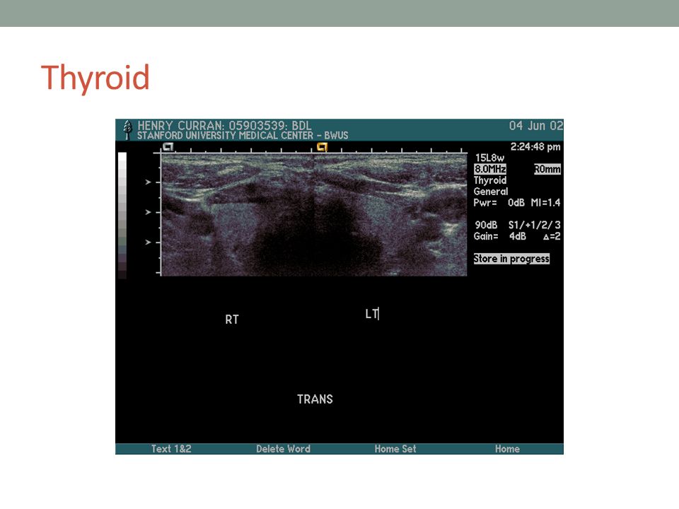
Thyroid

8
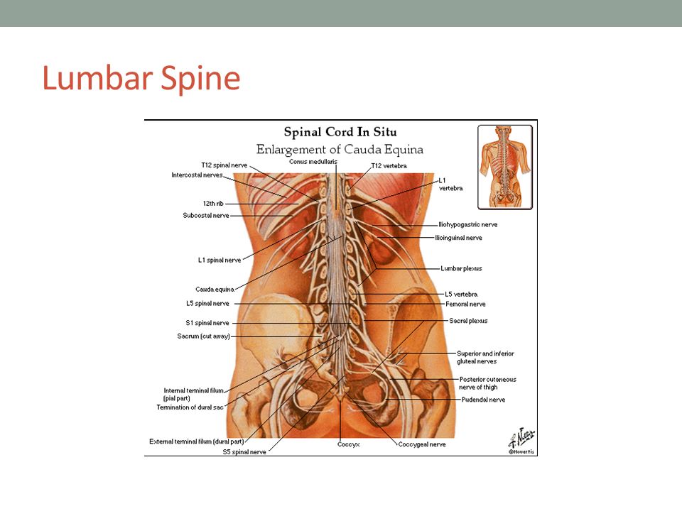
Spinal Vertebrae Breakfast at 7 Lunch at 12 Dinner at 5 Cervical roots: C1-C8 (all above their vertebrae except 8) Thoracic: T1-T12, Lumbar: L1-L5, Sacral: S1-S5
 Thoracic: T1-T12, Lumbar: L1-L5, Sacral: S1-S5")
9
Cervical Spine Anterior Posterior

10
Cervical spine

12
Thoracic Spine

13
Lumbar Spine

19
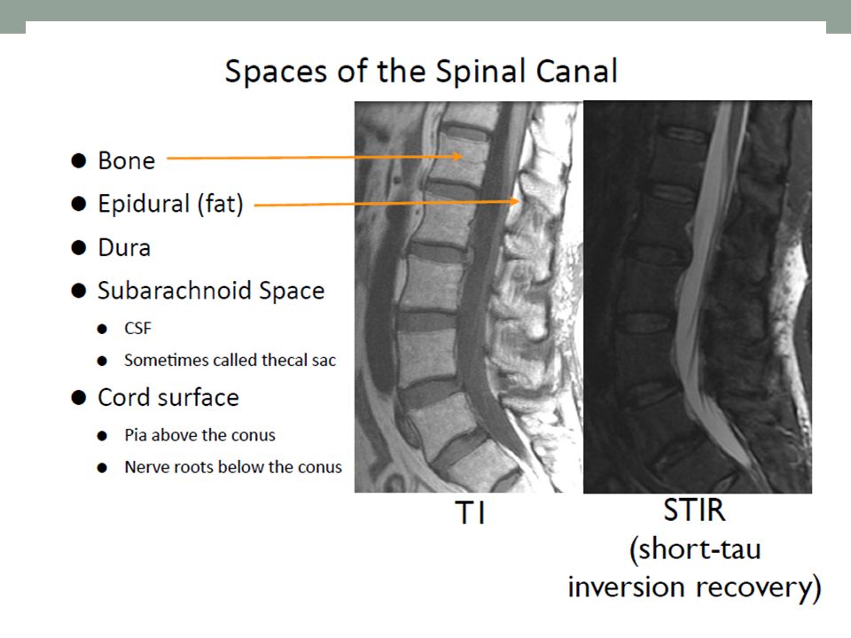
How does fat look in MRI? Without fat suppression, fat always appears bright Why? Short T1, normalT2 but high PD Fat precesses at a different frequency (3.5 ppm shift – how fast?) so it appears shifted in images This effect can be minimized by maximizing the strength of readout gradients (why?) How can we remove fat from the signal? Take advantage of difference in precession frequency Chemical saturation: Hit fat with selective 90º pulse, spoil the transverse signal, then do a normal measurement afterwards Short TI Inversion recover: After a 180º pulse, wait for the fat to recover to 0 (shorter T1), then measure the other signals
 so it appears shifted in images This effect can be minimized by maximizing the strength of readout gradients (why ) How can we remove fat from the signal. Take advantage of difference in precession frequency Chemical saturation: Hit fat with selective 90º pulse, spoil the transverse signal, then do a normal measurement afterwards Short TI Inversion recover: After a 180º pulse, wait for the fat to recover to 0 (shorter T1), then measure the other signals.")
20
T2 vs T2* decay When do we see T2* decay, when do we see T2 decay? T2* decay is observed after a 90º pulse if no other preparation is done (GRE sequence) T2 decay is observed if a 180º refocusing pulses is used to “unroll” any off-resonance defocusing (SE sequence) After the 180º pulse, must wait the same amount of time as we waited before the pulse until things have refocused
 T2 decay is observed if a 180º refocusing pulses is used to unroll any off-resonance defocusing (SE sequence) After the 180º pulse, must wait the same amount of time as we waited before the pulse until things have refocused.")
22
What sequences have we learned about? Simplest: GRE 2DFT Excite with 90º pulse, then read out K-space trajectory will be lines in the frequency encode direction, stepped in the phase encode direction between every TR Spin echo 2DFT Same as before, except now we add a 180º refocusing pulse after the 90º so that we’ll obtain T2 weighting instead of T2* 180º pulse occurs at TE/2 Spatial saturation Add a 90º pulse to excite a slab and then spoil it, before regular sequence This nulls the signal in the slab, so that only fresh spins flowing in will be visible

23
What sequences have we learned about? Fat suppression: Chem Sat Use a 90º selective pulse on the fat signal and spoil it, before regular sequence Ideally nulls the fat signal without effecting rest of image Fat suppression: STIR Use a 180º pulse to flip everything, then wait until the fat passes through 0 to do our readout Diffusion weighting Use strong bipolar (sums to zero) gradient, so that spins will dephase based on their movement during gradient Flow encoding Similar to diffusion weighting, except that we’re interested in much higher velocities (of bulk spins)
 gradient, so that spins will dephase based on their movement during gradient Flow encoding Similar to diffusion weighting, except that we’re interested in much higher velocities (of bulk spins).")
24
What sequences have we learned about? Fast spin echo Perform multiple 180º - readout – 180º - readout in a single TR Allows faster acquisition of SE image, but fat appears brighter EPI Instead of recording line by line, traverse 2DFT grid very quickly in single sequence Fast, but very susceptible to artifacts Spiral Instead of collecting k-space in grid like 2DFT, traverse k-space in a spiral, to be more efficient with gradients Efficient/fast, but susceptible to artifacts and leads to spatially variant resolution/blurring

Similar presentations


 Douglas C. Noll, Ph.D. Depts. of Biomedical Engineering and Radiology University of Michigan, Ann Arbor.>")


