Download presentation
Presentation is loading. Please wait.

2
Spinal cord

3
Traditional starting point for detailed consideration of CNS Uniform and simple organization of other parts of CNS Very important in day-to-day activities that we don’t even think about

4
Contains all neurons supplying the muscles used in bodily movements, as well as a major population of autonomic nerves Receives all sensory input from body and some from head- performs initial processing on most of this input

5
The spinal cord provides a crucial information conduit, connecting the brain with most of the body. It is the target of a number of disease processes, some of which (eg, spinal cord compression) are treatable but rapidly progressive if not treated.
 are treatable but rapidly progressive if not treated..")
6
Failure to diagnose some disorders of the spinal cord, such as spinal cord compression, can be catastrophic and may relegate the patient to a lifetime of paralysis. A knowledge of the architecture of the spinal cord and its coverings, and of the fiber tracts and cell groups that comprise it, is essential.

7
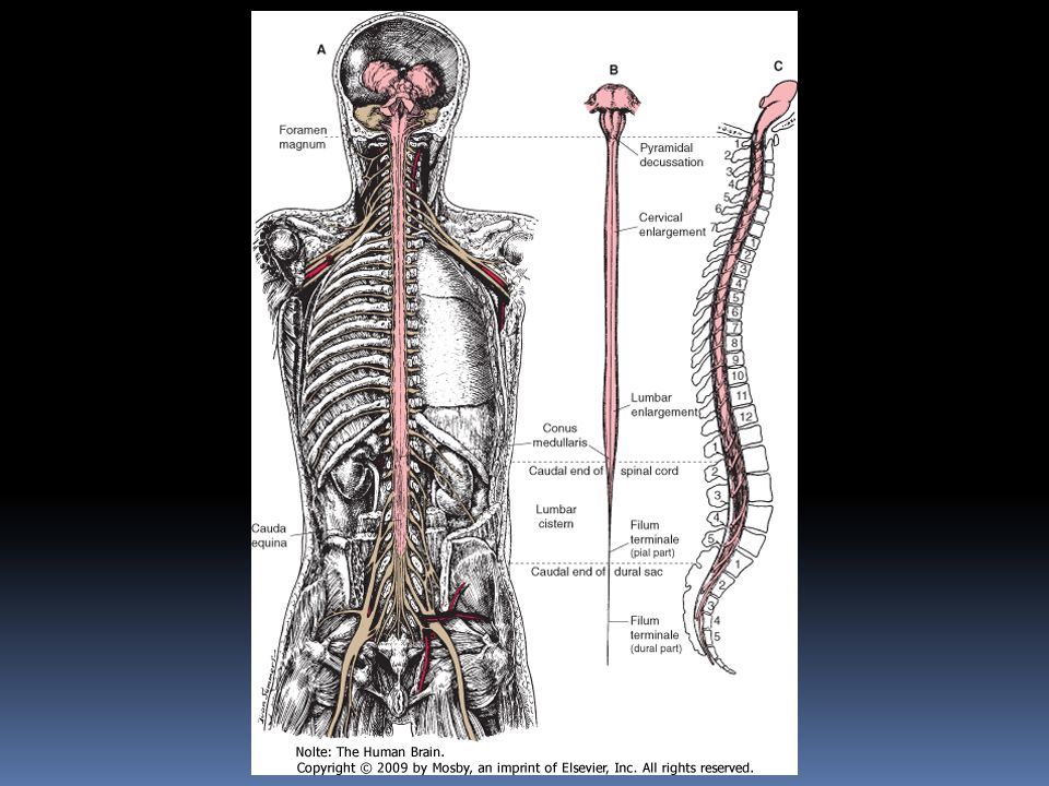
Basic facts Length 42-45 cm Maximum dia. 1 cm Weight 35 g Anatomically segmented in terms of attached nerve roots

8
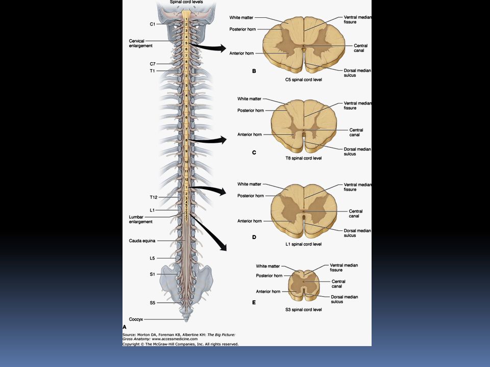
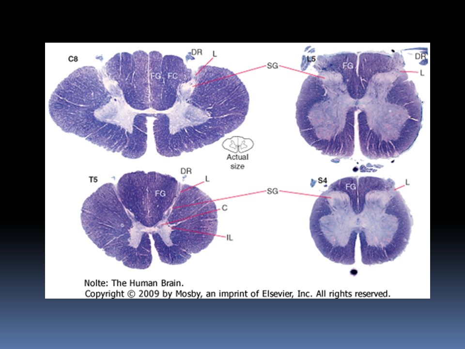
Spinal segment= portion of spinal cord giving rise to a spinal nerve 31 segments- 8 cervical, 12 thoracic, 5 lumbar, 1 coccygeal No external signs of segmentation

13
2 enlargements- cervical and lumbar Ends caudally in a pointed conus medullaris Enlargements contain large number of motor neurons Cervical enlargement is in C5-T1 segments- supplies upper limb Lumbar enlargement- L2 –S3 segments; supplies lower limbs

14
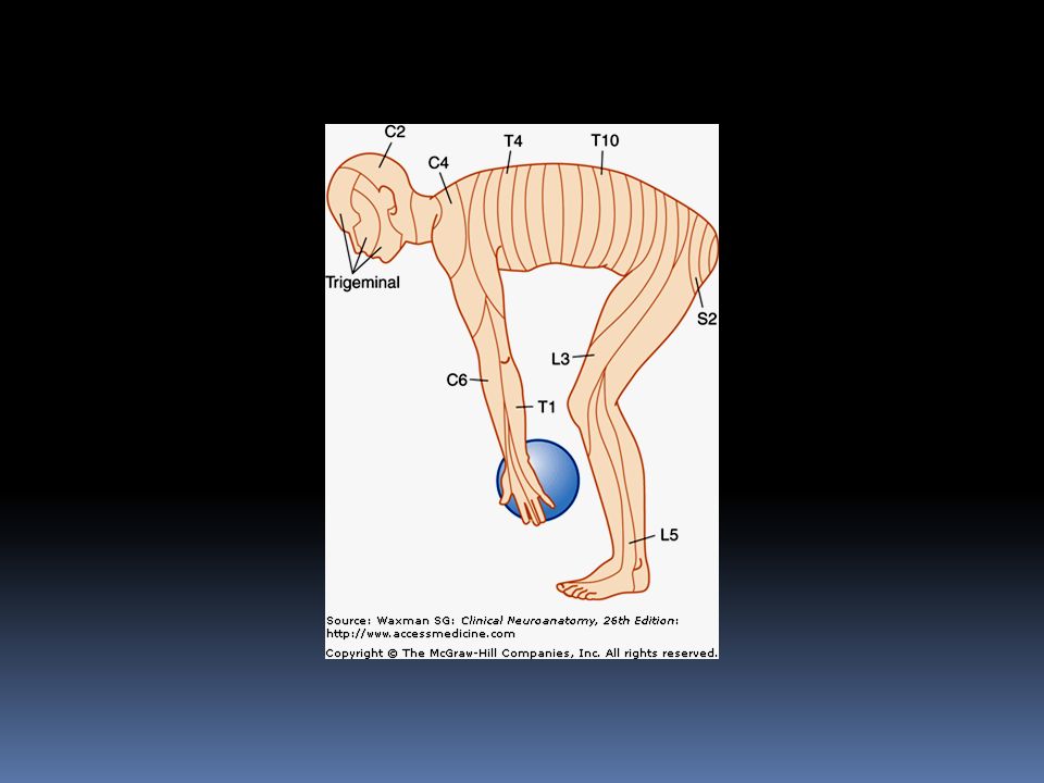
Spinal cord segments are related systematically to areas of skin and muscles The sensory component of each spinal nerve is distributed to a dermatome, a well-defined segmental portion of the skin in many patients there is no C1 dorsal root, there is no C1 dermatome

16
when a C1 dermatome does exist as an anatomic variant, it covers a small area in the central part of the neck, close to the occiput dermatomes for C5, C6, C7, C8, and T1 are confined to the arm C4 and T2 dermatomes are contiguous over the anterior trunk.

17
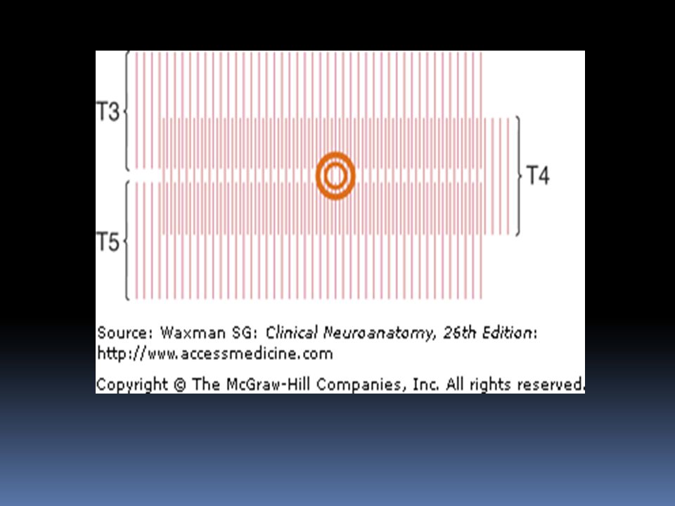
thumb, middle finger, and fifth digit are within the C6, C7, and C8 dermatomes, respectively Nipple-T4 Umbilicus – T 10

18
The territories of dermatomes tend to overlap, making it difficult to determine the absence of a single segmental innervation on the basis of sensory testing Knowledge of segmental skin and muscle innervation helps in diagnosis of injuries in or around spinal cord

20
Myotomes myotome refers to the skeletal musculature innervated by motor axons in a given spinal root Most muscles are innervated by motor axons that arise from several adjacent spinal roots. Nevertheless, lesions of a single spinal root, in many cases, can cause weakness and atrophy of a muscle

21
Segment-Pointer Muscles. RootMuscle C5 C6 C7 L3, L4 L5 S1 Deltoid Biceps Brachioradialis Triceps Quadriceps femoris Extensor hallucis longus Gastrocnemius

22
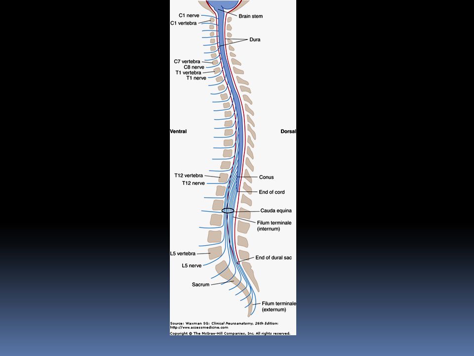
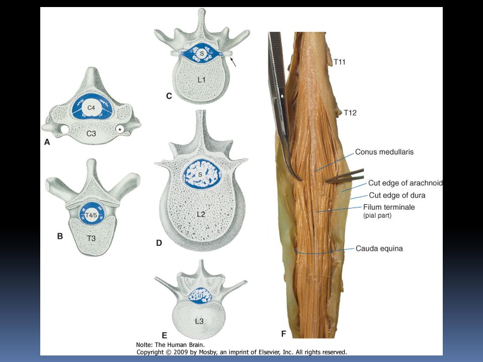
Correlation between length of spinal cord and vertebral column Spinal cord approaches adult length before vertebral canal Until 3 rd month of fetal life- same growth rates Later- body and vertebral column grow faster than spinal cord At birth spinal cord ends at L3 vertebra

23
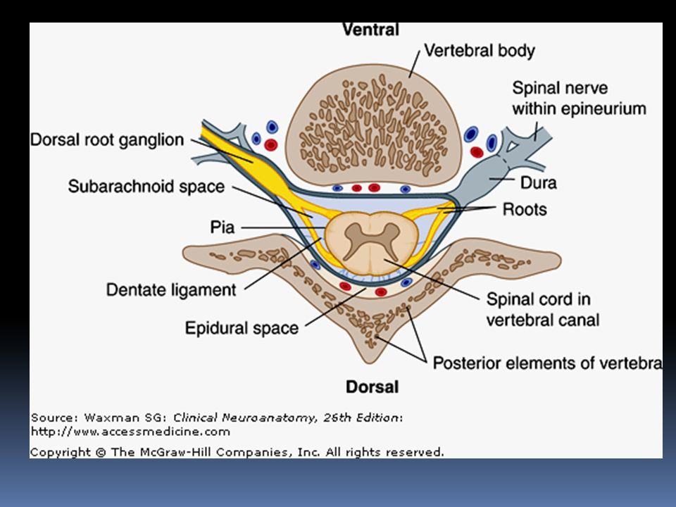
At few months of age- ends about the level of L1 vertebra Nerves exit through same intervertebral foramina as they did in fetal life Each dorsal root ganglion is at the level of appropriate foramen

24
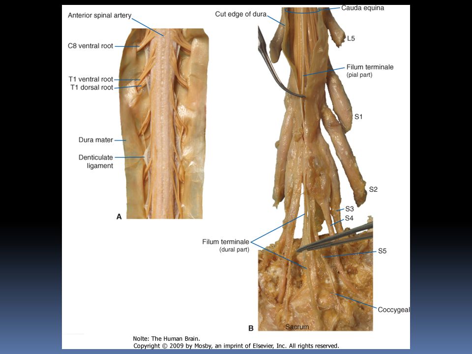
The lower nerve roots have to travel longer distances to reach their foramina of Below L1 -L2 level of vertebral column the collection of dorsal and ventral roots is called cauda equina

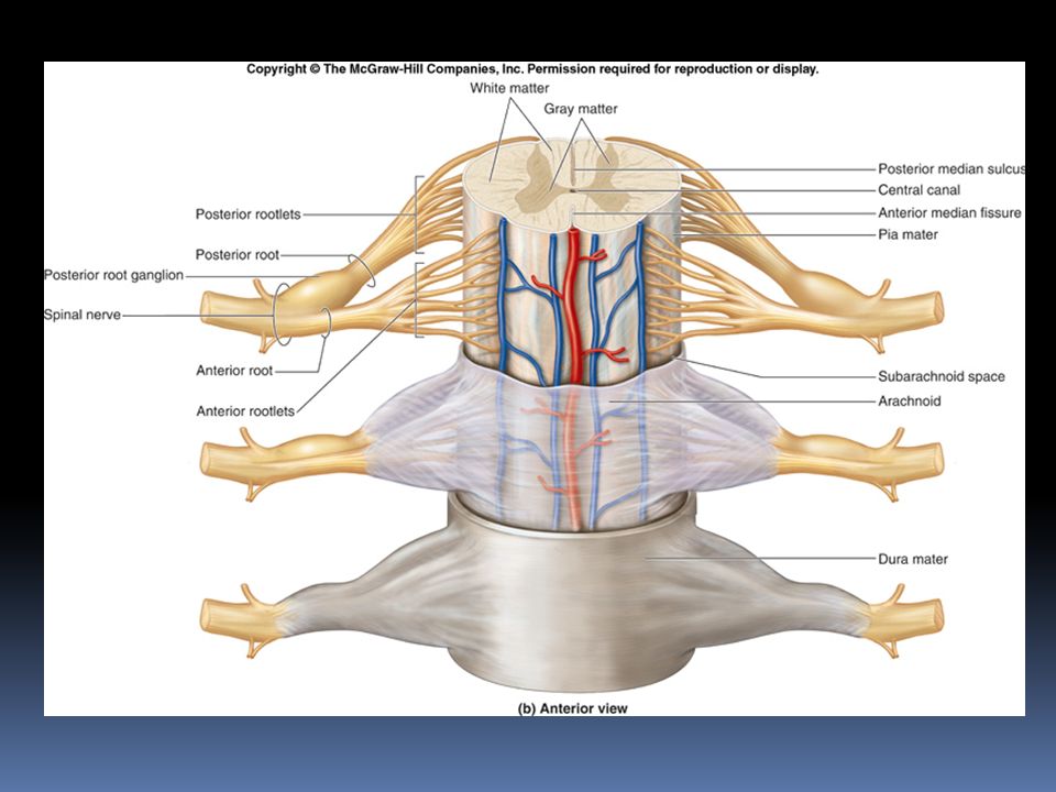
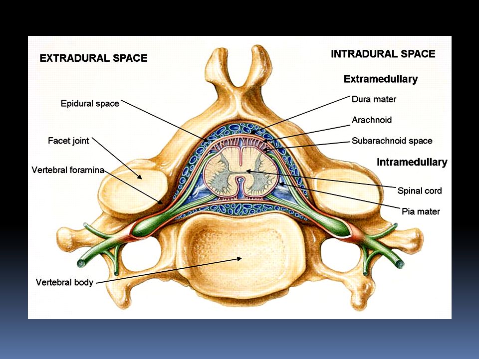
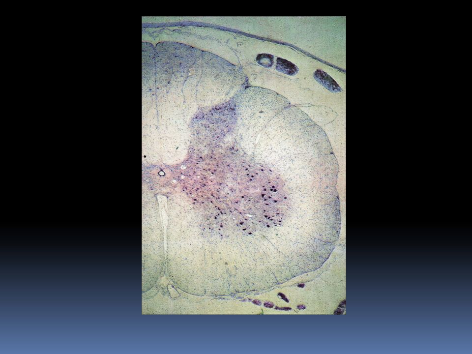
37
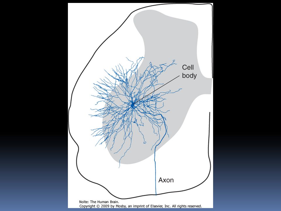
Spinal Gray Matter Is Regionally Specialized The anterior horn contains the cell bodies of the large motor neurons that supply skeletal muscle These alpha motor neurons, also referred to as lower motor neurons, are the only means by which the nervous system can exercise control over body movements, whether voluntary or involuntary;

38
a number of different parts and pathways of the nervous system can influence these lower motor neurons, but they alone can elicit muscle contraction.

39
Destruction of the lower motor neurons supplying a muscle or interruption of their axons therefore causes complete paralysis of that muscle. Lower motor neuron lesions cause paralysis of a type called flaccid paralysis, indicating that the muscle is limp and uncontracted.

43
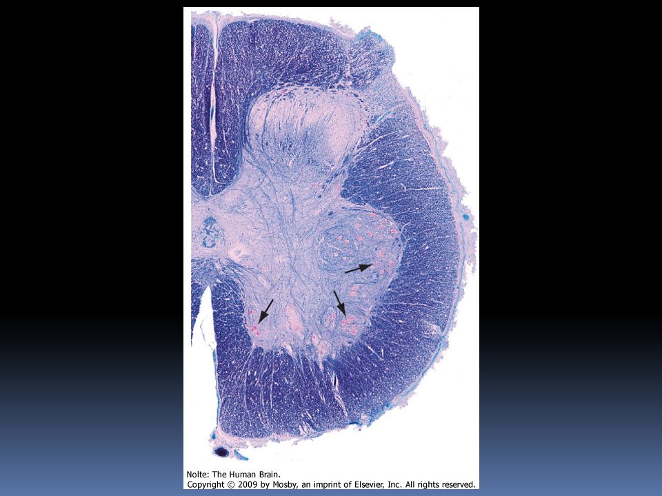
Two columns of motor neurons in the anterior horn of the cervical cord are recognized as separate entities. The spinal accessory nucleus extends from the caudal medulla to about C5.

44
The phrenic nucleus, containing the motor neurons that innervate the diaphragm, is located in the medial portion of the anterior horn in segments C3 to C5..

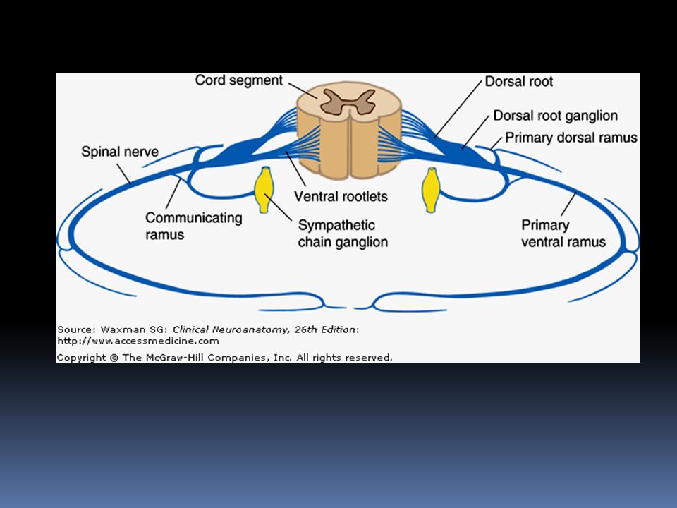
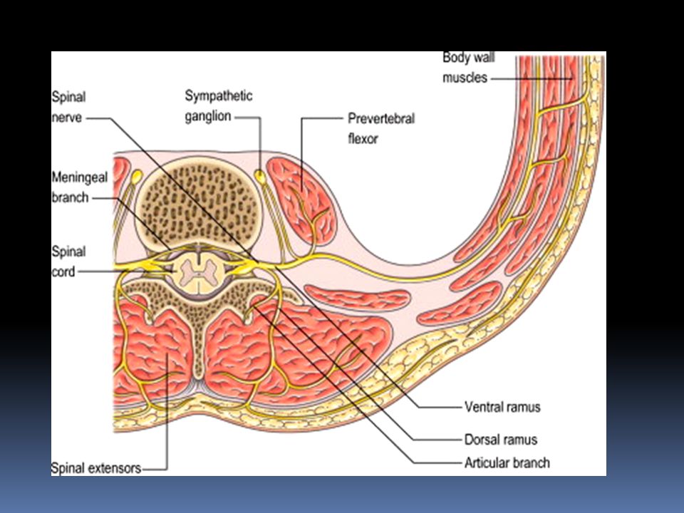
45
This makes injuries to the upper cervical spinal cord a matter of grave concern destruction of the descending pathways that control the phrenic nucleus and other respiratory motor neurons renders a patient unable to breathe

46
The gray matter that is intermediate to the anterior and posterior horns has some characteristics of both and also contains the spinal preganglionic autonomic neurons. In addition, at some levels it includes a distinctive region called Clarke's nucleus.

47
The preganglionic sympathetic neurons for the entire body lie in segments T1 through L3 most of them located in a column of cells called the intermediolateral cell column, which forms a pointy lateral horn on the spinal gray matter

48
Their axons leave through the ventral roots. Cells in a corresponding location in segments S2 to S4 constitute the sacral parasympathetic nucleus but do not form a distinct lateral horn.

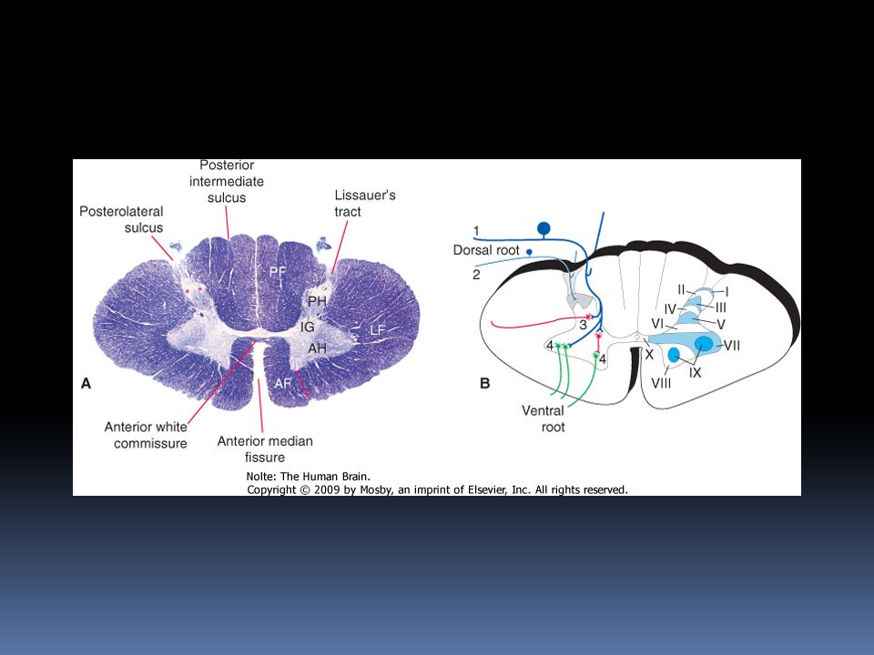
49
Their axons leave through the ventral roots and synapse on the postganglionic parasympathetic neurons for the pelvic viscera

50
Clarke's nucleus (or the nucleus dorsalis) rounded collection of large cells located on the medial surface of the base of the posterior horn from about T1 to L2. particularly prominent at lower thoracic levels

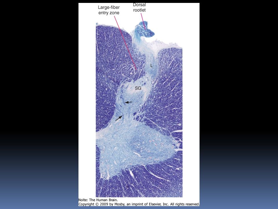
51
important relay nucleus for the transmission of information to the cerebellum may also play a role in forwarding proprioceptive information from the leg to the thalamus. Because of its prominent role in sensory processing, it is treated by many as part of the posterior horn.

53
The remainder of the intermediate gray matter is a collection of various projection neurons, sensory interneurons, and interneurons that synapse on motor neurons.

54
The posterior horn consists mainly of interneurons whose processes remain within the spinal cord and of projection neurons whose axons collect into long, ascending sensory pathways.

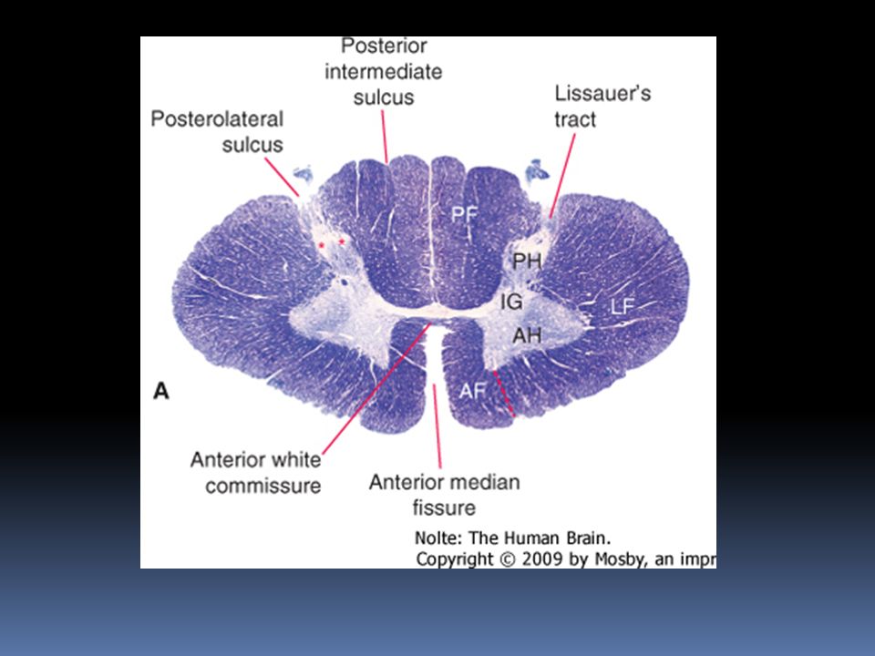
55
This area of gray matter contains two prominent parts, the substantia gelatinosa and the body of the posterior horn Both are present at all spinal levels. Between the substantia gelatinosa and the surface of the cord is a relatively pale-staining area of white matter called Lissauer's tract.*

57
This tract stains more lightly than the rest of the white matter ;contains the finely myelinated and unmyelinated fibers with which the substantia gelatinosa deals. The body of the posterior horn consists mainly of interneurons and projection neurons that transmit various types of somatic and visceral sensory information

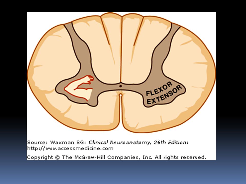
58
In this respect it functionally overlaps parts of the intermediate gray matter.

59
substantia gelatinosa distinctive region of gray matter that caps the posterior horn In myelin-stained preparations this region looks pale compared with the rest of the gray matter because it deals mostly with finely myelinated and unmyelinated sensory fibers that carry pain and temperature information.

60
Rexed’s laminae

63
Rexed's laminae Lamina I This thin marginal layer contains neurons that respond to noxious stimuli [pain, temperature] and send axons to the contralateral spinothalamic tract.
![Rexed s laminae Lamina I This thin marginal layer contains neurons that respond to noxious stimuli [pain, temperature] and send axons to the contralateral spinothalamic tract.](http://images.slideplayer.com/26/8567656/slides/slide_63.jpg "Rexed s laminae Lamina I This thin marginal layer contains neurons that respond to noxious stimuli [pain, temperature] and send axons to the contralateral spinothalamic tract.")
64
Lamina II- substantia gelatinosa made up of small neurons, some of which respond to noxious stimuli. Substance P, a neuropeptide involved in pathways mediating sensibility to pain, is found in high concentrations in laminas I and II.

65
Laminas III and IV [nucleus proprius] Their main input is from fibers that convey position and light touch sense. Lamina V contains cells that respond to both noxious and visceral afferent stimuli.
![ Laminas III and IV [nucleus proprius] Their main input is from fibers that convey position and light touch sense.](http://images.slideplayer.com/26/8567656/slides/slide_65.jpg " Lamina V contains cells that respond to both noxious and visceral afferent stimuli..")
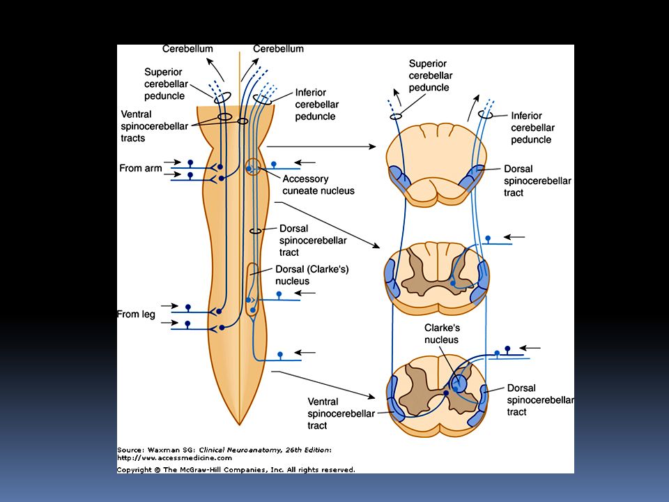
66
Lamina VI deepest layer,contains neurons that respond to mechanical signals from joints and skin.

67
Lamina VII contains the cells of the dorsal nucleus (Clarke's column) medially as well as a large portion of the ventral gray column. Clarke's column contains cells that give rise to the posterior spinocerebellar tract.

68
also contains the intermediolateral nucleus (or intermediolateral cell column) in thoracic and upper lumbar regions. Preganglionic sympathetic fibers project from cells in this nucleus, via the ventral roots and white rami communicantes, to sympathetic ganglia.

69
Laminas VIII and IX represent motor neuron groups in the medial and lateral portions of the ventral gray column The medial portion (also termed the medial motor neuron column) contains the LMNs that innervate axial musculature
 contains the LMNs that innervate axial musculature")
70
The lateral motor neuron column contains LMNs for the distal muscles of the arm and leg flexor muscles are innervated by motor neurons located close to the central canal extensor muscles are innervated by motor neurons located more peripherally

71
Lamina X represents the small neurons around the central canal or its remnants.

74
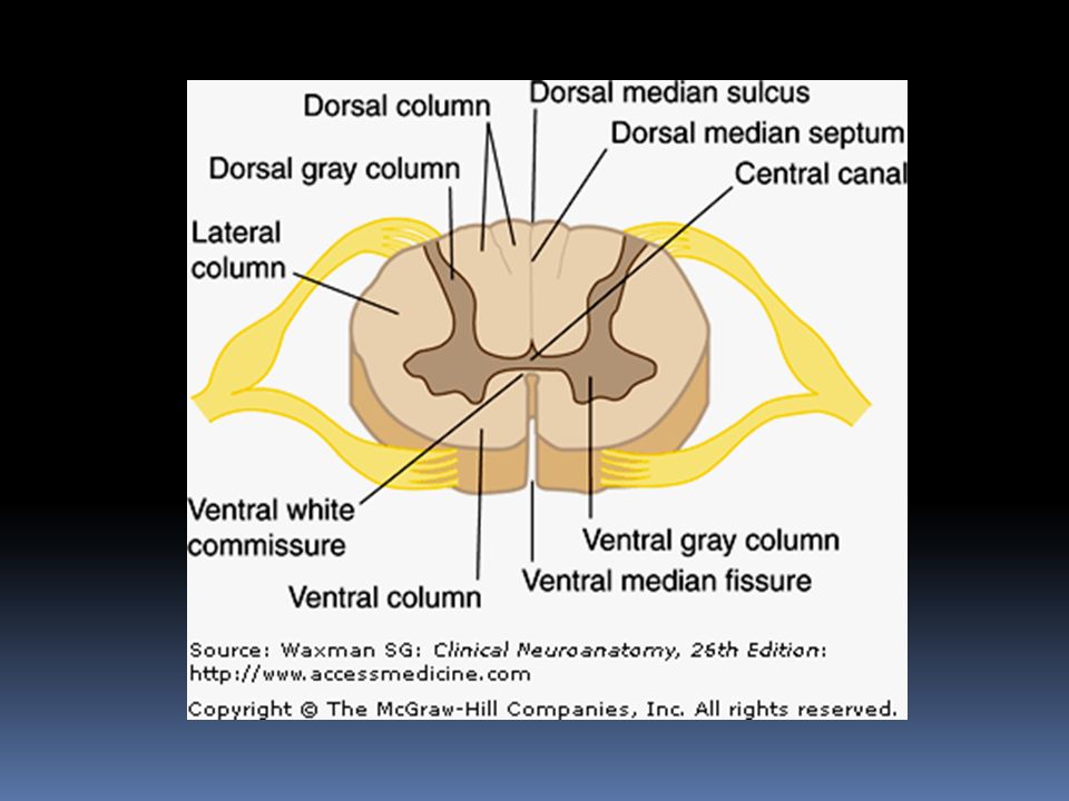
White Matter composed of myelinated and unmyelinated nerve fibers. The fast-conducting myelinated fibers form bundles (fasciculi) that ascend or descend for varying distances Fiber bundles with a common function are called tracts
 that ascend or descend for varying distances Fiber bundles with a common function are called tracts.")
75
The lateral and ventral white columns contain tracts that are not well delimited and may overlap in their cross sectional areas the dorsal column tracts are sharply defined by glial septa

76
Location of tracts

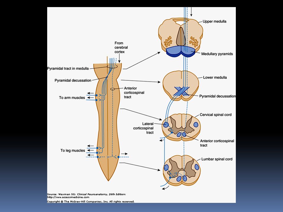
77
Descending tracts in the Spinal Cord. Lateral corticospinal (pyramidal) tract Fine motor function (controls distal musculature) Modulation of sensory functions
 tract Fine motor function (controls distal musculature) Modulation of sensory functions.")
79
Anterior corticospinal tract Vestibulospinal tract Gross and postural motor function (proximal and axial musculature Postural reflexes

80
Rubrospinal Reticulospinal Motor function Modulation of sensory transmission (especially pain) Modulation of spinal reflexes
 Modulation of spinal reflexes")
81
Descending autonomic Tectospinal Medial longitudinal fasciculus Modulation of autonomic functions Reflex head turning Coordination of head and eye movements

82
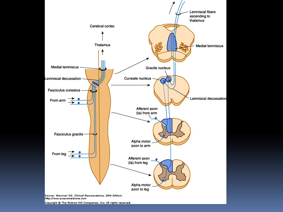
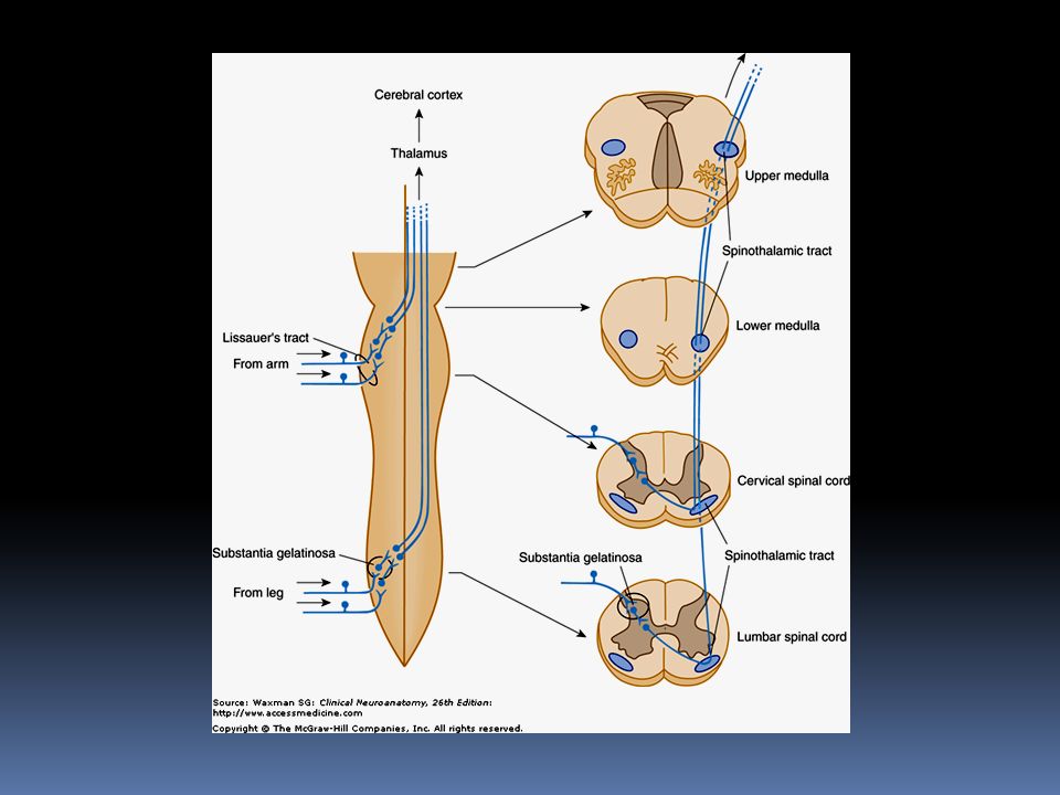
Ascending tracts Dorsal column system Spinothalamic tracts Fine touch, proprioception, two-point discrimination Sharp pain, temperature, crude touch

83
Dorsal spinocerebellar tract Ventral spinocerebellar Movement and position mechanisms

87
Somatotopic organization (segmental arrangement) in the spinal cord.
 in the spinal cord.")
88
Spinal Cord Circulation

89
Arteries-Anterior Spinal Artery formed by the midline union of paired branches of the vertebral arteries descends along the ventral surface of the cervical spinal cord, narrowing somewhat near T4.

90
Posterolateral Spinal Arteries arise from the vertebral arteries and course downward to the lower cervical and upper thoracic segments.

92
Radicular Arteries Some (but not all) of the intercostal arteries from the aorta supply segmental (radicular) branches to the spinal cord from T1 to L1. The largest of these branches, the great ventral radicular artery, also known as the arteria radicularis magna, or artery of Adamkiewicz, enters the spinal cord between segments T8 and L4

94
usually arises on the left and, in most individuals, supplies most of the arterial blood supply for the lower half of the spinal cord. occlusion in this artery is rare, it results in major neurologic deficits (eg, paraplegia, loss of sensation in the legs, urinary incontinence).
..")
95
Posterior Spinal Arteries paired arteries much smaller than the single large anterior spinal artery branch at various levels to form the posterolateral arterial plexus.

96
supply the dorsal white columns and the posterior portion of the dorsal gray columns.

Similar presentations


.>")
>")


 and Nerves. NERVOUS SYSTEM 1.Collect sensory input 2.Integrate sensory input 3.Motor output Functions of Nervous System.>")


