Download presentation
Presentation is loading. Please wait.

1
Skeletal Muscle Contraction
Dr. Wasif Haq

2
Introduction Muscles make 50% of total body mass.
40% by skeletal muscles & 10% combination of smooth & cardiac muscles. Sarcolemma: Cell membrane of muscle fiber. Ends fuse with tendons’ fiber to form muscle tendons inserting in bone. Sarcoplasm: Intracellular matrix of muscle fiber having suspended myofibrils. Abundant ATP & K+,P+,Mg++. Sarcoplasmic reticulum: Endoplasmic reticulum of muscles, store Ca++ needed for contraction. Sarcomere: Myofibrils comprise of smaller contractile units, smallest contractile unit of muscle fiber.

3
Structure of Myofibril.
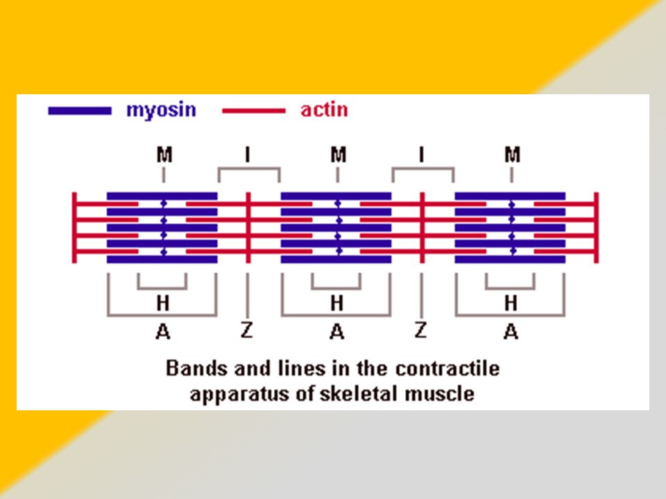
Sarcomeres comprise of series of alternating dark and light bands causing striated appearance. Dark band=A band. (Anisotropic) comprise of Myosin & ends of Actin. Lighter appearance in midsection called H-zone which is bisected by dark M line. Light band=I band (Isotropic) comprise of Actin only. Light band have middle Z line.
 comprise of Myosin & ends of Actin. Lighter appearance in midsection called H-zone which is bisected by dark M line. Light band=I band (Isotropic) comprise of Actin only. Light band have middle Z line.")
4
Structure of Myofilaments
Myofibrils contain myofilaments that in turn are composed of Actin and Myosin. Actin and Myosin ratio: 2:1 Myofibrils comprise of 1500 myosin (in center) attached to 3000 actin on each side. Portion of myofibril lying between 2 successive Z disc is Sarcomere.
 attached to 3000 actin on each side. Portion of myofibril lying between 2 successive Z disc is Sarcomere.")
5
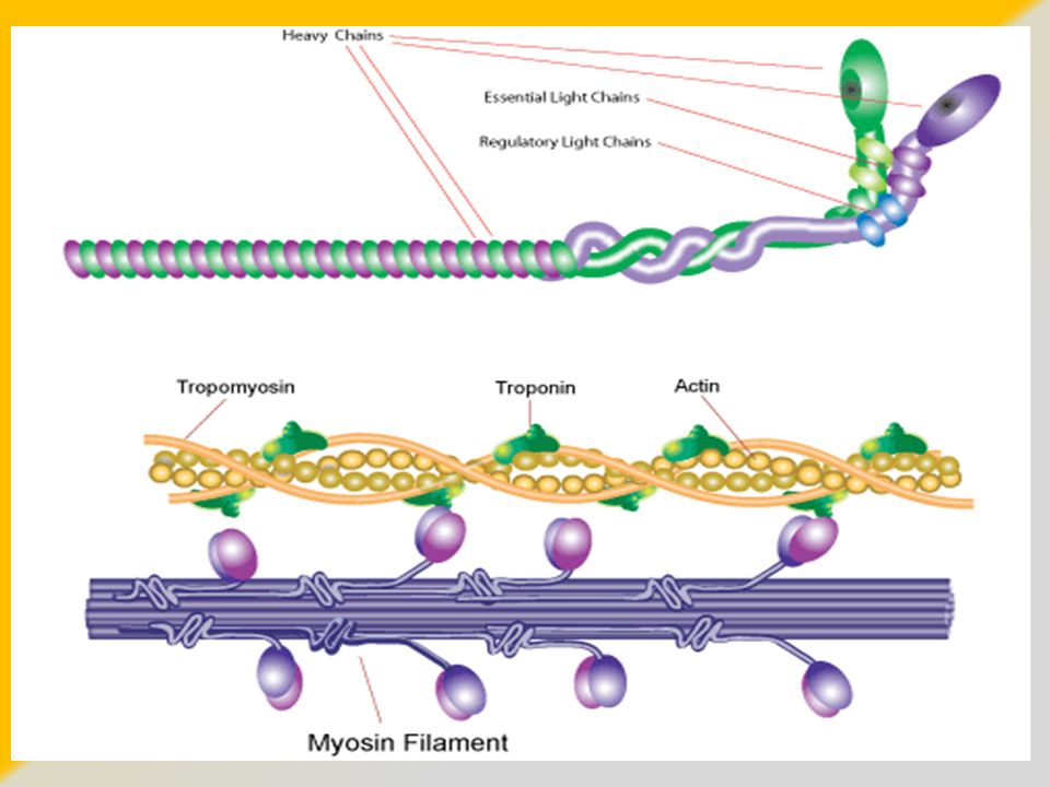
Myosin Myosin has 6 polypeptide chains, 4 light chains & 2 heavy chains. 2 heavy chains form double helix twisting forming tail. Ends of these chain folded to form head, thereby making 2 heads lying side by side at one end of double helix myosin.

7
Myosin Multiple tails unite to form body with many heads handing outwards on the sides of the body with a side hanging structure called arm that helps to extend the head away from the body. Cross bridges: Protruding arms & head together. Cross bridges are flexible at two points called hinges. 1. Origin of arm from body 2. Site of attachment of head to arm.

8
Myosin Hinged arms cause head to either extend far outward or come closer to the body. The hinged head in turn participate in actual contraction process. Head has ATPase enzyme cleaving ATP and thereby deriving energy needed to cause muscle contraction.

11
Actin Comprises of 3 proteins: Actin, Tropomyosin and Troponin
Actin twisted around each other in 2 chains like double helix structure made of F actin protein molecule, comprising of 1 active site resulting from union of 2 F actin strands. Tropomyosin: Attached with Actin. At resting phase, Troponin-Tropomyosin complex covers 7 active sites on actin thereby preventing myosin binding and eventually muscle contraction. Troponin: Attached with Tropomyosin. 3 subtypes. Troponin I: Affinity for Actin. Troponin T: Tropomyosin. Troponin C: Calcium ions, for contraction Ca++ uncover active sites.

12
Sliding Mechanism At resting phase, actin molecules are distant and apart from myosin and from each other. When contraction is needed, actin molecules are pulled closer to myosin and to each other, overlapping each other. Occurs because cross bridges of myosin become attached to active sites on actin, pulling the actin towards sarcomere. Changes in bands: Shortening of I & H band Z line brought closer to each other. Ca++ needed to facilitate binding between actin and myosin and ATP required to liberate energy.

14
Walk Along Theory In presence of Ca++ ions, head from cross bridges of myosin become attached to active sites on actin causing contraction. Walk along theory=Ratchet theory of contraction. Constant attachment and detachment of the head from active site on actin takes place, hence myosin head moves along the next active sites on actin in a sequence. Power stroke: The tilt of myosin head towards the arm causing dragging of the actin filament along. Followed by detachment of the head, returning to normal position and again binding with newer active site on actin. Fenn effect: Greater the amount of work to be performed by muscle, greater will be the amount of ATP to be cleaved.

15
Mechanism for muscle contraction
1. Action potential travels through motor nerve to endings on muscle fiber. 2. At the nerve ending, Acetylcholine; neurotransmitter is released. 3. Acetylcholine acts on Acetylcholine channels. 4. Inflow of Na+ ions occur causing depolarization. 5. Action potential travels through muscle fiber membrane. 6. Sarcoplasmic reticulum releases Ca++ 7. Ca++ inititate binding between actin and myosin. 8. Ca++ pumped back into Sarcoplasmic reticulum hence stopping contraction.

16
Extras Motor unit: All the muscle fibers innervated by single motor nerve. Muscle fatigue: Anaerobic glycolysis, lactic acid accumulation causing tissue injury. Hypertrophy: Increase in size of the muscle fiber. Hyperplasia: Increase in number of normal muscle fiber in normal arrangement. Rigor Mortis: Loss of ATP after death fails to separate cross bridges from actin causing muscles to remain contracted and remain rigid. Takes hours to resolve.

Similar presentations


 The Muscle Action Potential ( AP ) Muscle RMP = -90 mV ( same as in nerves.>")


