Download presentation
Presentation is loading. Please wait.

1
The Muscular System

2
or “Everything you ever wanted to know about Muscles, but were afraid to ask” !!!
This is the presentation on the muscular system for Human Anatomy and Physiology I at Oklahoma City Community College. I am Dennis Anderson.

3
How many muscles do we have???

4
Did you know that ? more than 50% of body weight is muscle ! And muscle is made up of proteins and water

6
The Muscular System Muscles are responsible for all movement of the body There are three basic types of muscle Skeletal Cardiac Smooth

7
Info About Muscles Only body tissue able to contract
create movement by flexing and extending joints Body energy converters (many muscle cells contain many mitochondria)
")
8
Muscles have Fascia- which is tough sheet like membrane

9
3 Types of Muscles

10
Three types of muscle Skeletal Cardiac Smooth

11
Classification of Muscle
Skeletal- found in limbs Cardiac- found in heart Smooth- Found in viscera Striated, multi- nucleated Striated, 1 nucleus Not striated, 1 nucleus voluntary involuntary

12
Characteristics of Muscle
Skeletal and smooth muscle are elongated Muscle cell = muscle fiber Contraction of a muscle is due to movement of microfilaments (protein fibers) All muscles share some terminology Prefixes myo and mys refer to muscle Prefix sarco refers to flesh
 All muscles share some terminology. Prefixes myo and mys refer to muscle. Prefix sarco refers to flesh.")
13
Shapes of Muscles Triangular- shoulder, neck Spindle- arms, legs
Flat- diaphragm, forehead Circular- mouth, anus

14
Skeletal Muscle Most are attached by tendons to bones
Cells have more than one nucleus (multinucleated) Striated- have stripes, banding Voluntary- subject to conscious control Tendons are mostly made of collagen fibers Found in the limbs Produce movement, maintain posture, generate heat, stabilize joints
 Striated- have stripes, banding. Voluntary- subject to conscious control. Tendons are mostly made of collagen fibers. Found in the limbs. Produce movement, maintain posture, generate heat, stabilize joints.")
15
Structure of skeletal muscle
Each cell (fibre) is long and cylindrical Muscle fibres are multi-nucleated Typically 50-60mm in diameter, and up to 10cm long The contractile elements of skeletal muscle cells are myofibrils
 is long and cylindrical. Muscle fibres are multi-nucleated. Typically 50-60mm in diameter, and up to 10cm long. The contractile elements of skeletal muscle cells are myofibrils.")
16
Skeletal muscle - Summary
Voluntary movement of skeletal parts Spans joints and attached to skeleton Multi-nucleated, striated, cylindrical fibres

17
Smooth Muscle No striations Spindle shaped Single nucleus
Involuntary- no conscious control Found mainly in the walls of hollow organs

18
Smooth muscle Lines walls of viscera
Found in longitudinal or circular arrangement Alternate contraction of circular & longitudinal muscle in the intestine leads to peristalsis

19
Structure of smooth muscle
Spindle shaped uni-nucleated cells Striations not observed Actin and myosin filaments are present( protein fibers)
")
20
Smooth muscle - Summary
Found in walls of hollow internal organs Involuntary movement of internal organs Elongated, spindle shaped fibre with single nucleus

21
Cardiac Muscle Striations Branching cells Involuntary
Found only in the heart Usually has a single nucleus, but can have more than one

22
Cardiac muscle Main muscle of heart Pumping mass of heart
Critical in humans Heart muscle cells behave as one unit Heart always contracts to it’s full extent

23
Structure of cardiac muscle
Cardiac muscle cells (fibres) are short, branched and interconnected Cells are striated & usually have 1 nucleus Adjacent cardiac cells are joined via electrical synapses (gap junctions) These gap junctions appear as dark lines and are called intercalated discs
 are short, branched and interconnected. Cells are striated & usually have 1 nucleus. Adjacent cardiac cells are joined via electrical synapses (gap junctions) These gap junctions appear as dark lines and are called intercalated discs.")
24
Cardiac muscle - Summary
Found in the heart Involuntary rhythmic contraction Branched, striated fibre with single nucleus and intercalated discs

25
Muscle Control Type of muscle Nervous control Type of control Example
Skeletal Controlled by CNS Voluntary Lifting a glass Skeletal Cardiac Regulated by ANS Involuntary Heart beating Smooth Controlled by ANS Involuntary Peristalsis

26
Types of Responses Twitch- Tetanus A single brief contraction
Not a normal muscle function Tetanus One contraction immediately followed by another Muscle never completely returns to a relaxed state Effects are compounded

27
Where Does the Energy Come From?
Energy is stored in the muscles in the form of ATP ATP comes from the breakdown of glucose during Cellular Respiration This all happens in the Mitochondria of the cell When a muscle is fatigued (tired) it is unable to contract because of lack of Oxygen
 it is unable to contract because of lack of Oxygen.")
28
Exercise and Muscles Isotonic- muscles shorten and movement occurs ( most normal exercise) Isometric- tension in muscles increases, no movement occurs (pushing one hand against the other)
")
29
How are Muscles Attached to Bone?
Origin-attachment to a immovable (fix) bone Insertion- attachment to an movable bone Muscles are always attached to at least 2 points Movement is attained due to a muscle moving an attached bone
 bone. Insertion- attachment to an movable bone. Muscles are always attached to at least 2 points. Movement is attained due to a muscle moving an attached bone.")
30
Muscle Attachments Insertion Origin
Muscles attach in at least two places in the body. *The origin is the attachment that moves the least. *The insertion is the attachment that moves the most. This diagram illustrates the origin and insertion of one of the neck muscles. Note the origin is on the clavicle and sternum. The insertion is on the skull. When the muscle contracts it will shorten the distance between the origin and insertion. The head will move when this muscle contracts. Remember the insertion is the end of the muscle that moves the most. Since the head moves the attachment on the head is called the insertion. The origin is generally on a larger body part will move the least. The chest does not move when this muscle contract. The bones in the chest are therefore the origin.

31
Types of Musculo-Skeletal Movement
Flexion You will need to know the action or movement performed for each of the muscles we study. The first type of movement is called flexion. Note the lower leg is being flexed in this diagram. During flexion the angle of joint is decreased. As the knee is flexed, the angle between the lower leg and the thigh is decreased. Flexion of the upper arm is also illustrated in this diagram. Here the angle between the arm and the frontal plane is decreased.

32
Extension Extension is the opposite of flexion. In extension the angle of a joint is increased. Extension of the lower leg causes an increased angle between the lower leg and the thigh.

33
Hyperextension Flexion and extension also apply to the neck. When a joint is extended past the anatomical position the movement is called hyperextension.

34
Abduction, Adduction & Circumduction
Abduction refers to moving away from the median plane of the body. Adduction is the opposite movement to abduction. It is moving toward the medial plane. Circumduction refers to inscribing a circle while moving a limb.

35
Rotation Rotation is turning a bone on its own axis. Moving the head back and forth to indicate “no” is an example of rotation. Note the difference between medial and lateral rotation. In lateral rotation the limb is rotated the lateral side of the body. Medial rotation rotates the limb toward the medial side of the body.

36
More Types of Movement……
Inversion- turn sole of foot medially Eversion- turn sole of foot laterally Pronation- palm facing down Supination- palm facing up Opposition- thumb touches tips of fingers on the same hand

37
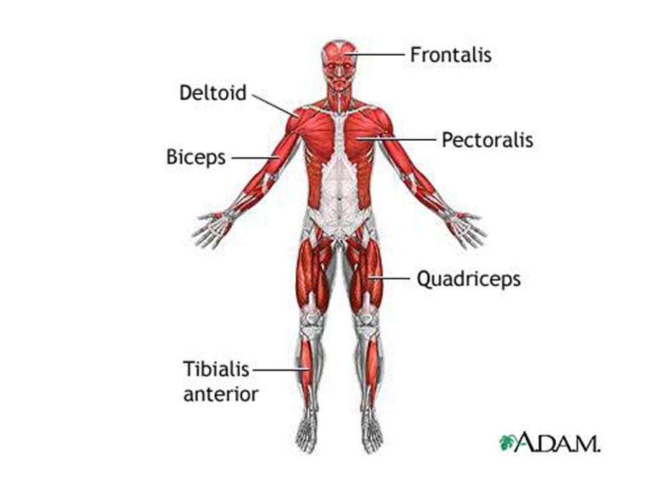
The Skeletal Muscles There are about 650 muscles in the human body
The Skeletal Muscles There are about 650 muscles in the human body. They enable us to move, maintain posture and generate heat. In this section we will only study a sample of the major muscles. There are about 650 muscles in the human body. They enable us to move, maintain posture and generate heat. In this unit will only study a sample of the major muscles.

38
Sternocleidomastoideus
Flexes and Rotates Head The first muscle we will learn is the sternocleidomastoideus. It is sometime called the sternocleitomastoid. It is the same neck muscle shown on the previous slide. * This muscle has two origins. The first origin is on the sternum manubrium. The second origin is on the clavicle. *The insertion is on the mastoid process of the skull. *Contraction of both sternocleidomastoideus muscles will flex the head. If just on of the muscles contracts, the head will rotate.

39
Extend Head, Adduct, Elevate or Depress Scapula
Trapezius Extend Head, Adduct, Elevate or Depress Scapula The trapezius is a large muscle in the upper back. It attaches to the skull, shoulder and vertebrae of the back. *When this muscle contracts it will cause the head to extend. It will also move the scapula. The direction the scapula moves depends on which part of the trapezius contracts. The trapezius may adduct elevate or depress the scapula.

40
Extend, Adduct & Rotate Arm Medially
Latissimus Dorsi Extend, Adduct & Rotate Arm Medially The latissimus dorsi is a large muscle in the back. It is often referred to as a lat. *It has origins on the vertebrae, ilium ribs and scapula. *The insertion is on the humerus. When it contracts it moves the humerus. *It can extend, adduct and rotate the arm medially. This is the main muscle used in movement such as pounding a nail with a hammer.

41
Abduct, Flex & Extend Arm
Deltoid Abduct, Flex & Extend Arm The deltoid covers the shoulder and has the shape of a delta. *It has origins on the scapula and clavicle. *The deltoid inserts on the deltoid tuberosity of the humerus. *Contraction of the deltoid will adduct the arm. If only the anterior fibers of the muscle contract it will flex the arm. Contraction of the posterior fibers will extend the arm.

42
Flexes, adducts & rotates arm medially
Pectoralis Major Flexes, adducts & rotates arm medially The pectoralis major is a large muscle in the pectoral region of the body. *It has origins on the clavicle and sternum. *The insertion is on the greater tubercle of the humerus. *Contraction of the pectoralis major will flex the arm. It will also adduct and rotate the arm medially. The pectoralis major is used in movements such a climbing, throwing and doing pushups.

43
Biceps Brachii Flexes Elbow Joint
The biceps brachii is located on the anterior side of the upper arm. It is often just called the biceps. There is a biceps femoris in the leg we will study shortly. *The biceps has two origins. One origin is on the corocoid process and the other on the Glenoid cavity of the scapula. The “bi” in biceps refers to the two origins. *It inserts on the radial tuberosity. *Contraction of the biceps will cause flexing at the elbow joint.

44
Triceps Brachii Extend Elbow Joint
The triceps is on the back of the upper arm. *It has three origins. Two origins are on the back of the humerus and one on the scapula. *The triceps inserts on the olecranon. *Movement of the triceps will extend the elbow joint.

45
Rectus Abdominus Flexes Abdomen
Rectus abdominus is a long muscle in the abdomen. *The muscle originates on the pubis. *It inserts on the xiphoid process of the sternum and also on cartilage of the ribs. *When rectus abdominus contracts it will flex the abdomen.

46
Diaphragm Inspiration
This is an inferior view of the diaphragm. This muscle separates the abdominal cavity from the thoracic cavity. *When it contracts it will cause inspiration.

47
Extends & Rotates Thigh Laterally
Gluteus Maximus Extends & Rotates Thigh Laterally The large muscle on the posterior side of the body at the top of each leg is the gluteus maximus. *The gluteus maximus originates on the ilium, sacrum and coccyx. *It inserts on the gluteal tuberosity of the femur. *This muscle will extend and rotate the thigh laterally.

48
Quadriceps femoris Muscle on the front of the thigh that extends the leg

49
Muscle properties and characteristics:
Excitability: irritability, the ability to respond to stimulus such as nerve impulse Contractibility: muscle fibers that are stimulated by nerves contract, or become short and thick, which causes movement Extensibility: the ability to be stretched Elasticity: allows the muscle to return to its original shape after it has contracted or stretched

50
Muscles attach by origin, insertion and by
Fascia: a tough, sheet-like membrane that covers and protects the tissue The deep muscles of the trunk and back, which are surrounded by the lumbodorsal fascia Muscles are partially contracted at all times, even when not in use Muscle tone- state of partial contraction Atrophy- loss of muscle tone when muscles are not used for a long period Contracture- a severe tightening of a flexor muscle resulting in bending of a joint Foot drop is a common contracture, but the fingers, wrists, knees, and other joints can also be affected

51
Diseases and Abnormal Conditions
Fibromyalgia: chronic, widespread pain in specific muscle sites Symptoms: muscle stiffness, numbness or tingling in the arms or legs, fatigue, sleep disturbances, headaches, and depression Cause is unknown, but stress, weather, and poor physical fitness affect the condition Treatment: directed toward pain relief and includes physical therapy, massage, exercise, stress reduction, and medication to relax muscles and relieve pain

52
Muscular Dystrophy A group of inherited diseases that lead to chronic, progressive muscle diseases that lead to muscle atrophy Usually appears in early childhood Most types result in total disability and early death Most common type: Duchenne muscular distrophy, which is caused by a genetic defect At birth the infant is healthy, as muscle cells die, the child loses the ability to move Onset usually occurs between 2 and 5 years of age By age 9-12, the child is confined to a wheelchair Eventually, the muscle weakness affects the heart and diaphragm, resulting in respiratory and/or cardiac failure that causes death. Life expectancy: late teens to the early twenties No cure, but physical therapy is used to slow progress

53
Myasthenia Gravis Chronic condition where nerve impulses are not properly transmitted to the muscles Leads to progressive muscular weakness and paralysis Affects respiratory muscles, can be fatal Cause is unknown, though is thought to be autoimmune (antibodies attacking the body’s own tissue) No cure; Treatment is supportive
 No cure; Treatment is supportive.")
54
Muscle Atrophy Inherited chronic progressive disorder in which results in muscle shrinking Results in complete disability Early death

55
Strain An overstretching of or injury to a muscle and/or tendon
Frequent site include the back, arms, and legs Cause: Prolonged or sudden muscle exertion Symptoms: myalgia (muscle pain), swelling, and limited movement Treatment: rest, muscle relaxants or pain medications, elevating the extremity, and alternating hot and cold applications
, swelling, and limited movement. Treatment: rest, muscle relaxants or pain medications, elevating the extremity, and alternating hot and cold applications.")
56
Muscle Spasms Muscle spasms, or cramps, are sudden, painful, involuntary muscle contractions Usually occur in the legs to feet and may result from overexertion, low electrolyte levels, or poor circulation Gentle pressure and stretching of the muscle are used to relieve the spasm

57
Draw where each of these muscles are on a poster paper.
For each describe the type of movement they perform…For example: Pectoralis- are in the upper chest and adducts and flexes the arms. Must be neat and have color Gastrocnemius Biceps brachii Triceps brachii Deltoid Sternocleidomastoid Rectus Abdominus Gluteus maximus Intercostals Latissimus dorsi Quadriceps femoris Tibialis anterior Trapezius Pectoralis

Similar presentations