Download presentation
Presentation is loading. Please wait.

1
The Nervous System: Neurons and Synapses
Chapter 7 The Nervous System: Neurons and Synapses

2
Nervous System CNS (central nervous system):
Brain Spinal cord PNS (peripheral nervous system): Cranial nerves Spinal nerves 2 Types of cells in nervous system: Neurons Supporting cells
: Cranial nerves. Spinal nerves. 2 Types of cells in nervous system: Neurons. Supporting cells.")
3
Neurons Structural and functional units of the nervous system.
Respond to physical and chemical stimuli. Produce and conduct electrochemical impulses. Release chemical regulators. Cannot divide by mitosis.

4
Neurons 3 Principal regions: Cell body (perikaryon):
Contains the nucleus, Nissl bodies (ER). Dendrites: Provide receptive area. Transmits electrical impulses to cell body. Axon: Conducts impulses away from cell body.
. Dendrites: Provide receptive area. Transmits electrical impulses to cell body. Axon: Conducts impulses away from cell body.")
5
Classification of Neurons
Functional: Based upon direction impulses conducted. Sensory or afferent: Conduct impulses from receptors to CNS. Motor or efferent: Conduct impulses out of CNS to effector. Association or interneurons: Located entirely within the CNS.

6
Classification of Neurons
Structural: # of processes that extend from cell body. Pseudounipolar: Short single process that branches like a T. Sensory neurons. Bipolar neurons: Have 2 processes. Retina of the eye. Multipolar: Have several dendrites and 1 axon. Most common type.

7
Supporting Cells 6 categories of supporting cells:
Schwaan cells (neurolemmocytes) Satellite cells (ganglionic gliocytes) Oligodendrocytes Microglia Astrocytes Ependymal cells
 Satellite cells (ganglionic gliocytes) Oligodendrocytes. Microglia. Astrocytes. Ependymal cells.")
8
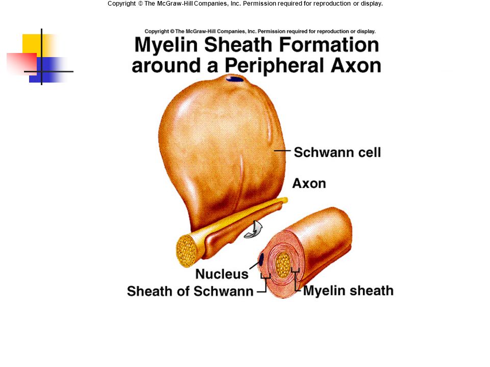
Supporting Cells Peripheral nervous system (PNS): Satellite cells:
Support neuron cell bodies within ganglia. Schwaan cells: Surround axons in PNS. Outer surface encased in basement membrane (neurilemma). Provide insulation. Nodes of Ranvier: Unmyelinated areas that produce nerve impulses.
. Provide insulation. Nodes of Ranvier: Unmyelinated areas that produce nerve impulses.")
9
Supporting Cells Central nervous system (CNS): Oligodendrocytes:
Form myelin sheaths. Insulation.

10
Supporting Cells CNS: Astrocytes: Most abundant glial cell.
Vascular processes cover capillaries. Stimulate tight junctions, contributing to blood-brain barrier. Regulate external environment of K+ and pH.

11
Supporting Cells CNS: Microglia: Ependymal cells:
Phagocytes, migratory. Ependymal cells: Secrete cerebrospinal fluid (CSF). Line ventricles.
. Line ventricles.")
12
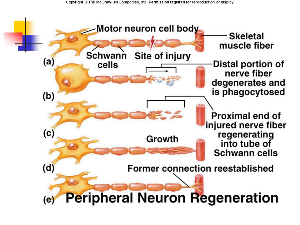
Nerve Regeneration PNS: Distal neuronal portion degenerates.
Schwann cells act as phagocytes. Schwann cells (basement membrane) form regeneration tube. Serves as guide for axon. Axon tip begins to grow towards destination.
 form regeneration tube. Serves as guide for axon. Axon tip begins to grow towards destination.")
14
Neurotrophins Nerve growth factors: Fetus: Adult:
Embryonic development of sensory neurons and sympathetic ganglia. Adult: Maintenance of sympathetic ganglia. Mature sensory neurons need for regeneration. Required to maintain spinal neurons. Sustain neurons that use dopamine.

15
Electrical Activity of Axons
All cells maintain resting membrane potential. Permeability of cell membrane: 80% electrochemical gradients of Na+ and K+. 20% Na+ / K+ ATP-ase pump. Excitability/irritability: Ability to produce and conduct electrical impulses.

16
Electrical Activity of Axons
Depolarization: Potential difference reduced (become more positive). Repolarization: Return to resting membrane potential (become more negative). Hyperpolarization: More negative than resting membrane potential.
. Repolarization: Return to resting membrane potential (become more negative). Hyperpolarization: More negative than resting membrane potential.")
17
Ion Gating in Axons Changes in membrane potential caused by ion flow through channels. Specific ion channels for Na+ and K+. Passive channels are always open. Voltage gated channels open in response to change in membrane potential.

18
Membrane Potential Copyright © The McGraw-Hill Companies, Inc. Permission required for reproduction or display. Basis of resting membrane potential: Selective permeability of plasma membranes. Strong permeability of potassium ions (K+). Weak permeability of sodium ions (Na+). Impermeability of large anions. Diffusion of ions down their concentration gradients. K+ diffuses down its concentration gradient and out of the cell. Positive charge leaves the cell causing the cell to become more negative. Na+ diffuses into the cell, but at a slower rate than K+ diffusion, thus cancelling some of the negative internal charge. Diffusion of ions down their electrical gradients; the electrical attraction of cations and anions to each other. As K+ diffuses out of the cell, making the cell more negative inside, K+ begins to move back into the cell towards the negative charge. (The positively charged potassium is attracted to the negatively charged cytosol.)
. Weak permeability of sodium ions (Na+). Impermeability of large anions. Diffusion of ions down their concentration gradients. K+ diffuses down its concentration gradient and out of the cell. Positive charge leaves the cell causing the cell to become more negative. Na+ diffuses into the cell, but at a slower rate than K+ diffusion, thus cancelling some of the negative internal charge. Diffusion of ions down their electrical gradients; the electrical attraction of cations and anions to each other. As K+ diffuses out of the cell, making the cell more negative inside, K+ begins to move back into the cell towards the negative charge. (The positively charged potassium is attracted to the negatively charged cytosol.)")
20
Voltage gated channels
anim0026-swf_voltage_ga.swf

21
Action Potentials Copyright © The McGraw-Hill Companies, Inc. Permission required for reproduction or display. The Action Potential The action potential consists of rapid dramatic changes in membrane potential that occur due to opening and closing of voltage-gated ion channels. It begins with a steady depolarization called the generator potential. If the generator potential reaches a critical voltage called the threshold, the membrane will continue to depolarize, followed by a period of repolarization and then a short period of hyperpolarization.

22
Action Potentials (AP)
Stimulus causes depolarization to threshold. Voltage gated (VG) Na+ channels open. Electrical and chemical (electrochemical) gradients inward. + feedback loop. VG K+ channels open. Electrochemical gradients outward. - feedback loop. Changes in membrane potential constitute AP. anim0027-rm_action_pote.rm
 Na+ channels open. Electrical and chemical (electrochemical) gradients inward. + feedback loop. VG K+ channels open. Electrochemical gradients outward. - feedback loop. Changes in membrane potential constitute AP. anim0027-rm_action_pote.rm.")
24
Action Potentials (AP)
Once AP completed, Na+ / K+ ATPase pump extrude Na+, and recover K+. All or none: When threshold reached, maximum potential change occurs. Coding for Intensity: Increased frequency of AP indicates greater stimulus strength. Recruitment: Activate more axons that have higher threshold.

25
Refractory Periods Absolute refractory period:
Axon membrane is incapable of producing another AP. Relative refractory period: Axon membrane can produce another action potential, but requires stronger stimulus.

26
Conduction of Nerve Impulses
Cable properties: Ability of neuron to transmit charge through cytoplasm. High internal resistance. An AP does not travel down the entire axon. Each AP is a stimulus to produce another AP in the next region of membrane with VG channels.

27
Conduction in an Unmyelinated Axon
Cable spread of depolarization with influx of Na+ depolarizes the adjacent membranes, propagating the AP. Conduction rate is slow.

28
Conduction in Myelinated Axon
Myelin prevents movement of Na+ and K+ through the membrane. Nodes of Ranvier contain VG Na+ and K+ channels. Saltatory conduction (leaps). Fast rate of conduction.
. Fast rate of conduction.")
Similar presentations


 Central Nervous System (CNS) Peripheral Nervous System (PNS) Peripheral Nervous.>")
 from one part of the body to another. ◦ Major regions.>")


