Download presentation
Presentation is loading. Please wait.

1
©1999 Timothy G. Standish Bielkoviny, enzýmy Protein Structure Július Cirák

2
©1999 Timothy G. Standish Polar Basic Acid ? Amine Different Amino Acid Classes OH O H H2NH2N C C R Generic Non-polar C C C OH H H O H+NH+N H H2NH2N C NH C C Histidine H H2NH2N C C H H O OH O C C Aspartic acid C C C OH H H O HS H H2NH2N Cysteine OH H H O H H H2NH2N C C C Alanine Protein Structure

3
©1999 Timothy G. Standish Levels Of Protein Organization Primary Structure - The sequence of amino acids in the polypeptide chain Secondary Structure - The formation of helices and pleated sheets due to hydrogen bonding between the peptide backbone Tertiary Structure - Folding of helices and sheets influenced by R group bonding Quaternary Structure - The association of more than one polypeptide into a protein complex influenced by R group bonding Protein Structure

4
©1999 Timothy G. Standish

5
Levels Of Protein Organization Primary Structure Met-Gly-Ala-Pro-His-Ile-Asp-Glu-Met-Ser-Thr-... The sequence of amino acids in the primary structure determines the folding of the molecule. Protein Structure

6
©1999 Timothy G. Standish Protein Secondary Structure The peptide backbone has areas of positive charge and negative charge These areas can interact with one another to form hydrogen bonds The result of these hydrogen bonds are two types of structures: helices pleated sheets Protein Structure

7
©1999 Timothy G. Standish + - Protein Secondary Structure: Helix C O OHCN H H H C HOH C H O CN H H H C HH C H N C O C H N O C C O C H N C H N C O C O C O C O C H N H N C H N Protein Structure

8
©1999 Timothy G. Standish + - Protein Secondary Structure: Helix C O OHCN H H H C HOH C H O CN H H H C HH C H N C O C H N O C C O C H N C H N C O C O C O C O C H N H N C H N Protein Structure

9
©1999 Timothy G. Standish Protein Secondary Structure: Helix R R R R R R R R R R R R R R R groups stick out from the helix influencing higher levels of protein organization Protein Structure

10
©1999 Timothy G. Standish Protein Secondary Structure: Pleated Sheet N H C O C H C C N O N H C O C H C C N O N H C O C H C C N O N H C O C H C C N O N H C O C H C C N O N H C O C H C C N O N H C O C H C C N O N H C O C H C C N O Protein Structure

11
©1999 Timothy G. Standish Protein Secondary Structure: Pleated Sheet N H C O C H C C N O N H C O C H C C N O N H C O C H C C N O N H C O C H C C N O N H C O C H C C N O N H C O C H C C N O N H C O C H C C N O N H C O C H C C N O N H C O C H C C N O N H C O C H C C N O N H C O C H C C N O N H C O C H C C N O Protein Structure

12
©1999 Timothy G. Standish Levels Of Protein Organization Tertiary Structure Tertiary structure results from the folding of helices and pleated sheets Factors influencing tertiary structure include: Hydrophobic interactions Hydrogen bonding Disulphide bridges Ionic bonds Protein Structure

13
©1999 Timothy G. Standish Globular and Fibrous e.g. haemoglobin 3º structure normally folds up in a ball hydrophilic R groups point outwards Hydrophobic R groups point inwards soluble metabolic functions e.g. collagen 2º structure does not fold up, form fibres not surrounded by hydrophilic R groups insoluble structural functions Protein Structure

14
©1999 Timothy G. Standish Hydrophobic interactions Protein Structure Valine OH O H H2NH2N C C H3CH3C CH 3 H C Proline OH O H H2N+H2N+ C C H2CH2C CH 2 H2CH2C OH O H H2NH2N C C H Glycine

15
©1999 Timothy G. Standish Hydrogen Bonding Asparagine H H2NH2N C C H H O OH O C NH 2 C Glutamine H H2NH2N C C H H O OH O C NH 2 C C H H N-------H (+) (-) O -------C slightly slightly Protein Structure
 (-) O C slightly slightly Protein Structure.")
16
©1999 Timothy G. Standish Disulphide bridges Cysteine C C C OH H H O HS H H2NH2N Cysteine C C C OH H H O HS H H2NH2N Cysteine C C C OH H H O S H H2NH2N Cysteine C C C OH H H O S H H2NH2N Protein Structure

17
©1999 Timothy G. Standish Ionic Bonds Glutamic acid H H2NH2N C C H H O OH O C O- C C H H Arginine OH O H H2NH2N C C H H C H H C C H H H N +H2N+H2N NH 2 C Protein Structure

18
©1999 Timothy G. Standish

19
e.g.G-3-P Dehydrogenase Tertiary Structure Picture source: SWISS-PROT Protein Structure

20
©1999 Timothy G. Standish Collagen is a fibrous protein made of 3 polypeptide helices held together by hydrogen bonding Every 3rd amino acid in the chain is a glycine (very small to let the chains lie close to each other) Collagen molecules are found side by side forming fbres The staggered ends help to give collagen fibres great tensile strength Protein Structure
 Collagen molecules are found side by side forming fbres The staggered ends help to give collagen fibres great tensile strength Protein Structure.")
21
©1999 Timothy G. Standish Levels Of Protein Organization Quaternary Structure Quaternary structure results from the interaction of independent polypeptide chains Factors influencing quaternary structure include: Hydrophobic interactions Hydrogen bonding The shape and charge distribution on amino acids of associating polypeptides Protein Structure

22
©1999 Timothy G. Standish

23
Haemoglobin Picture source: SWISS-PROT Protein Structure

24
©1999 Timothy G. Standish Haemoglobin Haemoglobin is a globular protein with a prosthetic ‘iron’ group In adults, hemoglobin is made up of 4 polypeptides (2 polypeptide chains and 2 polypeptide chains) Each polypeptide surrounds a prosthetic ‘haem’ group Hydrophobic interactions between side groups pointing inwards maintain the structure Hydrophilic side chains point outwards making it soluble Fe Protein Structure
 Each polypeptide surrounds a prosthetic ‘haem’ group Hydrophobic interactions between side groups pointing inwards maintain the structure Hydrophilic side chains point outwards making it soluble Fe Protein Structure.")
25
©1999 Timothy G. Standish G-3-P Dehydrogenase from Bacillus stearothermophilus Picture source: SWISS-PROT Protein Structure

26
©1999 Timothy G. Standish Sickle Cell Anaemia Protein Structure

27
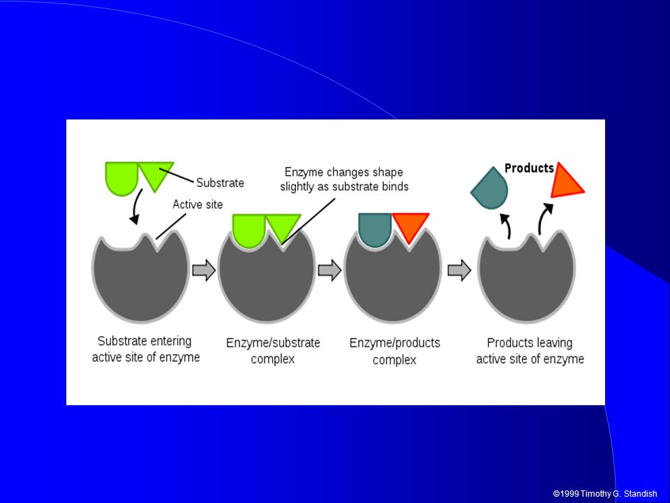
©1999 Timothy G. Standish E + S → ES → EP → E + P

28
©1999 Timothy G. Standish

Similar presentations

 Describe the structure of an amino acid and the formation and breakage of a peptide bond. (f) Explain the meaning.>")
 Monomer/Building Block Amino Acids (20 different.>")


 (P.G.T CHEM.) K.V. BALRAMPUR K.V. BALRAMPUR.>")


.>")

 Polymers made from chains of amino acids 20 amino acids used Linked by a peptide bond.>")

