Download presentation
Presentation is loading. Please wait.

1
Embryonic Neural Stem Cells & Potential Anesthetic-Induced Neurotoxicity Cheng Wang, MD., PhD Division of Neurotoxicity National Center of Toxicological Research (NCTR)/FDA The views provided in this presentation may not reflect those of the FDA
/FDA The views provided in this presentation may not reflect those of the FDA")
2
Representative Anesthetics 1) NMDA Antagonists: 2) GABA Agonists: Ketamine/PCP Midazolam Nitrous oxide Propofol Baclofen 3) Combination (NMDA antagonists and GABA agonists): Ketamine + Midazolam Nitrous oxide + Midazolam + Isoflurane (triple anesthetic drug protocol) 4) Narcotics: Fentanyl (Control) Fentanyl is an important control because its mechanism of action does not involve NMDA or GABA systems
 NMDA Antagonists: 2) GABA Agonists: Ketamine/PCP Midazolam Nitrous oxide Propofol Baclofen 3) Combination (NMDA antagonists and GABA agonists): Ketamine + Midazolam Nitrous oxide + Midazolam + Isoflurane (triple anesthetic drug protocol) 4) Narcotics: Fentanyl (Control) Fentanyl is an important control because its mechanism of action does not involve NMDA or GABA systems")
3
INTRODUCTION A great deal of concern has recently arisen regarding the safety of anesthesia in infants and children. There is mounting and convincing preclinical evidence in rodents and non-human primates that anesthetics in common clinical use are neurotoxic to the developing brain. The clinical relevance of anesthetic neurotoxicity is an urgent matter of public health. Recent advances in our understanding of stem cell biology and neuroscience have opened up new avenues of research for detecting anesthetic-induced neurotoxicity and developing potential protection/prevention strategies against anesthetic- induced neuronal injury.

6
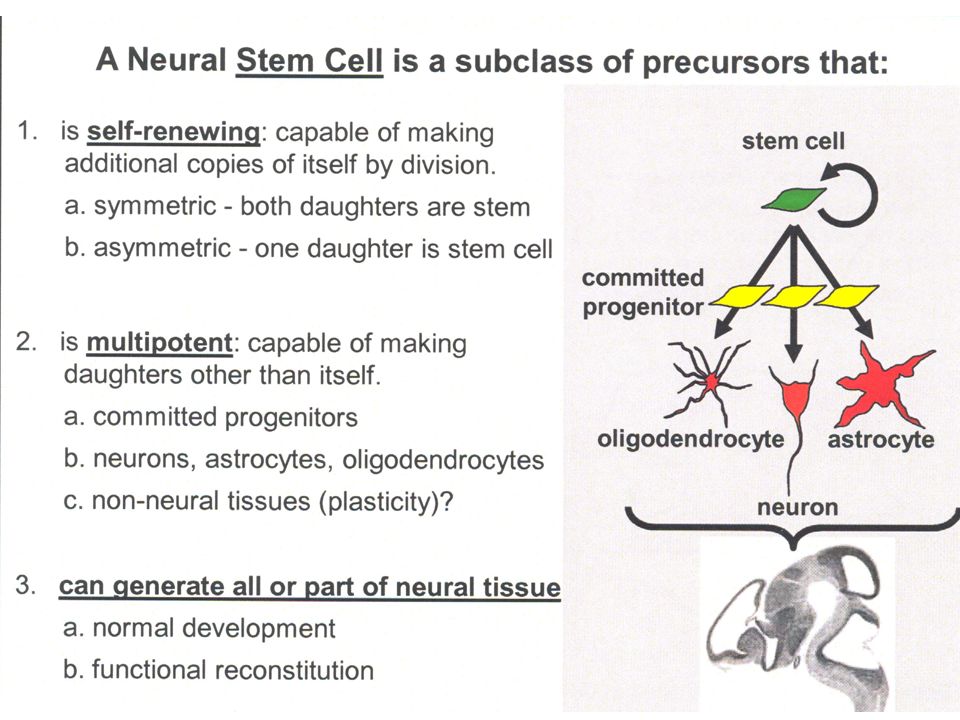
DIV 2 DIV 4 DIV 6 DIV 8 Rat Embryonic Neural Stem Cells Embryonic neural stem cells were harvested from cortical tissue of timed pregnancy embryonic day 14 Sprague-Dawley rats. plate cells at 96-well plate or petris at a concentration of one million cells per ml. Grow in serum-free N2 medium containing bFGF, EGF, NT 3 and PDGF (change medium every 3 days). perform experiments on Day In Vitro (DIV) 8.
. perform experiments on Day In Vitro (DIV) 8..")
7
A B Contrast phaseNestin Rat Embryonic Neural Stem Cell Culture (Day-In-Vitro 8)
")
8
A Nestin Rat Embryonic Neural Stem Cell Culture (Day-In-Vitro 8) B Nestin + EdU
 B Nestin + EdU")
9
Propofol 2,6-diisopropylphenol Propofol (marketed as Diprivan) is a hypnotic agent. Propofol, a GABA receptor agonist, is a widely used anesthetic agent for induction and maintenance of anesthesia for adults and children. Growing body of data suggest that exposure to anesthetics during certain periods of development has long-term deleterious effects. At the cellular level, there is evidence that anesthetic agents induce cell death, cause synaptic remodeling, and alter morphology of the developing brain.

10
Objective & Specific Aims Using embryonic neural stem cells, it is of considerable interest to determine: 1)How they respond to stress, e.g., prolonged (time-course) propofol exposure. 2)How propofol exposure (dose response) directs/signals these cells to undergo apoptosis or necrosis. 3)How their proliferation rate is affected by short- or prolonged anesthetic exposure. 4)How their fate, e.g., differentiation from progenitor cells to neurons or glial cells, is affected. 5)How the anti-oxidant agents affect anesthetic-induced neurotoxicity. 6)Clinically relevant protective strategies against anesthetic-induced developmental neural damage.
How propofol exposure (dose response) directs/signals these cells to undergo apoptosis or necrosis. 3)How their proliferation rate is affected by short- or prolonged anesthetic exposure. 4)How their fate, e.g., differentiation from progenitor cells to neurons or glial cells, is affected. 5)How the anti-oxidant agents affect anesthetic-induced neurotoxicity. 6)Clinically relevant protective strategies against anesthetic-induced developmental neural damage..")
11
LDH Release (Propofol; 24-hour Exposure)
")
12
MTT-Assay (Propofol; 24-hour Exposure) * * a
 * * a")
13
3-hour Exposure 6-hour Exposure Control Propofol (10 µM) propofol (50 µM) Propofol (100 µM) A B
 propofol (50 µM) Propofol (100 µM) A B")
14
Propofol Exposure (24-hour) AB ControlPropofol (50 µM; 24 hours)
 AB ControlPropofol (50 µM; 24 hours)")
15
AB ControlPropofol (50 µM) TUNEL-Assay
 TUNEL-Assay")
16
Scatter Plot (Sorting) Cellometer Vision ControlPropofol (50 µM; 24 hours)
 Cellometer Vision ControlPropofol (50 µM; 24 hours)")
17
EdU-DAPPI Staining Control Propofol (50 µM; 24 hrs) A E B F CD EdU DAPPI Edu-DAPPI
 A E B F CD EdU DAPPI Edu-DAPPI")
18
Neural Stem Cell Proliferation (Propofol; 24-hour Exposure) *
 *")
19
* Control Pro 3 hours Pro 6 hours Pro 24 hours Pro = Propofol (50 µM)
")
20
Assessment of Mitochondrial Membrane Potential Control Propofol-exposed

21
8-oxo dG ELISA-Assay (Propofol; 24-hour Exposure)
")
22
Control Propofol (50µM) Propofol+L-Ca L-Ca alone L-Ca = Acetyl-L-Carnitine (10 µM) *
 Propofol+L-Ca L-Ca alone L-Ca = Acetyl-L-Carnitine (10 µM) *")
23
SUMMARY Prolonged propofol exposure at clinically relevant concentration induces adverse effects on embryonic neural stem cells. Propofol-induced cell damage is most probably apoptotic in nature. Data from the EdU assay suggest that 24 h propofol (50 µM) can slow or stop neural stem cell division/proliferation. The presence of elevated levels of 8-oxo dG and its analogs in the culture medium suggest oxidative damage due to an increased generation of reactive oxygen species (ROS). Co-administration of acetyl-l-carnitine effectively blocks at least some of the toxicity of propofol, presumably by reducing ROS generation or increasing ROS scavenging.
 can slow or stop neural stem cell division/proliferation. The presence of elevated levels of 8-oxo dG and its analogs in the culture medium suggest oxidative damage due to an increased generation of reactive oxygen species (ROS). Co-administration of acetyl-l-carnitine effectively blocks at least some of the toxicity of propofol, presumably by reducing ROS generation or increasing ROS scavenging..")
24
Anion Cation unbalance ROS Anions Cations Anions Cations Anions Cations Propofol GABA- R caspases cell death cytochrome c Mitocondrion DNA Fragmentation Chromatin Condensation Disturbed Cell Cycle DFF40/CAD L-Carnitine Ca 2+ G1 S M G2 Excitatory-R Neural Stem Cell

25
ACKNOWLEDGEMENTS NCTR/FDA Fang Liu Natalya Sadovova Charles Fogle Xuan Zhang Shuliang Liu Deborah Hansen Merle G. Paule William Slikker Jr. Joseph Hanig David Jacobson-Kram William Rodriguez NICHD,CDER, NCTR CDER/FDA

Similar presentations


 ◦ TDwt (cells containing the TDAG 51 gene) ◦ TDko (cells lacking the TDAG 51 gene)>")


?>")
 In adult tissues cell death exactly balances cell division In apoptosis the cell destroys itself from within.>")

?>")


