Download presentation
Presentation is loading. Please wait.

1
In the mammalian cardiovascular system, the pulmonary and system circuits operate simultaneously. The two ventricles pump almost in unison While some blood is traveling to the lungs, the rest of the blood is flowing to the body. Double circulation in mammals depends on the anatomy and pumping cycle of the heart

2
To trace the double circulation pattern of the mammalian cardiovascular system, we’ll start with the pulmonary (lung) circuit.
 circuit.")
4
The pulmonary circuit carries blood from the heart to the lungs and back again. (1) The right ventricle pumps blood to the lungs via (2) the pulmonary arteries. As blood flows through (3) capillary beds in the right and left lungs, it loads O 2 and unloads CO 2. What causes O 2 and CO 2 to load and unload? Oxygen-rich blood returns from the lungs via the pulmonary veins to (4) the left atrium of the heart. Next, the oxygen-rich blood blows to (5) the left ventricle, as the ventricle opens and the atrium contracts.
 The right ventricle pumps blood to the lungs via (2) the pulmonary arteries. As blood flows through (3) capillary beds in the right and left lungs, it loads O 2 and unloads CO 2. What causes O 2 and CO 2 to load and unload. Oxygen-rich blood returns from the lungs via the pulmonary veins to (4) the left atrium of the heart. Next, the oxygen-rich blood blows to (5) the left ventricle, as the ventricle opens and the atrium contracts..")
5
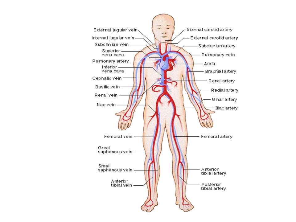
The left ventricle pumps oxygen-rich blood out to the body tissues through the systemic circulation. Blood leaves the left ventricle via (6) the aorta, which conveys blood to arteries leading throughout the body. The first branches from the aorta are the coronary arteries, which supply blood to the heart muscle. The next branches lead to capillary beds (7) in the head and arms. The aorta continues in a posterior direction, supplying oxygen-rich blood to arteries leading to (8) arterioles and capillary beds in the abdominal organs and legs. Within the capillaries, blood gives up much of its O 2 and picks up CO 2 produced by cellular respiration.
 the aorta, which conveys blood to arteries leading throughout the body. The first branches from the aorta are the coronary arteries, which supply blood to the heart muscle. The next branches lead to capillary beds (7) in the head and arms. The aorta continues in a posterior direction, supplying oxygen-rich blood to arteries leading to (8) arterioles and capillary beds in the abdominal organs and legs. Within the capillaries, blood gives up much of its O 2 and picks up CO 2 produced by cellular respiration..")
6
Venous return to the right side of the heart begins as capillaries rejoin to form venules and then veins. Oxygen-poor blood from the head, neck, and forelimbs is channeled into a large vein called (9) the anterior (or superior) vena cava. Another large vein called the (10) posterior (or inferior) vena cava drains blood from the trunk and hind limbs. The two venae cavae empty their blood into (11) the right atrium, from which the oxygen-poor blood flows into the right ventricle.
 the anterior (or superior) vena cava. Another large vein called the (10) posterior (or inferior) vena cava drains blood from the trunk and hind limbs. The two venae cavae empty their blood into (11) the right atrium, from which the oxygen-poor blood flows into the right ventricle..")
7
The mammalian heart is located beneath the breastbone (sternum) and consists mostly of cardiac muscle. The two atria have relatively thin walls and function as collection chambers for blood returning to the heart. The ventricles have thicker walls and contract much more strongly than the atria. Why do ventricles have thicker walls than the atria?

9
Fig. 42.5

10
A cardiac cycle is one complete sequence of pumping, as the heart contracts, and filling, as it relaxes and its chambers fill with blood. The contraction phase is called systole, and the relaxation phase is called diastole.

11
Cardiac output depends on two factors: the rate of contraction or heart rate (number of beats per second) and stroke volume, the amount of blood pumped by the left ventricle in each contraction. The average stroke volume for a human is about 75 mL. The typical resting cardiac output, about 5.25 L / min, is about equivalent to the total volume of blood in the human body. Cardiac output can increase about fivefold during heavy exercise. Heart rate can be measured indirectly by measuring your pulse - the rhythmic stretching of arteries caused by the pressure of blood pumped by the ventricles.

12
Four valves in the heart, each consisting of flaps of connective tissue, prevent backflow and keep blood moving in the correct direction. Between each atrium and ventricle is an atrioventricular (AV) valve which keeps blood from flowing back into the atria when the ventricles contract. Two sets of semilunar valves, one between the left ventricle and the aorta and the other between the right ventricle and the pulmonary artery, prevent backflow from these vessels into the ventricles while they are relaxing.
 valve which keeps blood from flowing back into the atria when the ventricles contract. Two sets of semilunar valves, one between the left ventricle and the aorta and the other between the right ventricle and the pulmonary artery, prevent backflow from these vessels into the ventricles while they are relaxing..")
13
The heart sounds we can hear with a stethoscope are caused by the closing of the valves. The sound pattern is “lub-dup, lub-dup, lub-dup.” The first heart sound (“lub”) is created by the recoil of blood against the closed AV valves. The second sound (“dup”) is the recoil of blood against the semilunar valves.
 is created by the recoil of blood against the closed AV valves. The second sound ( dup ) is the recoil of blood against the semilunar valves..")
14
A defect in one or more of the valves causes a heart murmur, which may be detectable as a hissing sound when a stream of blood squirts backward through a valve. Some people are born with heart murmurs. Others are due damage to the valves by infection. Most heart murmurs do not reduce the efficiency of blood flow enough to warrant surgery.

15
All blood vessels are built of similar tissues. The walls of both arteries and veins have three similar layers. On the outside, a layer of connective tissue with elastic fibers allows the vessel to stretch and recoil. A middle layer has smooth muscle and more elastic fibers. Lining the lumen of all blood vessels, including capillaries, is an endothelium, a single layer of flattened cells that minimizes resistance to blood flow. Structural differences of arteries, veins, and capillaries relate to their different functions Why would arteries and veins need to be elastic?

16
Structural differences relate to the different functions of arteries, veins, and capillaries. Capillaries lack the two outer layers and their very thin walls consist of only endothelium and its basement membrane, thus enhancing exchange.

18
Arteries have thicker middle and outer layers than veins. The thicker walls of arteries provide strength to accommodate blood pumped rapidly and at high pressure by the heart. Their elasticity (elastic recoil) helps maintain blood pressure even when the heart relaxes.
 helps maintain blood pressure even when the heart relaxes..")
19
The thinner-walled veins convey blood back to the heart at low velocity and pressure. Blood flows mostly as a result of skeletal muscle contractions when we move that squeeze blood in veins. Within larger veins, flaps of tissues act as one-way valves that allow blood to flow only toward the heart.

Similar presentations


>")
 Location: to the left of the midline in the Thoracic Cavity –Between the lungs and above the diaphragm Function: Pump blood.>")


 right semilunar valve (shown closed); to the pulmonary trunk right.>")


, nutrient molecules and waste materials (from the digestive system) 2.Regulates.>")