Download presentation
Presentation is loading. Please wait.

2
Hyaline: support and flexibility Articular cartilage Costal cartilage Laryngeal cartilage Tracheal cartilage Nasal cartilage Epiphyseal plate

3
Elastic : repeated bending External ear Epiglottis

4
Fibrocartilage: highly compressible Discs between the vertebrae Meniscus of knee Pelvic symphysis

5
Five functions of bone 1.Support 2.Protection 3.Movement 4.Mineral storage 5.Blood cell formation = hematopoiesis

6
Hematopoesis Infants have red marrow in medullary cavity Adults have red marrow in spongy bone and yellow marrow in medullary cavity

7
Parts of a Bone Periosteum- a 2 layer membrane around the diaphysis contains nerves and blood vessels The outside periosteum is tough protection The inside periosteum is osteogenic cells osteoblasts- bone germinators (builders) osteoclasts- bone breakers
 osteoclasts- bone breakers")
8
Endosteum- inside lining of the marrow cavity Also lines all the canals in bone

9
Type of bone (compact vs spongy) Compact bone- made of long cylinders called osteons Supplied with blood by Haversian canals up & down Linked by Volkmann’s canals across
 Compact bone- made of long cylinders called osteons Supplied with blood by Haversian canals up & down Linked by Volkmann’s canals across")
10
Structure of Bone

11
Spongy bone- trabeculae resist stress in adults, it contains marrow produces blood

13
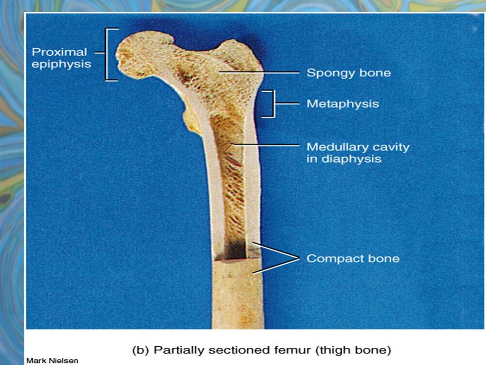
Longer than they are wide Diaphysis- shaft, surrounds medullary cavity Mostly compact bone Epiphysis- ends, spongy bone inside Compact bone outside Joint surface = articular cartilage Epiphyseal line = growth plate

15
2. Short Bones- roughly cube like Ex: wrist, ankle, sesamoid Mostly spongy, thin compact cover

16
3. Flat bones- flat, thin, curved Ex: ribs, skull Parallel compact bone surface, spongy inside

17
Irregular Bones 4.complicated shapes Ex: vertebrae, hip Mostly spongy bone enclosed by thin compact

18
Bones are made of organic and inorganic components Organic- cells: osteoblasts, osteoclasts, Osteoids: proteoglycans, collegen, fibers Inorganic- hydroxyapatites/ mineral salts

19
Calcium Calcium is necessary for: Nerve impulses Muscle contractions Blood coagulation Secretion of glands Cell division

20
Calcium is obtained in your diet and absorbed in the intestine under the control of vitamin D Diet should contain: Proteins, Vitamin C, Vitamin A, Vitamin B12, calcium, phosphorus, magnesium, and manganese for bone health Vitamin D is synthesized in skin

21
hhh

22
High Calcium Levels Change in plasma calcium homeostasis Detected by Thyroid Gland Which secretes Calcitonin Affects osteoblasts in bone tissue Which builds bone by depositing calcium Removes calcium from plasma Reduces plasma calcium levels

23
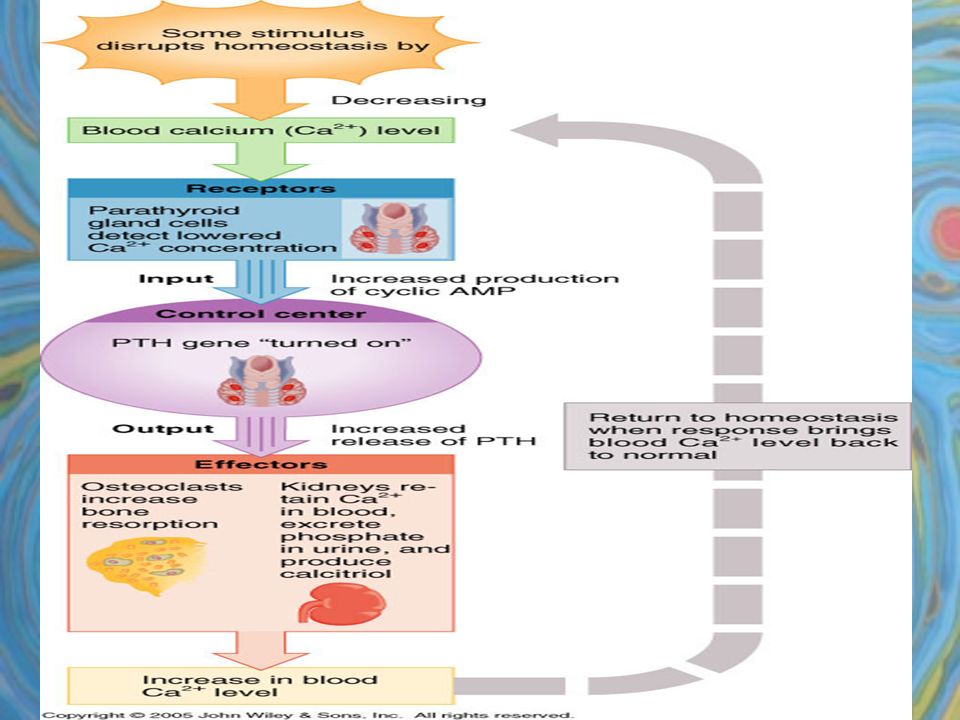
Low Calcium Levels A change in plasma calcium homeostasis Detected by the Parathyroid Gland Which secretes Parathyroid Hormone PTH Affects osteoclasts in bone tissue Which breaks down bone by removing calcium Deposits the calcium I the plasma Increases plasma calcium levels

25
Osteogenesis-bone creation Bone is always changing, growing, remodeling, and repairing Wolff’s Law- a bone will remodel in response to demands or forces placed on it ex: ballet dancer, weight lifter

26
Prenatal to Young Adult

27
Steps in ossification 1. starts as hyaline cartilage 2. bone replaces cartilage at periosteum 3. bone replaces cartilage at medulla 4. bone replaces cartilage at ends 5. only cartilage remaining is at ends (articular cartilage) and epiphyseal plate (growth plate) Steps in Ossification
 and epiphyseal plate (growth plate) Steps in Ossification.")
28
Calcium is absorbed from the intestine under control of Vitamin D Low calcium causes failure of many systems High calcium causes salt deposits in kidney, blood vessels

29
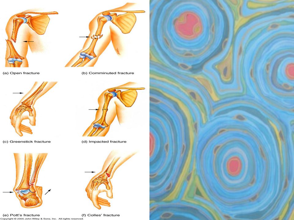
Fractures 1. Simple- clean closed break 2. Compound- broken ends break skin 3. Comminuted- fragmented (aged) 4. Compression- crushed 5. Depressed- pressed inward (skull) 6. Impacted- ends forced into each other ( the result of a fall) 7. Spiral- ragged twisted (sports) 8. Greenstick- break incomplete (child)
 4. Compression- crushed 5. Depressed- pressed inward (skull) 6. Impacted- ends forced into each other ( the result of a fall) 7. Spiral- ragged twisted (sports) 8. Greenstick- break incomplete (child).")
31
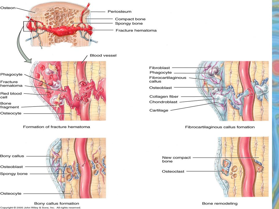
Steps in Repair 1. Hematoma 2. Fibrocartilaginous callus formation splints the broken bone 3. Bony (hard) callus 4. Remodeling
 callus 4. Remodeling.")
33
Osteoporosis –bone loss due to hormonal changes that interfere with calcium deposits in bone, leads to spine problems and breaks occurs at menopause

34
Rickets Childhood disorder caused by the lack of calcium and vitamin D in the diet. The bones are soft and do not support the weight, so they bend -bowed legs

35
Pagets Disease caused by the uneven deposit of calcium C C

Similar presentations


 Bone fractures are classified by: –The position of the bone ends after fracture –The completeness.>")


 2.Protection: skull, vertebrae,>")


