Download presentation
Presentation is loading. Please wait.

1
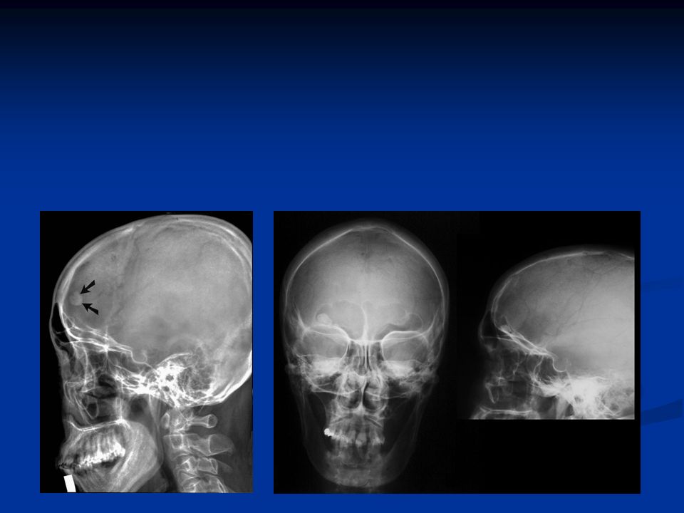
Osteoma Benign lesion of bone Age: 30-60 Location: m/c frontal sinuses Margin: narrow Periosteal Rxn: none Soft Tissue Mass: abscent

4
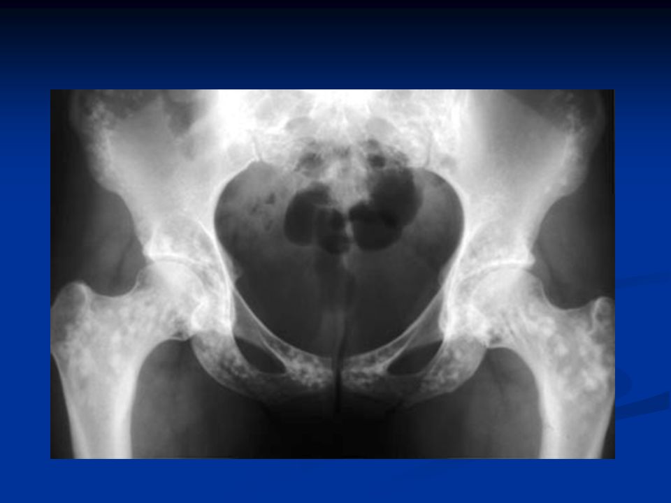
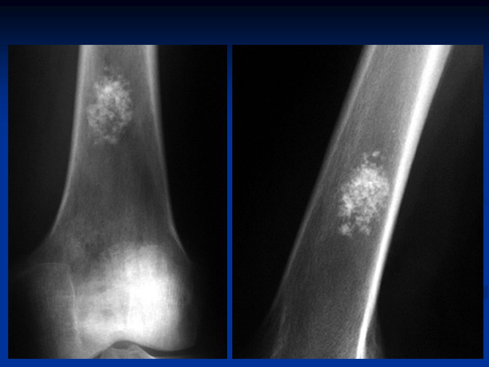
Osteopoikilosis Benign lesion of bone Age: rare before 3 Location: scattered diffusely through out metaepiphyseal regions of long bones, pelvic region, carpals/tarsals (think dots on a Dalmatian)
")
7
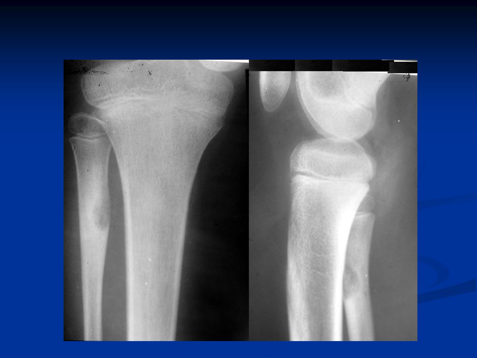
Osteoid Osteoma Age: 10-25 Nocturnal pain relieved by aspirin Location: 50-60% in femur (esp intertrochanteric region/neck) and tibia 20% bones of hands/feet Lumbar spine neural arch Oval/Round nidus <1cm surrounded by uniform sclerosis
 and tibia 20% bones of hands/feet Lumbar spine neural arch Oval/Round nidus <1cm surrounded by uniform sclerosis")
10
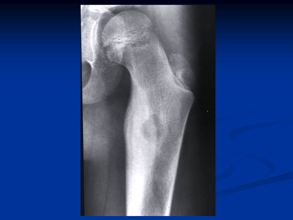
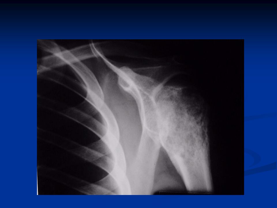
Osteoblastoma Age: 10-30 Location: spine, lumbar region m/c pedicale/lamina l/c TVP/SP, may involve long bones m/c lower extremities Expansile, mixed lytic/blastic appearance cortical thinning (eggshell) Nidus >2cm
 Nidus >2cm")
12
More photos of Osteoblastoma at the following link http://www.bluestormstudios.com/clientsites/bonecance r/index.php?db=content/Bonecancer&tbl=Bone_Tum ors&id=12

13
Osteosarcoma 2 nd m/c primary malignancy of bone! Age: m/c 10-25 (has been seen in pt of all ages) Location: 50-75% around knee 40% femur 16% tibia 15% humerus Metaphyseal lesions m/c, permative pattern of destruction, soft tissue extension, aggressive periosteal rxn (sunburst), cortical disruption, wide zone of transition, can show mixed pattern of osteolysis & sclerosis (cumulus cloud appearance)
 Location: 50-75% around knee 40% femur 16% tibia 15% humerus Metaphyseal lesions m/c, permative pattern of destruction, soft tissue extension, aggressive periosteal rxn (sunburst), cortical disruption, wide zone of transition, can show mixed pattern of osteolysis & sclerosis (cumulus cloud appearance).")
20
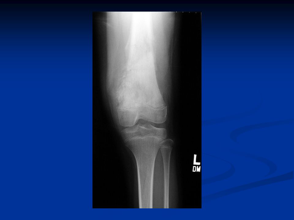
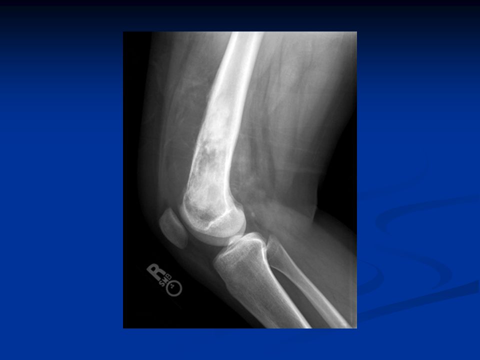
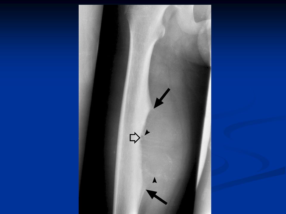
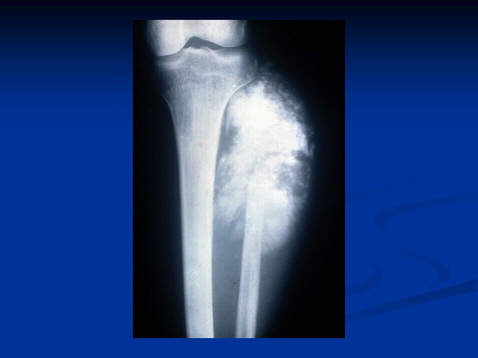
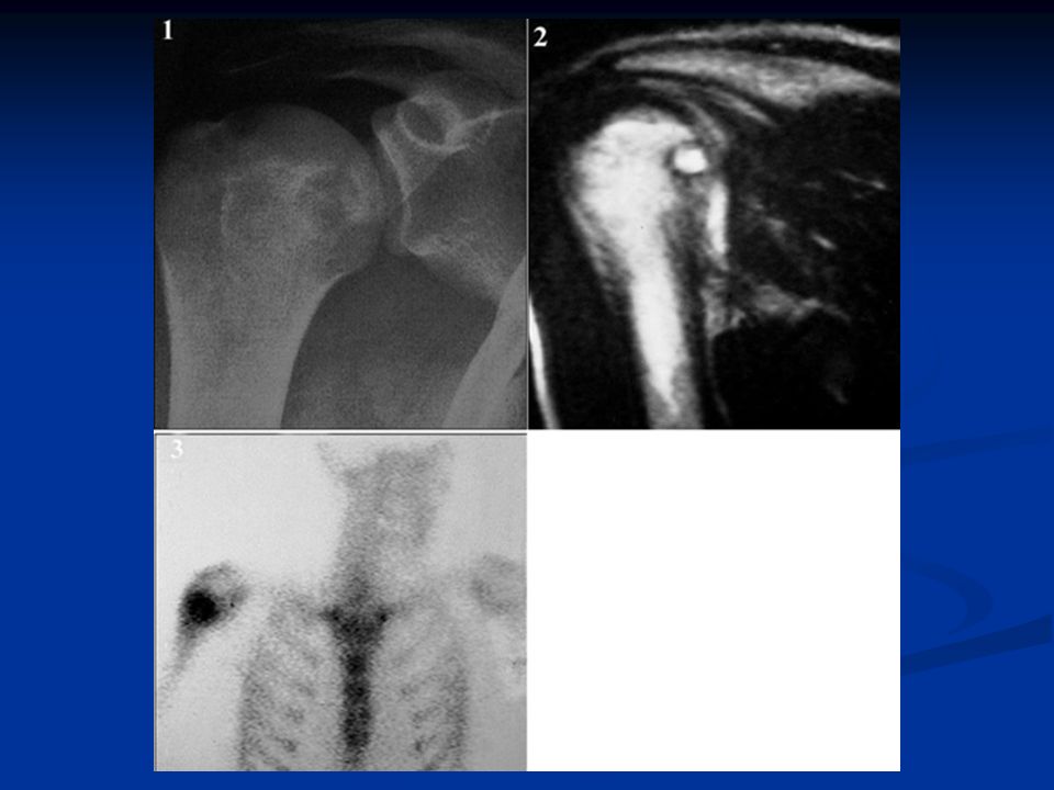
Osteochondroma (cartilage forming tumors) M/C benign tumor of bone Age: 70-80% encountered before 20 YOA Location: any bone that develops by endochondral ossification 30% femur 20% humerus 17% tibia spine/ribs can be involved Metaphyseal/diametaphyseal osteocartilaginous out growth with a pedunculated or sessile base and a cartilage cap (think cauliflower) oriented away from joint articulation
 M/C benign tumor of bone Age: 70-80% encountered before 20 YOA Location: any bone that develops by endochondral ossification 30% femur 20% humerus 17% tibia spine/ribs can be involved Metaphyseal/diametaphyseal osteocartilaginous out growth with a pedunculated or sessile base and a cartilage cap (think cauliflower) oriented away from joint articulation")
21
Note the broad, flat base, sessile

22
AP ViewLateral View (tibia)
")
23
AP view T2 weighted MRI

25
Enchondroma M/C benign tumor of the hand Age: 10-30 Location: 40-65% in the hand, proximal phalanges, met heads 25% long bones, more often in lower extremity Well defined/geographic, expansile, medullary lz, with some Calcification, endosteal erosion causing cortical thinning. Calcification usually appears stippled or punctate.

26
PA view (note fx)Lateral
Lateral")
28
Note fx

30
Ollier’s Disease

31
Maffucci’s Syndrome

32
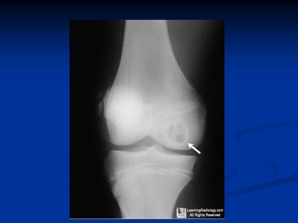
Chondroblastoma Has to involve EPIPHESIS! May span growth plate 2 nd M/C benign tumor of patella Age: 10-25 Location: Poximal & distal ends of femur & trochanters Proximal tibia Proximal humerus Ilia medullary based epiphyseal lz, can show metaphyseal extension Round/oval shaped geographic, lytic lz, can have stippled or punctuate calcifications (50%) & periostitis (30-50%)
 & periostitis (30-50%).")
36
Note this one spans growth plate

39
Chondrosarcoma 3 rd M/C primary malignancy of bone Age: 40-60 Location: 50% of cases pelvis or proximal femur Bones around knee Humerus scapula & ribs Large, aggressive, expansile metaphyseal lz, with wide zone of transition, cortical dissolution, erratic calcifications stippled, fluffy or arcs & rings in appearance, periosteal rxn laminated/spiculated. Soft tissue mass

41
Not the best image

42
CT of humerus from previous slide

43
Note ‘fluffy’ appearing calcifications

44
Soft tissue mass calcifications

45
MRI from previous slide of soft tissue mass

46
Fibrous Cortical Defect FCD common between 4-8 yoa FCD common between 4-8 yoa Long bones of lower extremity 90% of cases, areas around knee account for 55% of all FCD & NOF Long bones of lower extremity 90% of cases, areas around knee account for 55% of all FCD & NOF FCD metaphyseal based but run parallel to long access of bone FCD metaphyseal based but run parallel to long access of bone

47
Non-ossifying Fibroma NOF common 8-20 yoa NOF common 8-20 yoa Larger than FCD at 2-7cm usually in metaphysis region of tibia with cortical expansion/thinning, bubbly appearance Larger than FCD at 2-7cm usually in metaphysis region of tibia with cortical expansion/thinning, bubbly appearance

48
Fibrosarcoma Can occur at any age but m/c 30- 50 yoa Can occur at any age but m/c 30- 50 yoa 70% of cases involve long bones, m/c femur 40%, tibia 16% 70% of cases involve long bones, m/c femur 40%, tibia 16% Meta/diametaphyseal moth eaten pattern, cortical dissolution, with soft tissue mass. Periosteal rxn is rare. Meta/diametaphyseal moth eaten pattern, cortical dissolution, with soft tissue mass. Periosteal rxn is rare.

49
Fibrosarcoma

50
Giant Cell Tumor M/C in 20-30 yoa, females m/c M/C in 20-30 yoa, females m/c 75% long tubular bones, 50% involve knee region or distal radius 75% long tubular bones, 50% involve knee region or distal radius M/C tumor of patella M/C tumor of patella Sacrum (m/c benign tumor of sacrum) & vertebral body involvement has been see Sacrum (m/c benign tumor of sacrum) & vertebral body involvement has been see Metaphyseal origin with epiphysis extention Metaphyseal origin with epiphysis extention
 & vertebral body involvement has been see Sacrum (m/c benign tumor of sacrum) & vertebral body involvement has been see Metaphyseal origin with epiphysis extention Metaphyseal origin with epiphysis extention")
51
GCT

52
GCT

53
Simple (Unicameral) Bone Cyst M/C <14 yoa M/C <14 yoa 75% occur in proximal humerus & femur, upper extremity more common 75% occur in proximal humerus & femur, upper extremity more common Active lesions up against growth plate Active lesions up against growth plate Latent cyst found in diaphysis Latent cyst found in diaphysis Fallen-fragment sign Fallen-fragment sign
 Bone Cyst M/C <14 yoa M/C <14 yoa 75% occur in proximal humerus & femur, upper extremity more common 75% occur in proximal humerus & femur, upper extremity more common Active lesions up against growth plate Active lesions up against growth plate Latent cyst found in diaphysis Latent cyst found in diaphysis Fallen-fragment sign Fallen-fragment sign")
54
SBC

55
ABC I think we all are familiar with ABC I think we all are familiar with ABC 5-20 yoa 5-20 yoa M/C benign tumor of the Clavical M/C benign tumor of the Clavical Will show fluid levels on MRI Will show fluid levels on MRI Spinal Lesions can be seen Spinal Lesions can be seen Spine Diff Dx: GCT, Osteoblastoma, hemangioma Spine Diff Dx: GCT, Osteoblastoma, hemangioma

56
Hemangioma m/c >40 yoa females m/c >40 yoa females m/c benign tumor of spine m/c benign tumor of spine Bright on T1 & T2 MRI Bright on T1 & T2 MRI Corduroy-cloth appearance on xray Corduroy-cloth appearance on xray

57
Hemanogioma Left side of the v-body Left side of the v-body

58
Hemangioma

59
Lipoma 5-70 yoa m/c 4 th decade 5-70 yoa m/c 4 th decade Rarest of all primary tumors of bone Rarest of all primary tumors of bone Fibula, femur, tibia, calcaneus locations common Fibula, femur, tibia, calcaneus locations common “Target” sign on plain film central sclerosis surrounded by lucent area “Target” sign on plain film central sclerosis surrounded by lucent area

60
Lipoma

61
Lipoma

62
Chordoma Notochord remnants Notochord remnants m/c 40-70 yoa m/c 40-70 yoa Only primary malignant tumor know to cross intervetebral disc into sequential segments Only primary malignant tumor know to cross intervetebral disc into sequential segments Sacrococcygeal area m/c 50-60% Sacrococcygeal area m/c 50-60% Spheno-occipital (clivus) 25-40% Spheno-occipital (clivus) 25-40% C2 occasionally involved C2 occasionally involved Large soft tissue mass, cortical distruction Large soft tissue mass, cortical distruction
 25-40% Spheno-occipital (clivus) 25-40% C2 occasionally involved C2 occasionally involved Large soft tissue mass, cortical distruction Large soft tissue mass, cortical distruction")
63
Chordoma

64
Chordoma

65
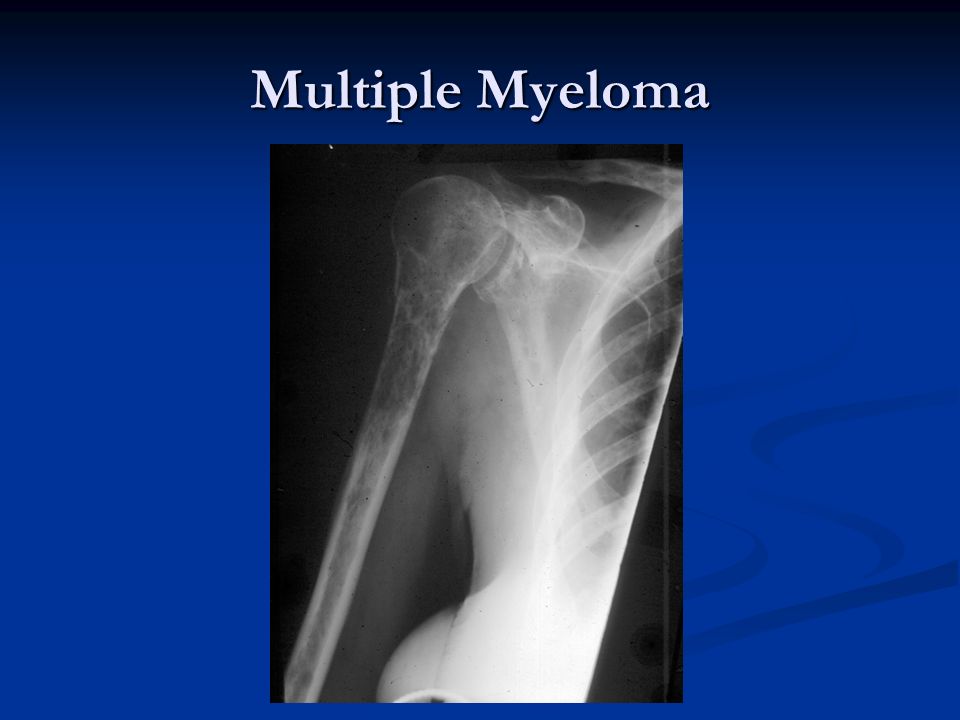
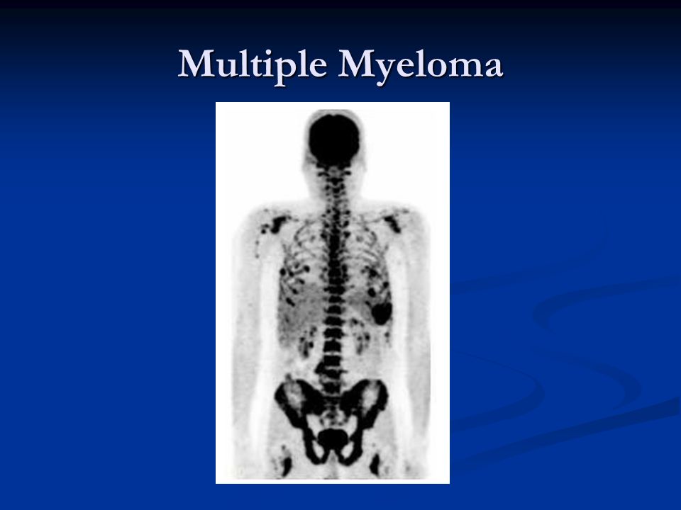
Multiple Myeloma

68
EG

69
EG

70
Padget’s

71
All images are property of their respective owners. All images are property of their respective owners.

Similar presentations


>")


 normal cell of origin Most are classified.>")


. General considerations Primary bone tumors are much less than secondary tumors. All age groups affected,>")


