Download presentation
Presentation is loading. Please wait.

1
Tissue Repair

2
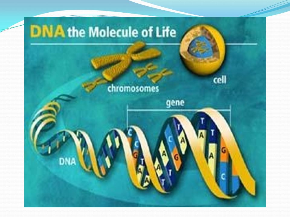
Somatic Cell Division Interphase – 95% of cell cycle Organelle duplication, DNA replication, Growth

3
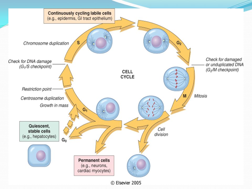
G1 Phase Metabolically active Organelle duplication, but no DNA replication Duration variable – short in embryonic and cancer cells Prepares for S phase Cells that remain in G1 for a long time = G0 (permanent tissues, such as neural tissue)
")
4
S Phase Committed to cell division once this starts DNA and centrosome replication Semi-conservative replication of DNA: two identical daughter genomes

5
G2 Phase Growth continues Enzymes and proteins synthesized for cell division Determining Cell Stage Cells at different stages of the cell cycle can also be distinguished by their DNA content

6
Mitotic (M) Phase mitosis plus cytokinesis Mitosis: Prophase Metaphase Anaphase Telophase
 Phase mitosis plus cytokinesis Mitosis: Prophase Metaphase Anaphase Telophase")
7
Regulation of the Cell Cycle Cell Cycle Lengths Vary by cell type: Embryonic cells 1-Stem cells (e.g., blood cells and epithelial cells) 2-Sperm cells G1 prolonged in stable or permanent cells (called G0) G1 rapid or non-existent in rapidly-dividing cells
 2-Sperm cells G1 prolonged in stable or permanent cells (called G0) G1 rapid or non-existent in rapidly-dividing cells")
8
Embryonic cells Cell growth not part of cell cycle All energy goes into DNA synthesis So G1 lacking and G2 quite short Each round of division subdivides original cytoplasm into smaller and smaller cells, Until adult cell size is reached

9
Cell-Cycle Checkpoints G1 checkpoint In yeast, called start In animal cells, called restriction point G2 checkpoint Located at boundary between G2 and M phase Proper completion of DNA synthesis required before cell can initiate mitosis Spindle Assembly Checkpoint Boundary between metaphase and anaphase All chromosomes must be properly attached to the spindle

11
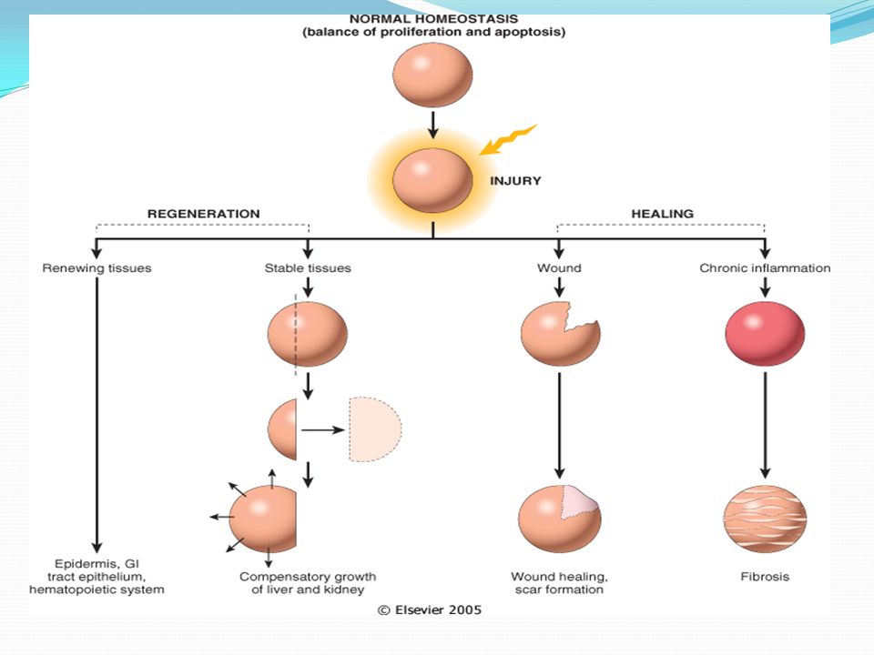
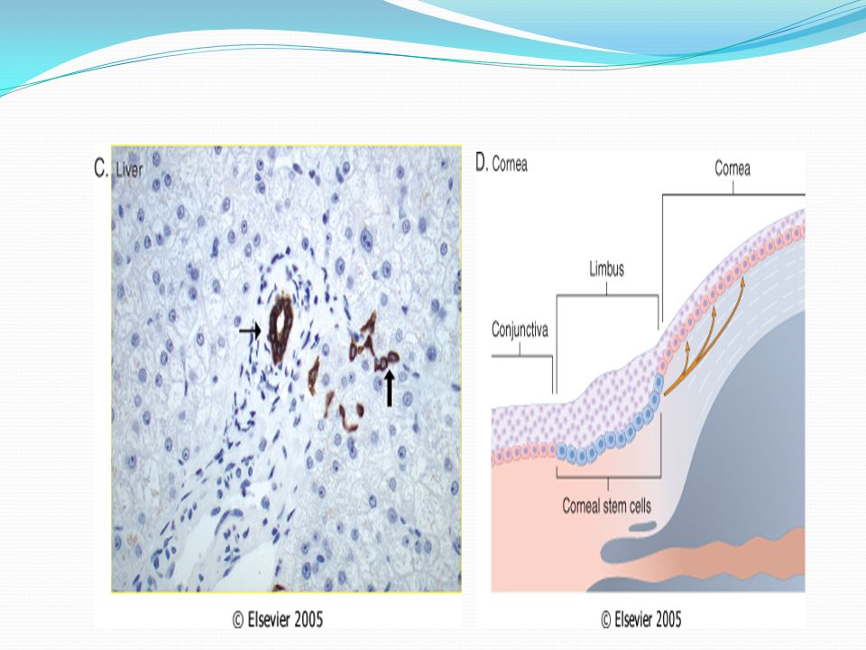
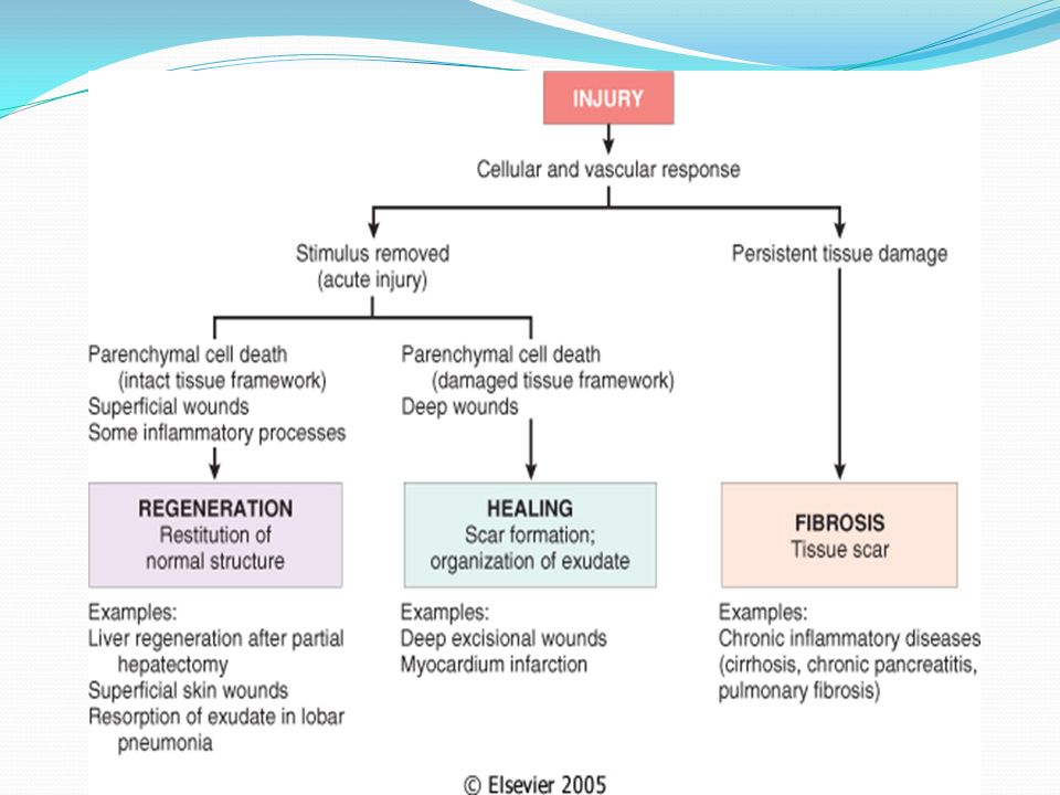
Regeneration refers to growth of cells and tissues to replace lost structures, such as the growth of an amputated limb in amphibians. In mammals, whole organs and complex tissues rarely regenerate after healing

12
Healing is usually a tissue response to a wound (commonly in the skin), to inflammatory processes in internal organs, or to cell necrosis in organs incapable of regeneration
, to inflammatory processes in internal organs, or to cell necrosis in organs incapable of regeneration")
13
Regeneration requires an intact connective tissue scaffold. By contrast, healing with scar formation occurs if the extracellular matrix (ECM) framework is damaged, causing alterations of the tissue architecture.
 framework is damaged, causing alterations of the tissue architecture..")
14
Repair processes are critical for the maintenance of normal structure and function and survival of the organism. The healing of skin wounds is just the most common example of repair processes. However, in healthy tissues, repair, in the form of regeneration or healing, occurs after practically any insult that causes tissue destruction.

17
Tissue Repair

22
Epidermal Growth Factor (EGF) and Transforming Growth Factor-α (TGF-α). Hepatocyte Growth Factor (HGF)/scatter factor Vascular Endothelial Growth Factor (VEGF). Platelet-Derived Growth Factor (PDGF). Fibroblast Growth Factor (FGF Wound repair: FGFs participate in macrophage, fibroblast, and endothelial cell migration in damaged tissues and migration of epithelium to form new epidermis TGF-β and Related Growth Hematopoiesis:FGFs have been implicated in the differentiation of specific lineages of blood cells and development of bone marrow stroma. Transforming Growth Factors. Effects of TGF-β on mesenchymal cells it generally stimulates the proliferation of fibroblasts and smooth muscle cells. TGF-β is a potent fibrogenic agent that stimulates fibroblast chemotaxis, enhances the production of collagen, fibronectin, and proteoglycans. It inhibits collagen degradation by decreasing matrix proteases and increasing protease inhibitor activities. TGF-β is involved in the development of fibrosis in a variety of chronic inflammatory conditions particularly in the lungs, kidney, and liver. TGF-β has a strong anti-inflammatory effect.
/scatter factor Vascular Endothelial Growth Factor (VEGF). Platelet-Derived Growth Factor (PDGF). Fibroblast Growth Factor (FGF Wound repair: FGFs participate in macrophage, fibroblast, and endothelial cell migration in damaged tissues and migration of epithelium to form new epidermis TGF-β and Related Growth Hematopoiesis:FGFs have been implicated in the differentiation of specific lineages of blood cells and development of bone marrow stroma. Transforming Growth Factors. Effects of TGF-β on mesenchymal cells it generally stimulates the proliferation of fibroblasts and smooth muscle cells. TGF-β is a potent fibrogenic agent that stimulates fibroblast chemotaxis, enhances the production of collagen, fibronectin, and proteoglycans. It inhibits collagen degradation by decreasing matrix proteases and increasing protease inhibitor activities. TGF-β is involved in the development of fibrosis in a variety of chronic inflammatory conditions particularly in the lungs, kidney, and liver. TGF-β has a strong anti-inflammatory effect..")
25
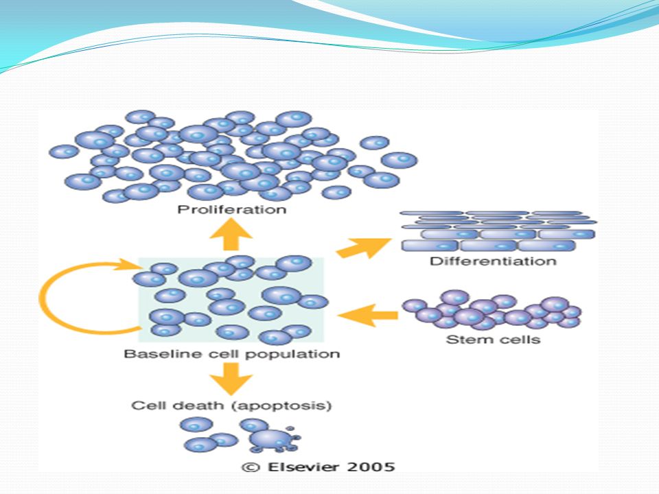
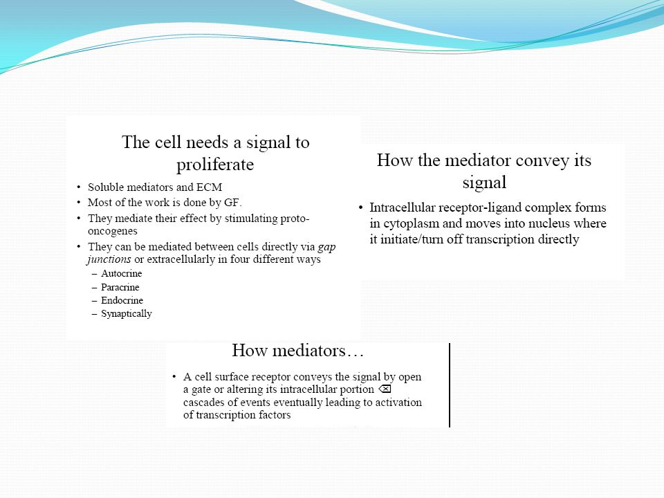
The replication of cells is generally stimulated by growth factors or by signaling from ECM components through integrins To enter the cycle, quiescent cells first must go through the transition from G 0 to G 1, the first decision step, which functions as a gateway to the cell cycle Checkpoint activation delays the cell cycle and triggers DNA repair mechanisms

26
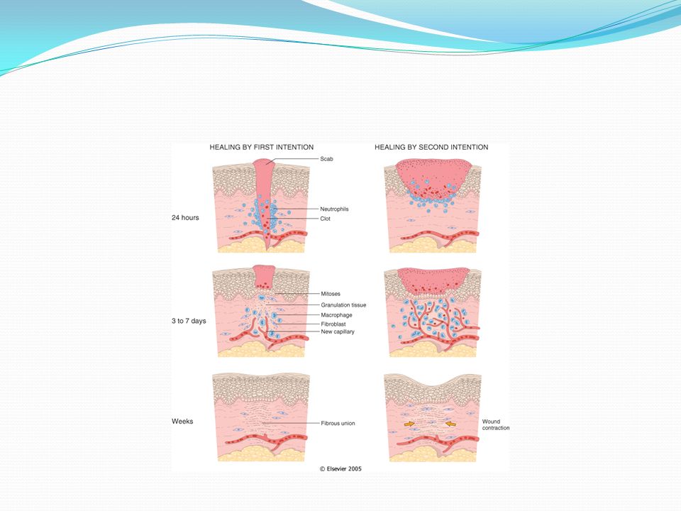
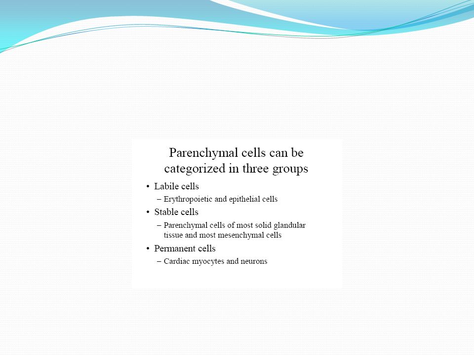
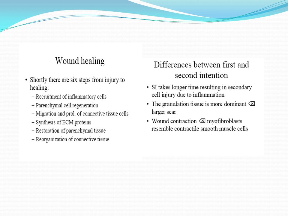
The goal of the repair process is to restore the tissue to its original state. The inflammatory reaction set in motion by the injury contains the damage, eliminates the damaging stimulus, removes injured tissue Some tissues can be completely reconstituted after injury, such as the repair of bone after a fracture or the regeneration of the surface epithelium in a cutaneous wound. For tissues that are incapable of regeneration, repair is accomplished by connective tissue deposition, producing a scar. This term is most often used in connection to wound healing in the skin, but it is also used to describe the replacement of parenchymal cells by connective tissue, as in the heart after myocardial infarction. If damage persists, inflammation becomes chronic, and tissue damage and repair may occur concurrently. Connective tissue deposition in these conditions is usually referred to as fibrosis.

27
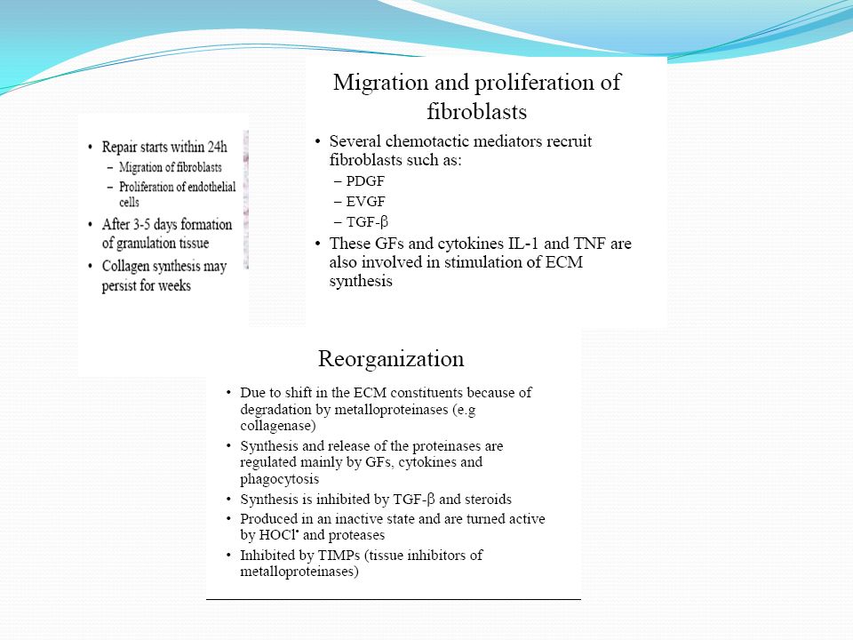
Repair begins early in inflammation. Fibroblasts and vascular endothelial cells begin proliferating to form a specialized type of tissue that is the hallmark of healing, granulation tissue. Pink, soft, granular appearance on the surface of wounds, but it is the histologic features that are characteristic: The formation of new small blood vessels (angiogenesis) and the proliferation of fibroblasts New granulation tissue is often edematous.
 and the proliferation of fibroblasts New granulation tissue is often edematous..")
30
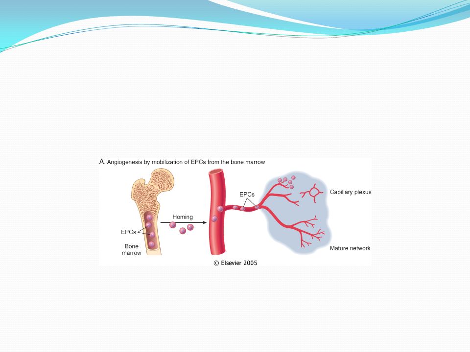
Angiogenesis from Pre-Existing Vessels Angiogenesis from Endothelial Precursor Cells Growth Factors and Receptors Involved in Angiogenesis

31
Vasodilation in response to nitric oxide and VEGF-induced increased permeability of the pre-existing vesselnitric oxide Proteolytic degradation of the BM of the parent vessel by metalloproteinases and disruption of cell-to-cell contact between endothelial cells of the vessel by plasminogen activator Migration of endothelial cells toward the angiogenic stimulus Proliferation of endothelial cells, just behind the leading front of migrating cells Maturation of endothelial cells, which includes inhibition of growth and remodeling into capillary tubes Recruitment of periendothelial cells (including pericytes for small capillaries and vascular smooth muscle cells for larger vessels) to support the endothelial tubes and form the mature vessel.
 to support the endothelial tubes and form the mature vessel.")
32
SCAR FORMATION Growth factors and cytokines released at the site of injury induce fibroblast proliferation and migration into the granulation tissue framework of new blood vessels and loose ECM that initially forms at the repair site. (1) emigration and proliferation of fibroblasts in the site of injury (2) deposition of ECM, and (3) tissue remodeling. Fibroblast Migration and Proliferation Migration of fibroblasts to the site of injury and their subsequent proliferation are triggered by multiple growth factors, including TGF-β, PDGF, EGF, FGF, and the cytokines IL-1 and TNF Macrophages are important cellular constituents of granulation tissue, clearing extracellular debris, fibrin, and other foreign material at the site of repair. Fibroblast migration and proliferation, increased synthesis of collagen and fibronectin, and decreased degradation of ECM by metalloproteinases TGF-β is also chemotactic for monocytes and causes angiogenesis in vivo, possibly by inducing macrophage influx. ECM Deposition and Scar Formation : As repair continues, the number of proliferating endothelial cells and fibroblasts decreases. (PDGF, FGF, TGF-β) and cytokines (IL-1, IL-13) Net collagen accumulation, however, depends not only on increased collagen synthesis but also on decreased degradation. The replacement of granulation tissue with a scar involves transitions in the composition of the ECM. The balance between ECM synthesis and degradation results in remodeling of the connective tissue framework-an important feature of both chronic inflammation and wound repair.
 emigration and proliferation of fibroblasts in the site of injury (2) deposition of ECM, and (3) tissue remodeling. Fibroblast Migration and Proliferation Migration of fibroblasts to the site of injury and their subsequent proliferation are triggered by multiple growth factors, including TGF-β, PDGF, EGF, FGF, and the cytokines IL-1 and TNF Macrophages are important cellular constituents of granulation tissue, clearing extracellular debris, fibrin, and other foreign material at the site of repair. Fibroblast migration and proliferation, increased synthesis of collagen and fibronectin, and decreased degradation of ECM by metalloproteinases TGF-β is also chemotactic for monocytes and causes angiogenesis in vivo, possibly by inducing macrophage influx. ECM Deposition and Scar Formation : As repair continues, the number of proliferating endothelial cells and fibroblasts decreases. (PDGF, FGF, TGF-β) and cytokines (IL-1, IL-13) Net collagen accumulation, however, depends not only on increased collagen synthesis but also on decreased degradation. The replacement of granulation tissue with a scar involves transitions in the composition of the ECM. The balance between ECM synthesis and degradation results in remodeling of the connective tissue framework-an important feature of both chronic inflammation and wound repair..")
35
Components of the ECM Collagens for tensile strength. Elastin important in large vessels, uterus, skin and ligament Proteoglycans and hyaluronan forms hydrated gels. Proteoglycans also store bFGF Adhesive Proteoglycans connects ECM- ECM and ECM-cell integrins. The ECM - cell interaction can activate the same pathways as growth factors

36
The surgeon told his patient that woke up after having been operated: "I'm afraid we're going to have to operate you again. Because, you see, I forgot my rubber gloves inside you." "Well, if it's just because of them, I'd rather pay for them if you just leave me alone."

37
Questions

50
1) Which of the following is associated with acute inflammation? a) Neutophils b) Macrophages c) Lymphocytes d) Tissue fibrosis e) Tissue necrosis
 Neutophils b) Macrophages c) Lymphocytes d) Tissue fibrosis e) Tissue necrosis.")
51
4-7Mediators such as histamine, thrombin, and platelet activating factor (PAF) stimulate the redistribution of which of the following from its normal intracellular stores in granules (Weibel-Palade bodies) to the cell surface? a) P-selectin b) E-selectin c) ICAM-1 d) VCAM-1 e) CD31 4.6) Lymphocyte homing to high endothelial venules
 P-selectin b) E-selectin c) ICAM-1 d) VCAM-1 e) CD31 4.6) Lymphocyte homing to high endothelial venules.")
52
2) Acute inflammation may be triggered by infections, trauma, physical or chemical agents, tissue necrosis, foreign bodies, and immune reactions. Which of the following is NOT seen in acute inflammation? a) Alterations in vascular caliber b) Decrease in blood flow c) Structural changes in the microvasculature (edema) d) Plasma proteins and leukocytes leaving the circulation e) Leukocyte focusing to eliminate the offending agent
 Alterations in vascular caliber b) Decrease in blood flow c) Structural changes in the microvasculature (edema) d) Plasma proteins and leukocytes leaving the circulation e) Leukocyte focusing to eliminate the offending agent.")
53
Match the endothelial molecule (leukocyte adhesion molecule) with its major role: 4.1) Rolling, adhesion to activated endothelium (neutrophils, T cells) 4.2) Adhesion (eosinophils, monocytes, lymphocytes) 4.3) Leukocyte migration through endothelium 4.4) Rolling (neutrophils, monocytes, lymphocytes) 4.5) Adhesion, arrest, transmigration (all leukocytes) 4.6) Lymphocyte homing to high endothelial venules a) ICAM-1 b) P-selectin c) E-selectin d) VCAM-1 e) GlyCam-1 f) CD31 (PECAM)
 with its major role: 4.1) Rolling, adhesion to activated endothelium (neutrophils, T cells) 4.2) Adhesion (eosinophils, monocytes, lymphocytes) 4.3) Leukocyte migration through endothelium 4.4) Rolling (neutrophils, monocytes, lymphocytes) 4.5) Adhesion, arrest, transmigration (all leukocytes) 4.6) Lymphocyte homing to high endothelial venules a) ICAM-1 b) P-selectin c) E-selectin d) VCAM-1 e) GlyCam-1 f) CD31 (PECAM)")
54
4-8 Cytokine activation of endothelium (with neutrophils) involves which of the following? a) IL-1 and IL-2 b) IL-3 c) IL-4 and IL-5 d) TH-1 cells and TH-2 cells e) IL-1 and TNF
 IL-1 and IL-2 b) IL-3 c) IL-4 and IL-5 d) TH-1 cells and TH-2 cells e) IL-1 and TNF.")
55
5-Which of the following is NOT a general principle of the chemical mediators of inflammation? a) Mediators originate either from plasma or from cells b) The production of active mediators is triggered by microbial products or by host proteins c) Most mediators perform their biologic activity by initially binding to specific receptors on target cells d) One mediator can stimulate the release of other mediators by target cells themselves e) Mediators can act on one or few target cell types f) Once activated and released from the cell, most of these mediators last a long time (long-lived) g) Most mediators have the potential to cause harmful effects
 Mediators originate either from plasma or from cells b) The production of active mediators is triggered by microbial products or by host proteins c) Most mediators perform their biologic activity by initially binding to specific receptors on target cells d) One mediator can stimulate the release of other mediators by target cells themselves e) Mediators can act on one or few target cell types f) Once activated and released from the cell, most of these mediators last a long time (long-lived) g) Most mediators have the potential to cause harmful effects.")
56
7-2-NO causes vasodilation by what mechanism? a) Constriction of smooth muscle b) Relaxation of smooth muscle c) Constriction of striated muscle d) Relaxation of striated muscle
 Constriction of smooth muscle b) Relaxation of smooth muscle c) Constriction of striated muscle d) Relaxation of striated muscle.")
57
8- Which of the following is NOT true regarding contribution to inflammation? a) Lysosomal constituents increase vascular permeability and tissue damage b) Oxygen free radicals amplify the cascade that elicits the inflammatory response c) Neuropeptides help initiate and propagate the inflammatory response d) The response to hypoxia decreases vascular permeability e) The response to necrotic cells is pro-inflammatory
 Lysosomal constituents increase vascular permeability and tissue damage b) Oxygen free radicals amplify the cascade that elicits the inflammatory response c) Neuropeptides help initiate and propagate the inflammatory response d) The response to hypoxia decreases vascular permeability e) The response to necrotic cells is pro-inflammatory.")
58
10-1Which of the following appears, histologically, as an eosinophilic meshwork of threads or sometimes as an amorphous coagulum? a) Serous inflammation b) Fibrinous inflammation c) Suppurative or purulent inflammation d) Ulcers
 Serous inflammation b) Fibrinous inflammation c) Suppurative or purulent inflammation d) Ulcers.")
59
10-2Which of the following is characterized by the production of large amounts of pus consisting of neutrophils, necrotic cells, and edema fluid? a) Serous inflammation b) Fibrinous inflammation c) Suppurative or purulent inflammation d) Ulcers
 Serous inflammation b) Fibrinous inflammation c) Suppurative or purulent inflammation d) Ulcers.")
60
10-3Which of the following is marked by the outpouring of a thin fluid that, depending on the size of injury, is derived from either the plasma or the secretions of mesothelial cells lining the peritoneal, pleural, and pericardial cavities? a) Serous inflammation b) Fibrinous inflammation c) Suppurative or purulent inflammation d) Ulcers
 Serous inflammation b) Fibrinous inflammation c) Suppurative or purulent inflammation d) Ulcers.")
61
1-Which of the following is NOT capable of regeneration? a) Epithelial tissue b) Cardiac tissue c) Skin d) Liver e) Kidney
 Epithelial tissue b) Cardiac tissue c) Skin d) Liver e) Kidney.")
62
2-1 Which of the following is the correct order of the cell cycle? a) G1 => S => G2 => M b) G2 => S => G1 => M c) G1 => M => G2 => S d) G2 => M => G1 => S e) G1 => G2 => S => M f) G1 => G2 => M => S
 G1 => S => G2 => M b) G2 => S => G1 => M c) G1 => M => G2 => S d) G2 => M => G1 => S e) G1 => G2 => S => M f) G1 => G2 => M => S.")
63
2-2 Which part of the cell cycle has the most redundancies, is tightly regulated by proteins called cyclins, and associated enzymes called cyclin- dependent kinases (CDKs)? a) Between G0 and G1 b) Between G1 and S c) Between S and G2 d) Between G2 and M e) Between M and G1
 Between G0 and G1 b) Between G1 and S c) Between S and G2 d) Between G2 and M e) Between M and G1.")
64
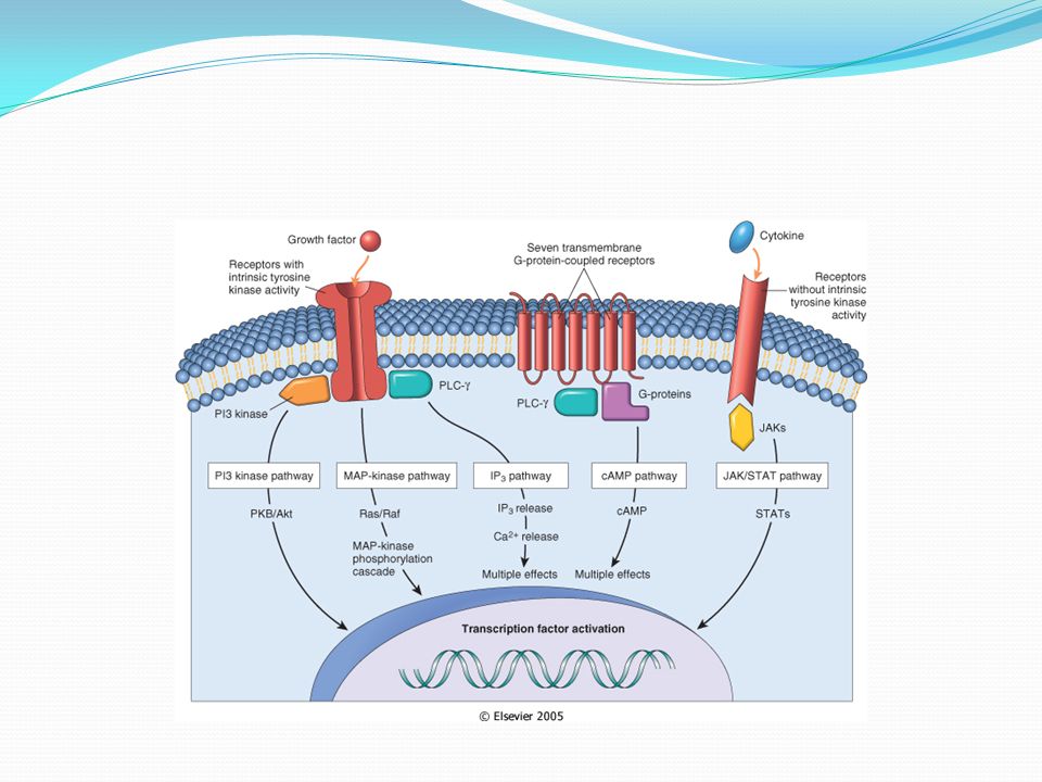
9-1 Which of the following describes the JAK/STAT signal transduction pathway? a) Receptors with intrinsic tyrosine kinase activity b) Receptors lacking intrinsic tyrosine kinase activity that recruit kinases c) Seven transmembrane G-protein-coupled receptors (GPCRs) d) Steroid hormone receptors
 Receptors with intrinsic tyrosine kinase activity b) Receptors lacking intrinsic tyrosine kinase activity that recruit kinases c) Seven transmembrane G-protein-coupled receptors (GPCRs) d) Steroid hormone receptors.")
65
9-2 Which of the following signal transduction pathways includes cytokines such as interleukin-2 (IL- 2)? a) Receptors with intrinsic tyrosine kinase activity b) Receptors lacking intrinsic tyrosine kinase activity that recruit kinases c) Seven transmembrane G-protein-coupled receptors (GPCRs) d) Steroid hormone receptors
 Receptors with intrinsic tyrosine kinase activity b) Receptors lacking intrinsic tyrosine kinase activity that recruit kinases c) Seven transmembrane G-protein-coupled receptors (GPCRs) d) Steroid hormone receptors.")
66
9-3 Which of the following is associated with the cAMP signal transduction pathway? a) Receptors with intrinsic tyrosine kinase activity b) Receptors lacking intrinsic tyrosine kinase activity that recruit kinases c) Seven transmembrane G-protein-coupled receptors (GPCRs) d) Steroid hormone receptors
 Receptors with intrinsic tyrosine kinase activity b) Receptors lacking intrinsic tyrosine kinase activity that recruit kinases c) Seven transmembrane G-protein-coupled receptors (GPCRs) d) Steroid hormone receptors.")
67
11-1 Which of the following extracellular matrix (ECM) fibrous structural proteins is the most common protein in the animal world and is composed of a triple helix of three polypeptide chains? a) Collagen b) Elastin c) Fibrillin d) Elastic fibers
 Collagen b) Elastin c) Fibrillin d) Elastic fibers.")
68
11-2 The ECM contains cell adhesion molecules (CAMs). Which of the following families of CAMs are generally involved in calcium-dependent homotypic interactions? a) Immunoglobulin b) Cadherins c) Integrins d) Selectins
 Immunoglobulin b) Cadherins c) Integrins d) Selectins.")
69
11-5 Which of the following families of CAMs function in adhesion of leukocytes to endothelial cells? a) Immunoglobulin b) Cadherins c) Integrins d) Selectins
 Immunoglobulin b) Cadherins c) Integrins d) Selectins.")
70
13-1 Which of the following growth factors appears to be the most important in scar formation during the fibroblast migration and proliferation stage? a) EFG b) PDGF c) FGF d) TNS e) TGF
 EFG b) PDGF c) FGF d) TNS e) TGF.")
71
13-2 Which of the following is the most true regarding scar formation (net collagen accumulation)? a) Depends on increased collagen synthesis only b) Depends on decreased degradation only c) Depends on either increased collagen synthesis or decreased degradation d) Depends on both increased collagen synthesis and decreased degradation e) Depends on neither increased collagen synthesis nor decreased degradation
 Depends on increased collagen synthesis only b) Depends on decreased degradation only c) Depends on either increased collagen synthesis or decreased degradation d) Depends on both increased collagen synthesis and decreased degradation e) Depends on neither increased collagen synthesis nor decreased degradation.")
73
90. Amyloidosis a. Appears as extracellular basophilic hyaline material b. Can be stained with Congo Red dye c. Show an apple green birefringence in polarised light d. Is a complication of medullary carcinoma of the thyroid e. Is a complication of Hodgkin's disease

Similar presentations


 1999 by W.B. Saunders Company.>")


