Download presentation
Presentation is loading. Please wait.

1
http://insilico.ehu.es/tm.php http://www.basic.northwestern.edu/biotools/oligocalc.htm http://protein.bio.puc.cl/cardex/servers/melting/sup_mat/servers_list.htmll

2
Labelling probes and primers In the cases of Northern and Southern blots probes are pieces of single stranded DNA that are complimentary to the single stranded target on the membrane. Short oligonucleotides or primers are used for labeling sections for light, fluorescent or electron microscopy and also in amplification reactions How are probes made? 1.synthetically 2.cDNA derived from RNA by using reverse transcriptase 3.Isolated from genomic DNA using PCR or restriction digest followed by PCR 4.Isolated from many copies of plasmids following digestion and gel isolation 5.Isolated from a specialized viral vector that makes single stranded DNA Probes and primers vary in size from 20 to 1000s of bases Probes for Southerns tend to be at least 100bp long

3
Probes for a blot must be tagged so we can detect them when bound to the target on the membrane Common labels include radio-isotopes e.g. 32P, fluorescent molecules e.g. acridine orange, and colour generating enzymes e.g. alkaline phosphatase When the probe is generated one or more nucleotides needs to be labeled For detection of probes in electron microscopy the label needs to be electron dense e.g. gold

4
Methods of labeling probes 1. 5’ end labeling Use an enzyme such as T4 polynucleotide kinase to catalyse the transfer of a gamma phosphate from ATP to the 5’OH of the probe. Works for ss and ds DNA A -P-P-*P 5’OH-----------------3’OH → 5’ *P------------------3’OH + ADP

5
2. 3’ end labeling Use TdT to add homopolymer extensions at the 3’OH of a probe. Works for ss and ds DNA

6
3. PCR based labeling Probe is amplified by PCR in the presence of labeled nucleotides, which are incorporated into newly amplified DNA this method generates more probe and labels it, unlike most other methods which require large quantities of DNA to label.

7
4. Nick translation based labeling Dnase 1 nicks the DNA (cuts phosphodiester bonds) DNA polymerase (with a 5’ to 3’ exonuc act) replaces nucleotides with new dNTPs, one or more of which is labeled.
 DNA polymerase (with a 5’ to 3’ exonuc act) replaces nucleotides with new dNTPs, one or more of which is labeled..")
8
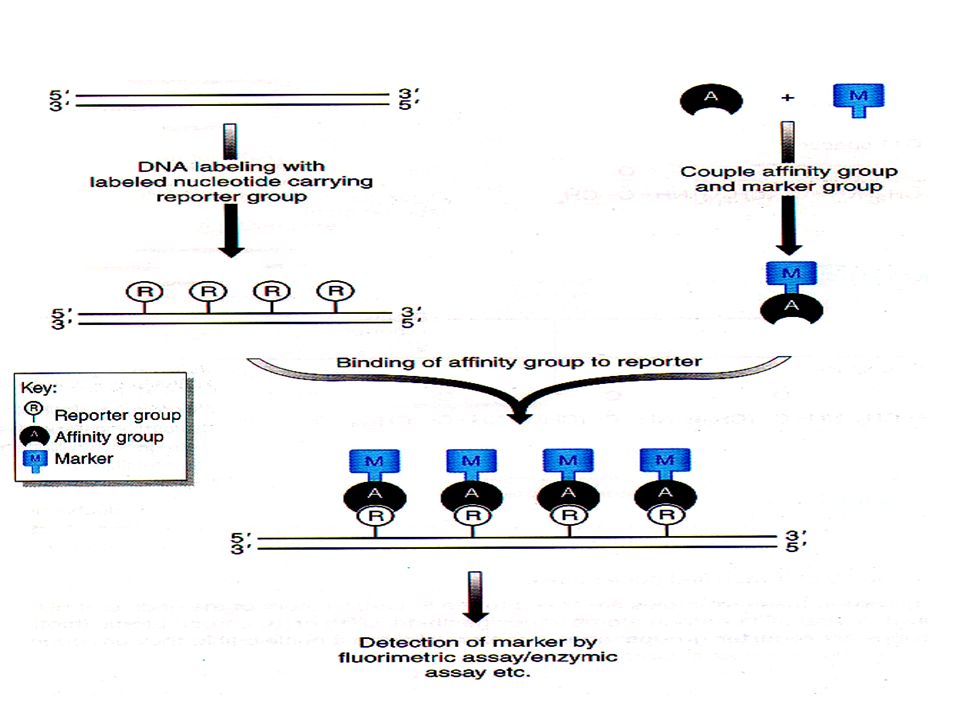
Using Biotin as a tag Biotin is a naturally occurring B vitamin, normally used in energy metabolism. Avidin and Strepavidin are proteins that normally act as B vitamin scavengers and bind Biotin. Avidin and Strepavidin bind with a very high affinity and we can use this to detect biotin labeled probes. The avidin or strepavidin can be attached to an enzyme or a fluorescent label. This gives you a choice as to how you will visualize your probe. Biotin can be incorporated as biotinylated dNTPs in a PCR reaction or end labeling or added as biotinylated dNTPs in a nick translation reaction. The required visualization agent, fluorescence, gold, enzyme is then added bound to strepavidin and binds to the biotinylated nucleotides.

10
The protein recognizing the group can be a specific antibody, e.g. the dioxygenin system or any other ligand that has a very high affinity for a specific group e.g. Biotin-strepavidin. The marker can be detected in various ways e.g. if it carries a fluorescent dye it can be detected in a fluorimetric assay it can be an enzyme such as alkaline phosphatase which can be coupled with an enzyme assay yielding a product that can be measured colorimetrically

Similar presentations


 Analysis of DNA (Sequencing) Chemical Synthesis.>")


 BIOTECHNOLOGY Lecture 7 5th May, 2006 PhD Course.>")


 FISH 2) “Restriction mapping” 3) Southern analysis : DNA 4) Northern analysis: RNA tells size tells which tissues or conditions.>")


