Download presentation
Presentation is loading. Please wait.

1
Abdominal Vasculature SONO 131 – Lecture #4

2
Vascular Anatomy Arterioles Artery Heart Capillaries Venules Vein

3
Vascular Anatomy Vessel Walls – Tunica intima – Tunica media – Tunica adventitia – Vasa vasorum Arteries Veins

4
Circulatory Anatomy Aorta – Ascending – Arch – Descending – Thoracic – Abdominal

5
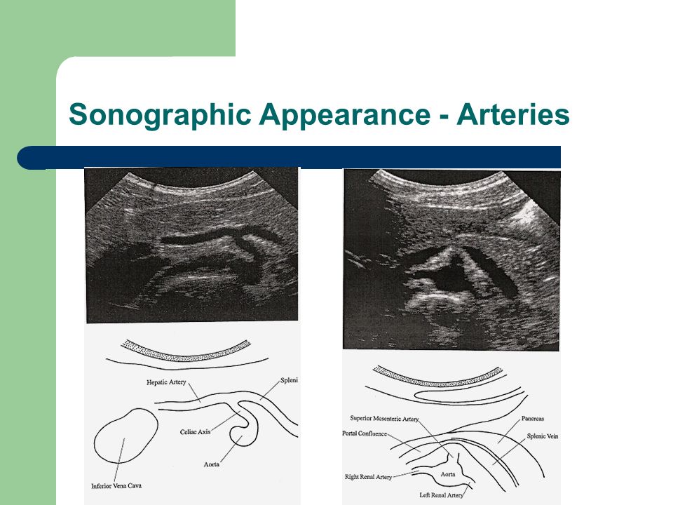
Anterior Branches Abdominal Aorta Celiac Trunk [Axis] – Common hepatic Hepatic – Right and Left Hepatic Gastroduodenal – Left Gastric – Splenic Left gastroepiploic Short gastric artery Several smaller splenic arteries Great pancreatic artery Hepatic – Left and right hepatic SMA – Inferior pancreatic – Duodenal – Colic – Ileocolic – intestinal IMA – Left colic – Sigmoid – Superior rectal
![Anterior Branches Abdominal Aorta Celiac Trunk [Axis] – Common hepatic Hepatic – Right and Left Hepatic Gastroduodenal – Left Gastric – Splenic Left gastroepiploic Short gastric artery Several smaller splenic arteries Great pancreatic artery Hepatic – Left and right hepatic SMA – Inferior pancreatic – Duodenal – Colic – Ileocolic – intestinal IMA – Left colic – Sigmoid – Superior rectal](http://images.slideplayer.com/26/8442894/slides/slide_5.jpg "Anterior Branches Abdominal Aorta Celiac Trunk [Axis] – Common hepatic Hepatic – Right and Left Hepatic Gastroduodenal – Left Gastric – Splenic Left gastroepiploic Short gastric artery Several smaller splenic arteries Great pancreatic artery Hepatic – Left and right hepatic SMA – Inferior pancreatic – Duodenal – Colic – Ileocolic – intestinal IMA – Left colic – Sigmoid – Superior rectal")
6
Celiac Trunk

7
Mesenteric Arteries

8
Lateral & Dorsal Branches Lateral – Phrenic Paired arteries – Renal Right & Left – Gonadal Dorsal – Lumbar 4 on each side of aorta

9
Renal Arteries

10
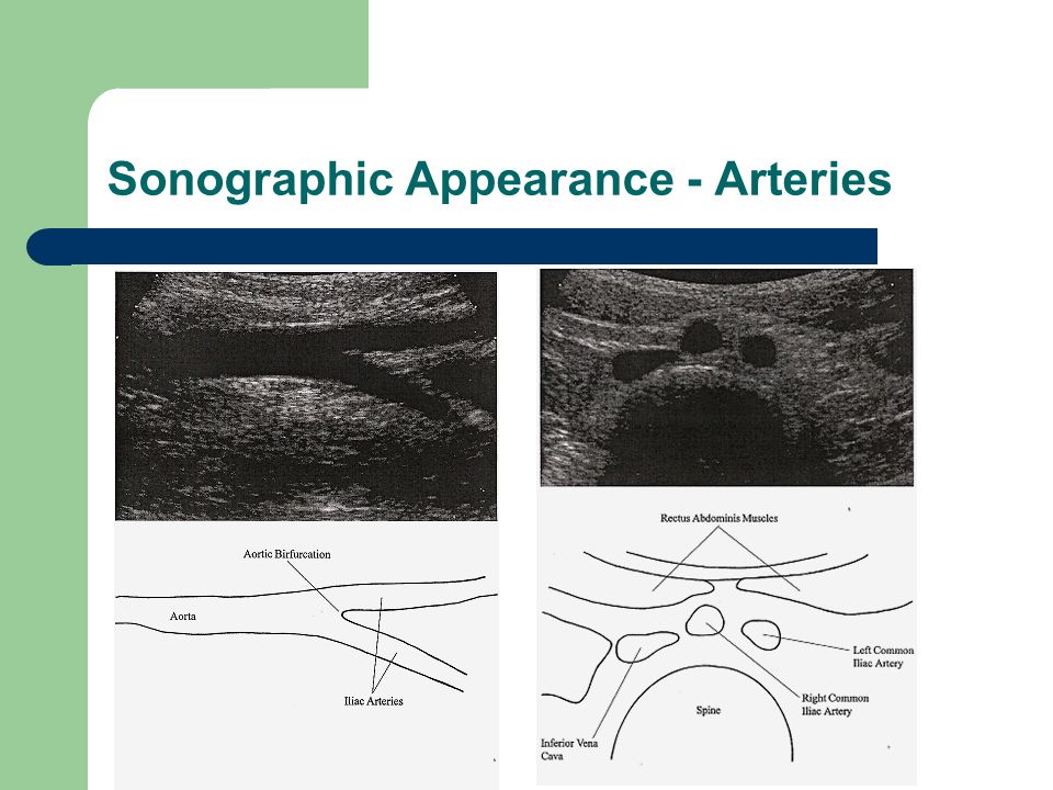
Sonographic Appearance - Arteries

13
Abdominal Artery Summary Abdominal aorta – Celiac Axis Splenic artery Hepatic artery Left gastric artery – SMA Inferior pancreatioduatenal Branches to the colon – Renal Arteries Level L1 – 2 – IMA – Aortic bifurcation Right & Left Common Iliac arteries

14
Abdominal Venous System

15
Inferior Vena Cava IVC = union of common iliac veins Tributaries to IVC – 3 anterior hepatic veins – 3 lateral Right suprarenal veins Renal veins Right testicular or ovarian vein – 5 lateral abdominal wall veins inferior phrenic + lumbar – 3 veins of origin common iliac + median sacral Drains – Abdominal organs – Abdominal structures – Lower extremities

16
Lateral Abdominal Veins Suprarenal Veins – Right & Left Renal Veins – Right & Left Gonadal Veins – Testicular or Ovarian

17
Anterior Abdominal Veins Hepatic Veins – Right – Middle – Left

18
Portal Venous System Portal Vein Splenic Vein Superior Mesenteric Vein Inferior Mesenteric Vein

19
Sonographic Appearance - Veins

20
Abdominal Vein - Summary IVC – Right and left common iliac veins – Renal veins – Hepatic veins Portal vein – Splenic & SMV – Right & left portal vein – Porta hepatis – Hepatopetal flow

21
Clinical Indications Abdominal Vasculature Imaging Arterial – Suspect aortic aneurysm – Possible ateriovenous fistula – Possible mesenteric ischemia Venous – Leg swelling – Portal hypertension

22
Arterial Abnormalities Atherosclerosis - altering of intimal lining of artery by focal accumulation of lipids, complex carbohydrates, blood and blood products, fibrous tissue and or calcium deposits – Cause – No known cause, but progression linked hyperlipidemia, hypertension, cigarette smoking and diabetes mellitus – Signs, Symptoms – None until significant stenosis – Sonographic Appearance – Luminal irregularities, tortuosity, vessel wall calcification

23
Aorta Aneurysm True Dilation of artery due to wall weakness Lined by all 3 components of artery wall – Fusiform – Saccular False Lined by outer layers of aortic wall or clot – Dissecting – Pseudoaneurysm

24
Aneurysm - Patient Presentation Causes – Atherosclerosis – Trauma – Syphilis – Marfan’s syndrome – Mycotic (Infective) Clinical Features – Abdominal or back pain – Abdominal bruit – Pulsatile abdominal mass – Impaired distal arterial flow
 Clinical Features – Abdominal or back pain – Abdominal bruit – Pulsatile abdominal mass – Impaired distal arterial flow")
25
Dissecting Aortic Aneurysm Type I – Ascending Aorta – Aortic Arch – Most dangerous [spiral] Type II – Marfan’s Syndrome – Ascending Aorta – Aortic Arch Type III – Descending Aorta – Abdominal Aorta
![Dissecting Aortic Aneurysm Type I – Ascending Aorta – Aortic Arch – Most dangerous [spiral] Type II – Marfan’s Syndrome – Ascending Aorta – Aortic Arch Type III – Descending Aorta – Abdominal Aorta](http://images.slideplayer.com/26/8442894/slides/slide_25.jpg "Dissecting Aortic Aneurysm Type I – Ascending Aorta – Aortic Arch – Most dangerous [spiral] Type II – Marfan’s Syndrome – Ascending Aorta – Aortic Arch Type III – Descending Aorta – Abdominal Aorta")
26
Aortic Aneurysm

27
Ultrasound Presentation

28
Endovascular Stent Graft Medtronic Aneurx Cook Zentih Gore Excluder

29
Stent Graft Therapy

30
Completed Deployment Contralateral Iliac Leg

31
Stent Graft Therapy Pre – Stent PlacementPost – Stent Placement

32
Stent Graft Therapy Pre – Stent Placement Post – Stent Placement

33
Aortic Rupture Risk Factors Diameter Rapid expansion > 0.6 cm / year Family history Hypertension COPD, Current Smoking Shape: Eccentric > Saccular> Fusiform

34
Inflammatory Aneurysms Aneurysm enveloped by a dense fibrotic reaction Uncommon – 5 to 20% of aneurysms Uncertain cause Clinically like other aneurysms

35
Branch Vessel Aneurysm Splenic – Most common – Usually multiple & occur in main splenic trunk – Life threatening Hepatic – 2 nd most common – Right hepatic arterial branch – Common cause – systemic infection, arteriosclerosis, blunt trauma – Silent or asymptomatic

36
Branch Vessel Aneurysm SMA – Rarest [1 in 12,000] – Cause - cystic medial necrosis (mycotic aneurysm) – Intestinal angina & postprandial abdominal pain – General abdominal pain, fever Renal Artery – Low incidence – approximately 20% – Symptoms – palpable mass, hypertension, blood in urine, flank pain
![Branch Vessel Aneurysm SMA – Rarest [1 in 12,000] – Cause - cystic medial necrosis (mycotic aneurysm) – Intestinal angina & postprandial abdominal pain – General abdominal pain, fever Renal Artery – Low incidence – approximately 20% – Symptoms – palpable mass, hypertension, blood in urine, flank pain](http://images.slideplayer.com/26/8442894/slides/slide_36.jpg "Branch Vessel Aneurysm SMA – Rarest [1 in 12,000] – Cause - cystic medial necrosis (mycotic aneurysm) – Intestinal angina & postprandial abdominal pain – General abdominal pain, fever Renal Artery – Low incidence – approximately 20% – Symptoms – palpable mass, hypertension, blood in urine, flank pain")
37
Vascular Stenosis Vessel lumen narrowed Post stenotic dilatation Increased velocities in area of stenosis Down stream changes – Turbulence – Decreased velocities – Slowed acceleration during systole – Relative elevation of diastolic velocities

38
Abdominal Artery Evaluation Doppler flow patterns [Angle corrected @ 60] – Aorta Proximal – high systolic / low diastolic flow Distal – triphasic flow – Celiac Axis Spectral broadening Unchanged after meals – Hepatic Artery Spectral broadening
![Abdominal Artery Evaluation Doppler flow patterns [Angle 60] – Aorta Proximal – high systolic / low diastolic flow Distal – triphasic flow – Celiac Axis Spectral broadening Unchanged after meals – Hepatic Artery Spectral broadening](http://images.slideplayer.com/26/8442894/slides/slide_38.jpg "Abdominal Artery Evaluation Doppler flow patterns [Angle 60] – Aorta Proximal – high systolic / low diastolic flow Distal – triphasic flow – Celiac Axis Spectral broadening Unchanged after meals – Hepatic Artery Spectral broadening")
39
Arterial Flow Characteristics

40
Renal Artery Stenosis – Associated with uncontrollable hypertension Up to 6% of all hypertensive patients have renal artery stenosis as underlying cause – Decreased glomerular filtration rate – Ischemic renal damage – Atherosclerotic plaque within first 2 cm – Fibromuscular dysplasia – lesions in distal 2/3 of renal artery

41
Renal Artery Stenosis

42
Mesentery Artery Stenosis Lack of adequate blood supply due to underlying vascular compromise – Mesenteric atherosclerotic disease – Embolic phenomenon Individuals at risk: – Smoking, coronary disease, PAD, chronic renal disease, diabetes mellitus Symptoms: – Progressive postprandial pain, weight loss, change in bowel habits, epigastrc bruit

43
Venous Flow Characteristics

44
Venous Abnormalities Vena Caval Obstruction Tumors of the IVC Portal Venous Thrombosis Portal Venous Hypertension

45
Vena Caval Obstruction IVC site of clot or tumor Greenfield filter – Reduce risk of clot embolizing

46
Renal Vein Thrombosis

47
Hepatic Venous Abnormalities Budd-Chiari Syndrome – Occlusion of some or all of the hepatic veins or occlusion of IVC – Clinically – Ascites, right upper quadrant pain, hepatomegaly – Sonographically – sluggish flow in IVC & hepatic veins

48
Portal Venous Abnormalities Thrombosis Hypertension

49
Portosystemic Shunts

50
Surgical end-to-side or side-to-side anastomosis of portal vein and IVC or TIPS

51
TIPS [Transjugular Interhepatic Portosystemic Shunts]
![TIPS [Transjugular Interhepatic Portosystemic Shunts]](http://images.slideplayer.com/26/8442894/slides/slide_51.jpg "TIPS [Transjugular Interhepatic Portosystemic Shunts]")
52
Abdominal Vasculature Review

Similar presentations