Download presentation
Presentation is loading. Please wait.

1
The Special Senses Vision - 3
Professor A.M.A Abdel Gader MD, PhD, FRCP (Lond., Edin), FRSH (London) Professor of Physiology, College of Medicine & King Khalid University Hospital Riyadh, Saudi Arabia
, FRSH (London) Professor of Physiology, College of Medicine & King Khalid University Hospital. Riyadh, Saudi Arabia.")
3
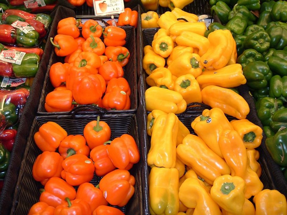
Color (Photopic) Vision
‘Young - Helmholtz theory’ ‘The Trichromatic theory’

10
History of color vision
Newton (1704) used a prism to show that sunlight was composed of light with all colors in the rainbow. He defined it as the spectrum.
 used a prism to show that. sunlight was composed of light with all. colors in the rainbow. He defined it as the spectrum.")
11
History of color vision
Primary colors: Thomas Young 1807: primary colors: when mixed >>> white or any other color

12
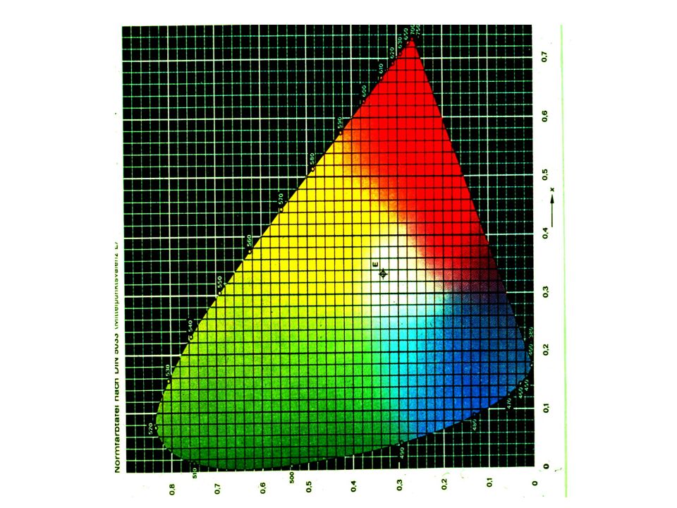
Mixing colors

14
Photopic vision (CONES)
Helmholtz : The three primary colors are perceived by three photoreceptor pigments (with broad absorption curves) White light is produced by mixing three colours
 White light is produced by mixing three colours.")
15
Cone wavelength ranges
M L Relative absorption 400 500 600 700 Wavelength (nm)
")
16
Photopic vision (CONES)
Cone pigments: three kinds 565 535 440

17
Photopic vision Young Helmholtz theory
Color vision is subserved by three types of cones, each containing a photoreceptor pigment most sensitive to one primary color Cones (contain red-sensitive pigment) Cones (contain green-sensitive pigment) Cones (contain blue-sensitive pigment) in the fovea centralis
 Cones (contain green-sensitive pigment) Cones (contain blue-sensitive pigment) in the fovea centralis.")
18
Cone wavelength ranges
M L Relative absorption 400 500 600 700 Wavelength (nm)
")
19
Color Blindness Weakness or total blindness in detecting a primary color: Definitions: Trichromats: see the 3 1ry colors Dichromats: blind to one 1ry color Monochromats: have color pigment

20
Color Blindness –cont. Prot …… Red Deuter …. Green Trit …… Blue
Anamoly …weakness Protanamoly Deuteranamoly Trichromats Tritanamoly

21
Color Blindness –cont. Anamoly …weakness Anopia …. Total loss
Protanopia Deuteranopia Dichromats Tritanopia

22
Trichromatic/dichromatic color vision
In its most severe forms, colorblindness is caused by the absence of one of the cone visual pigments. Shown here, the spectral sensitivities of the cone pigments in color normal trichromats are compared with those of a color blind person. Also compare the spectrum as it appears to a color normal person with the illustration of how it might look to a red-green color blind person. Red-green color blindness is common--about 4-5% of the population (8-10% men red-green colorblind). Dichromats – missing one whole group of photopigments (cat and dogs – dichromats) a. Missing M pigments = deuteranopes b. Missing L pigments = protanopes Monochromats – missing two groups of photopigments Moderate colorblindness – anomolous trichromats; 3 different color pigments but from only 2 of the photopigment groups a. 2 different L-type pigments = deuteranomalous b. 2 different M photopigments = protanomalous More information on this topic can be found elsewhere (Nathans et al., 1992; Sharpe et al., 1998; Sharpe et al., 1999; Stockman et al., 2000). Here, we provide a brief summary of those areas that are relevant to cone spectral sensitivity measurements made in dichromats in order to determine the "normal" M- and L-cone spectral sensitivities. Persons with red-green defects have difficulty distinguishing between reds, greens and yellows but can discriminate between blues and yellows. Protanopes often can name red and green correctly because green looks lighter to them than red. The M- and L-cone photopigment genes lie in a head to tail tandem array on the q-arm of the X-chromosome. Each gene consists of six coding regions, called exons, which are transcribed to produce the opsin. Because the M- and L-cone photopigment genes are highly homologous and adjacent to one another, intragenic recombination between them is common and can lead to the production of hybrid or fusion genes, some of which code for anomalous pigments. Each hybrid gene can be identified by the site, usually between exons, at which the fusion occurs. For example, L3M4 indicates a hybrid gene in which exons 1 to 3 derive from an L-cone pigment gene and exons 4 to 6 from an M-cone pigment gene. Because exons 1 and 6 in the L- and M-cone pigment genes are identical, a L1M2 hybrid pigment gene encodes a de facto M-cone photopigment. The classification of hybrid genes, and genes in general, is complicated by polymorphisms in the normal population, the most common of which is the frequent substitution of alanine by serine at codon 180 in exon 3. Of 304 genotyped Caucasian males, we estimate that 56% have the serine variant [identified as L(S180)] and 44% the alanine variant [identified as L(A180)] for their L-cone gene (see Table 1, which summarizes data from Winderickx et al., 1993; Neitz & Neitz, 1998; Sharpe et al., 1998; and from Schmidt, Sharpe, Knau & Wissinger, personal communication). The L-cone polymorphism, and its distribution in the normal population, must be considered when estimating the "normal" L-cone spectral sensitivities. In contrast, the M(A180) versus M(S180) polymorphism for the M-cone pigment is much less frequent: 94% (Winderickx et al., 1993) or 93% (Neitz & Neitz, 1998) of males have the M(A180) variant. The spectral sensitivity of the photopigment that is encoded by the L2M3(A180) hybrid gene is practically indistinguishable from the photopigment encoded by the normal M(A180) [(or L1M2(A180)] cone pigment gene, its lmax being only 0.2 nm (Merbs & Nathans, 1992) or 0.0 nm (Asenjo, Rim & Oprian, 1994) or insignificantly different (Sharpe et al., 1998) from that of the M(180) cone pigment. Thus, spectral sensitivities from protanopes carrying either L1M2(A180) or L2M3(A180) genes in their opsin gene array can be reasonably combined to estimate the normal M-cone spectral sensitivities, as was done by Stockman and Sharpe (2000). Dichromats with single photopigment genes in the M- and L-cone pigment gene array [e.g., L(A180), L(S180), L1M2(A180) or L2M3(A180)] are especially useful for measuring normal cone spectral sensitivities, since they should possess only a single longer wavelength photopigment. Dichromats with multiple photopigment genes are less useful, unless the multiple genes produce photopigments with the same or nearly the same spectral sensitivities: for example, L1M2(A180)+M(A180) or L2M3(A180)+M(A180). Trichromatic/dichromatic color vision
. Dichromats – missing one whole group of photopigments (cat and dogs – dichromats) a. Missing M pigments = deuteranopes. b. Missing L pigments = protanopes. Monochromats – missing two groups of photopigments. Moderate colorblindness – anomolous trichromats; 3 different color pigments but from only 2 of the photopigment groups. a. 2 different L-type pigments = deuteranomalous. b. 2 different M photopigments = protanomalous. More information on this topic can be found elsewhere (Nathans et al., 1992; Sharpe et al., 1998; Sharpe et al., 1999; Stockman et al., 2000). Here, we provide a brief summary of those areas that are relevant to cone spectral sensitivity measurements made in dichromats in order to determine the. normal M- and L-cone spectral sensitivities. Persons with red-green defects have difficulty distinguishing between reds, greens and yellows but can discriminate between blues and yellows. Protanopes often can name red and green correctly because green looks lighter to them than red. The M- and L-cone photopigment genes lie in a head to tail tandem array on the q-arm of the X-chromosome. Each gene consists of six coding. regions, called exons, which are transcribed to produce the opsin. Because the M- and L-cone photopigment genes are highly homologous and adjacent. to one another, intragenic recombination between them is common and can lead to the production of hybrid or fusion genes, some of which code for. anomalous pigments. Each hybrid gene can be identified by the site, usually between exons, at which the fusion occurs. For example, L3M4 indicates a. hybrid gene in which exons 1 to 3 derive from an L-cone pigment gene and exons 4 to 6 from an M-cone pigment gene. Because exons 1 and 6 in the L- and M-cone pigment genes are identical, a L1M2 hybrid pigment gene encodes a de facto M-cone photopigment. The classification of hybrid genes, and genes in general, is complicated by polymorphisms in the normal population, the most common of which is the. frequent substitution of alanine by serine at codon 180 in exon 3. Of 304 genotyped Caucasian males, we estimate that 56% have the serine variant. [identified as L(S180)] and 44% the alanine variant [identified as L(A180)] for their L-cone gene (see Table 1, which summarizes data from Winderickx. et al., 1993; Neitz & Neitz, 1998; Sharpe et al., 1998; and from Schmidt, Sharpe, Knau & Wissinger, personal communication). The L-cone polymorphism, and its distribution in the normal population, must be considered when estimating the normal L-cone spectral sensitivities. In contrast, the M(A180) versus M(S180) polymorphism for the M-cone pigment is much less frequent: 94% (Winderickx et al., 1993) or 93% (Neitz. & Neitz, 1998) of males have the M(A180) variant. The spectral sensitivity of the photopigment that is encoded by the L2M3(A180) hybrid gene is practically indistinguishable from the photopigment. encoded by the normal M(A180) [(or L1M2(A180)] cone pigment gene, its lmax being only 0.2 nm (Merbs & Nathans, 1992) or 0.0 nm (Asenjo, Rim. & Oprian, 1994) or insignificantly different (Sharpe et al., 1998) from that of the M(180) cone pigment. Thus, spectral sensitivities from protanopes. carrying either L1M2(A180) or L2M3(A180) genes in their opsin gene array can be reasonably combined to estimate the normal M-cone spectral. sensitivities, as was done by Stockman and Sharpe (2000). Dichromats with single photopigment genes in the M- and L-cone pigment gene array [e.g., L(A180), L(S180), L1M2(A180) or L2M3(A180)] are. especially useful for measuring normal cone spectral sensitivities, since they should possess only a single longer wavelength photopigment. Dichromats. with multiple photopigment genes are less useful, unless the multiple genes produce photopigments with the same or nearly the same spectral sensitivities: for example, L1M2(A180)+M(A180) or L2M3(A180)+M(A180). Trichromatic/dichromatic color vision.")
23
Color Blindness –cont. Prevalence: males ………….8% females …. 0.4%
Inheritance: sex-linked due abnormal gene in the X chromosome

25
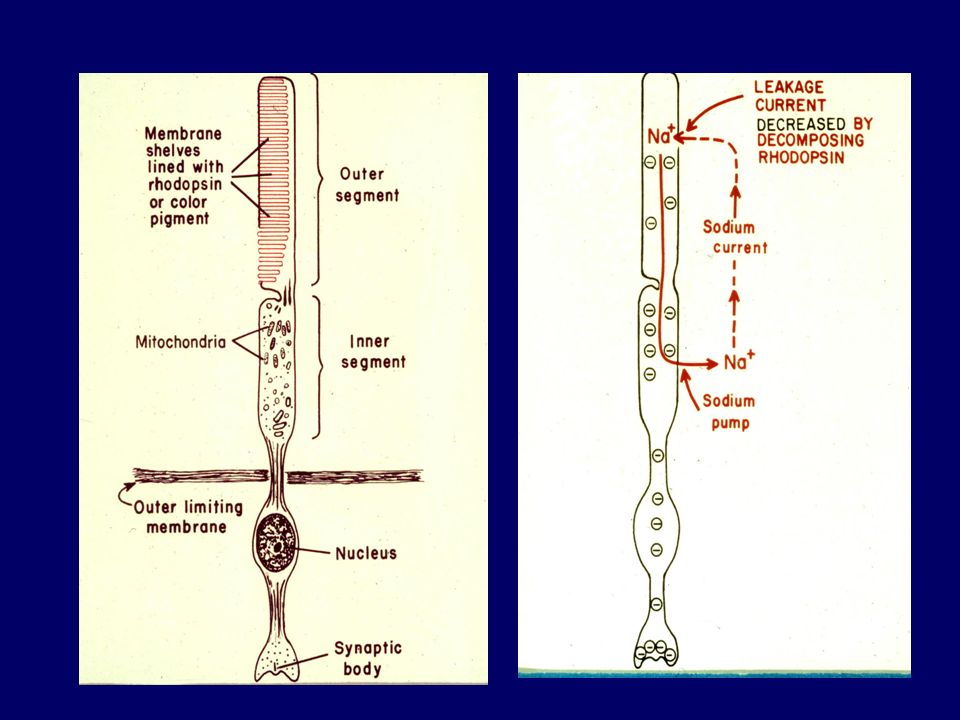
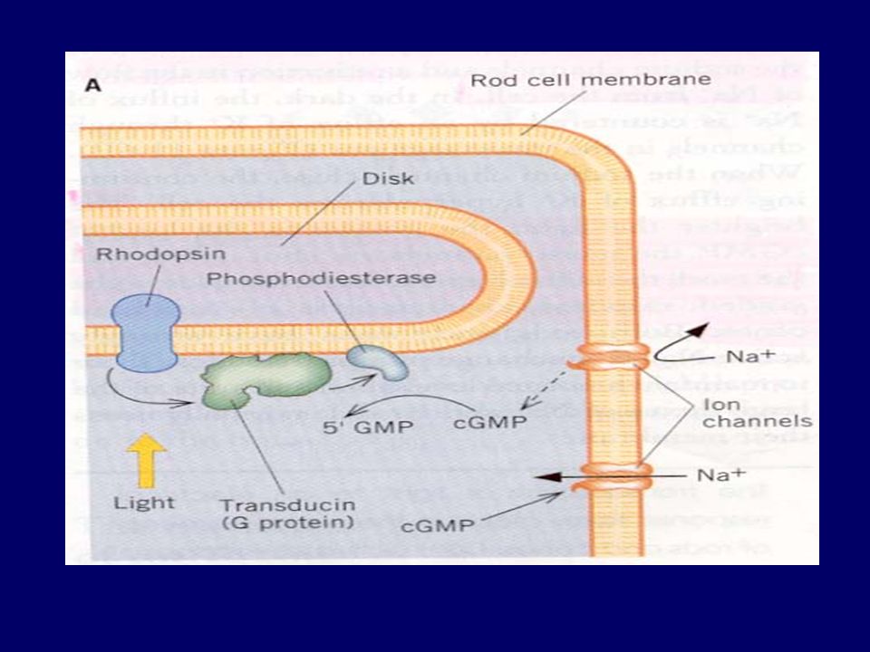
Electrophysiology of Vision

26
Hyperpolarization of receptor Action potential in optic nerve fibres
Light Change in photopigment Metarhodopsin II Activation of transducin Activation of phophodiesterase Decrease IC cyclic GMP Closure of Na channels Hyperpolarization of receptor Action potential in optic nerve fibres

28
Photoreception

30
Bleaching and Regeneration of Visual Pigments
Figure 17.15

32
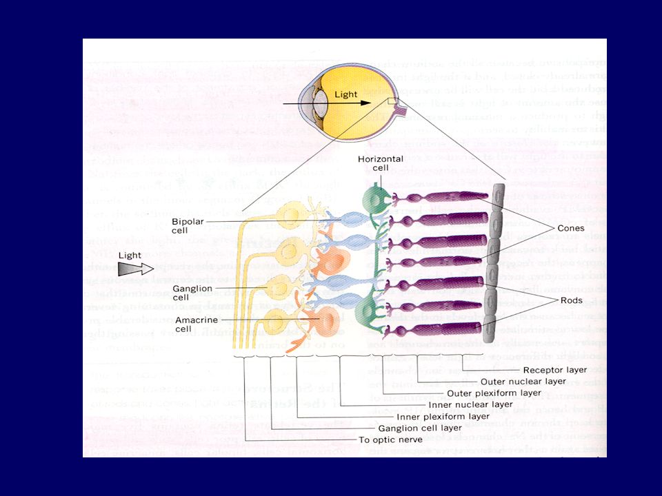
Electrophysiology of Vision
Electric recording in Retinal cells: Bipolar cells: Hyper- & Depolarization Horizental cells: Hyper- & Depolarization Amacrine cells: Depolarizing potential Ganglion cells:Depolarizing potential

Similar presentations


 Dept.>")