Download presentation
Presentation is loading. Please wait.

1
Chapter 15 B The Ear

2
The External Ear Auricle Surrounds entrance to external acoustic meatus Protects opening of canal Provides directional sensitivity External acoustic meatus Ends at tympanic membrane (eardrum) Ceruminous glands Tympanic membrane Is a thin, semitransparent sheet Separates external ear from middle ear
 Ceruminous glands Tympanic membrane Is a thin, semitransparent sheet Separates external ear from middle ear")
3
The Ear [INSERT FIG. 17.20] The Anatomy of the Ear.
![The Ear [INSERT FIG ] The Anatomy of the Ear.](http://images.slideplayer.com/26/8429517/slides/slide_3.jpg "The Ear [INSERT FIG ] The Anatomy of the Ear.")
4
The Ear The Middle Ear Also called tympanic cavity Communicates with nasopharynx via auditory tube Permits equalization of pressures on either side of tympanic membrane Encloses and protects three auditory ossicles Malleus (hammer) Incus (anvil) Stapes (stirrup)
 Incus (anvil) Stapes (stirrup)")
5
The Ear

6
Vibration of Tympanic Membrane Converts arriving sound waves into mechanical movements Auditory ossicles conduct vibrations to inner ear Tensor tympani muscle Stiffens tympanic membrane Stapedius muscle Reduces movement of stapes at oval window

7
The Ear The Inner Ear Contains fluid called endolymph Bony labyrinth surrounds and protects membranous labyrinth Subdivided into Vestibule Semicircular canals Cochlea

8
The Ear

9
The Inner Ear Vestibule Encloses saccule and utricle Receptors provide sensations of gravity and linear acceleration Semicircular canals Contain semicircular ducts Receptors stimulated by rotation of head Cochlea Contains cochlear duct (elongated portion of membranous labyrinth) Receptors provide sense of hearing
 Receptors provide sense of hearing")
10
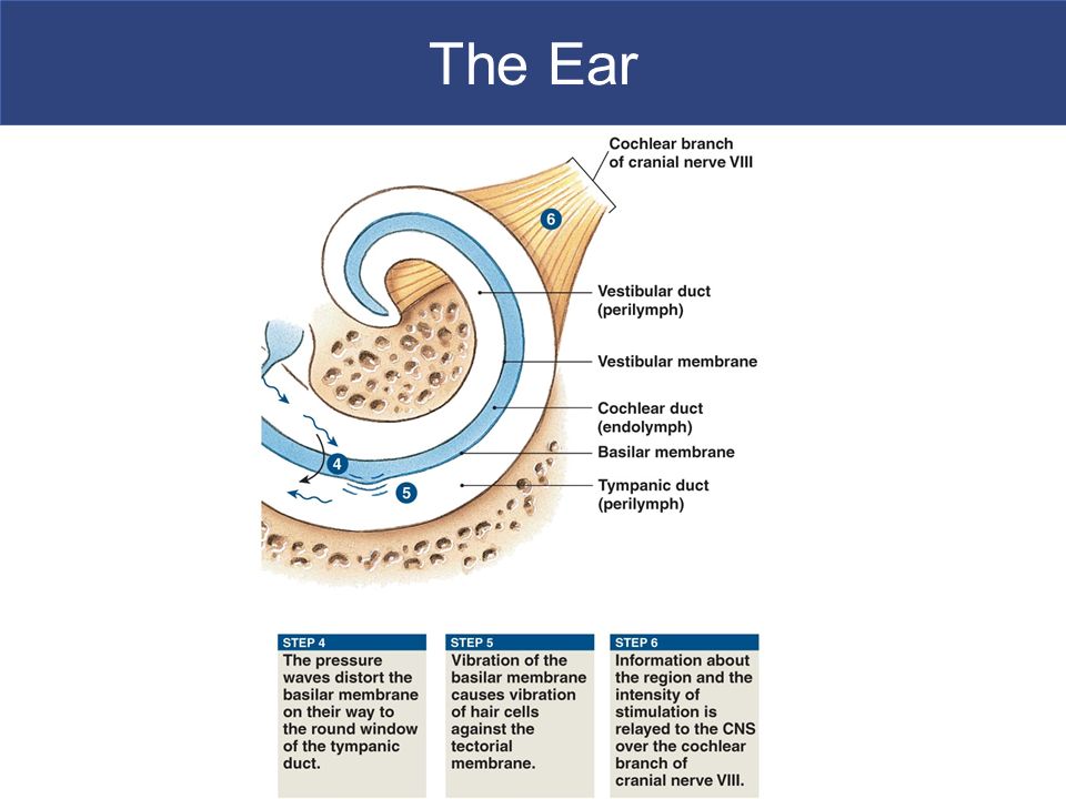
The Ear The Inner Ear Round window Thin, membranous partition Separates perilymph from air spaces of middle ear Oval window Formed of collagen fibers Connected to base of stapes

11
The Ear Stimuli and Location Sense of gravity and acceleration From hair cells in vestibule Sense of rotation From semicircular canals Sense of sound From cochlea

12
The Ear The Semicircular Ducts Are continuous with utricle Each duct contains Ampulla with gelatinous cupula Associated sensory receptors Stereocilia – resemble long microvilli: –Are on surface of hair cell Kinocilium – single, large cilium

13
The Ear The Semicircular Ducts.

14
The Ear The Semicircular Ducts.

15
The Ear The Semicircular Ducts.

16
The Ear The Utricle and Saccule Provide equilibrium sensations Are connected with the endolymphatic duct, which ends in endolymphatic sac Maculae Oval structures where hair cells cluster Statoconia Densely packed calcium carbonate crystals on surface of gelatinous mass Otolith (ear stone) = gel and statoconia
 = gel and statoconia")
17
The Ear The Saccule and Utricle.

18
The Ear

19
Pathways for Equilibrium Sensations Vestibular receptors Activate sensory neurons of vestibular ganglia Axons form vestibular branch of vestibulocochlear nerve (VIII) Synapse within vestibular nuclei
 Synapse within vestibular nuclei")
20
The Ear Pathways for Equilibrium Sensations.

21
The Ear Hearing Cochlear duct receptors Provide sense of hearing

22
The Ear The Structure of the Cochlea.

23
The Ear Diagrammatic and Sectional Views of the Cochlear Spiral.

24
The Ear Hearing Auditory ossicles Convert pressure fluctuation in air into much greater pressure fluctuations in perilymph of cochlea Frequency of sound: –determined by which part of cochlear duct is stimulated Intensity (volume): –determined by number of hair cells stimulated
: –determined by number of hair cells stimulated")
25
The Ear Hearing Cochlear duct receptors Basilar membrane: –separates cochlear duct from tympanic duct –hair cells lack kinocilia –stereocilia in contact with overlying tectorial membrane »is attached to inner wall of cochlear duct

26
The Ear The Organ of Corti.

27
The Ear The Organ of Corti.

28
The Ear The Nature of Sound.

29
The Ear

31
Auditory Pathways Cochlear branch Formed by afferent fibers of spiral ganglion neurons: –enters medulla oblongata –synapses at dorsal and ventral cochlear nuclei –information crosses to opposite side of brain Figure 17–31

32
The Ear

33
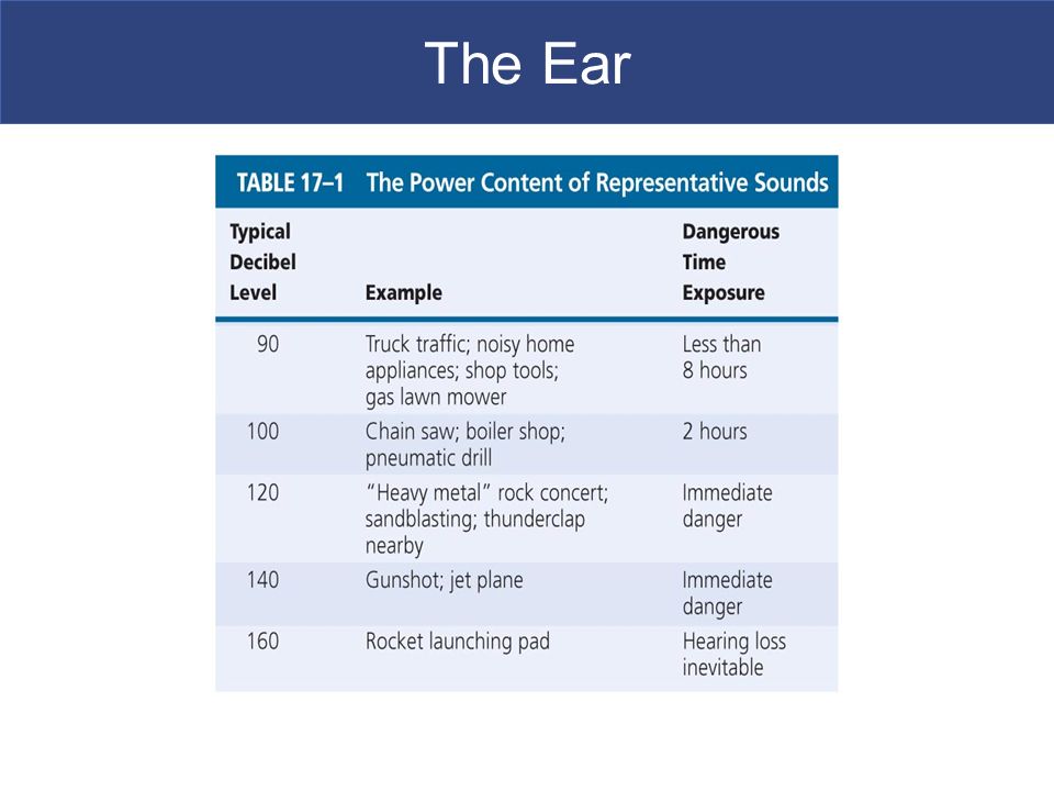
Hearing Range From softest to loudest represents trillion-fold increase in power Never use full potential Young children have greatest range

34
The Ear

36
With age, damage accumulates Tympanic membrane gets less flexible Articulations between ossicles stiffen Round window may begin to ossify

Similar presentations
 SYSTEM>")


