Download presentation
Presentation is loading. Please wait.

1
The Peripheral Auditory System George Pollak Section of Neurobiology

2
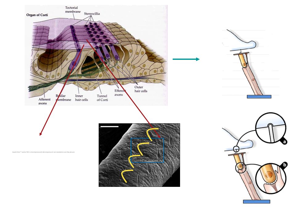
stereocillia of inner hair cells stereocillia of outer hair cells Hair cells, the transducers of the auditory system, and how they work 1

3
Organ of Corti Basilar membrane Hair cells, the transducers of the auditory system, and how they work. 1 stereocillia of inner hair cells stereocillia of outer hair cells

4
Organ of Corti Basilar membrane

7
stereocilia on one hair cell

9
9

13
Hi K + Lo Na + Hi Na + Lo K + Hi K + Lo Na + E k = 58 log K out K in = 0mV Potential difference between Endolymph and cell interior E k = 58 log K out K in = ~-70mV Potential difference between Perilymph and cell interior endolymph perilymph

14
-45 mV

15
Hi K + low Na + Hi K + low Na + Hi K + low Na + Hi Na + low K + Hi Na + low K +

16
small leakage of K + into cell -45 mV K + into cell -70 mV No K + into cell

17
Next, we are going to build a cochlea

18
Basilar membrane Stapes

19
Sound is changed from a pressure wave in the air into mechanical movements on the basilar membrane

23
round window

24
Traveling waves on basilar membrane round window oval window

25
The structure of the basilar membrane causes it to perform a frequency to place transformation

26
Basilar Membrane has continuously changing dimensions along its length Base responds maximally to high frequencies Apex responds maximally to low frequencies Stiff Narrow and thick flexible wide and thin

28
Basilar membrane converts frequency to a place of maximal response

29
Frequency-to-Place Transformation in the Cochlea

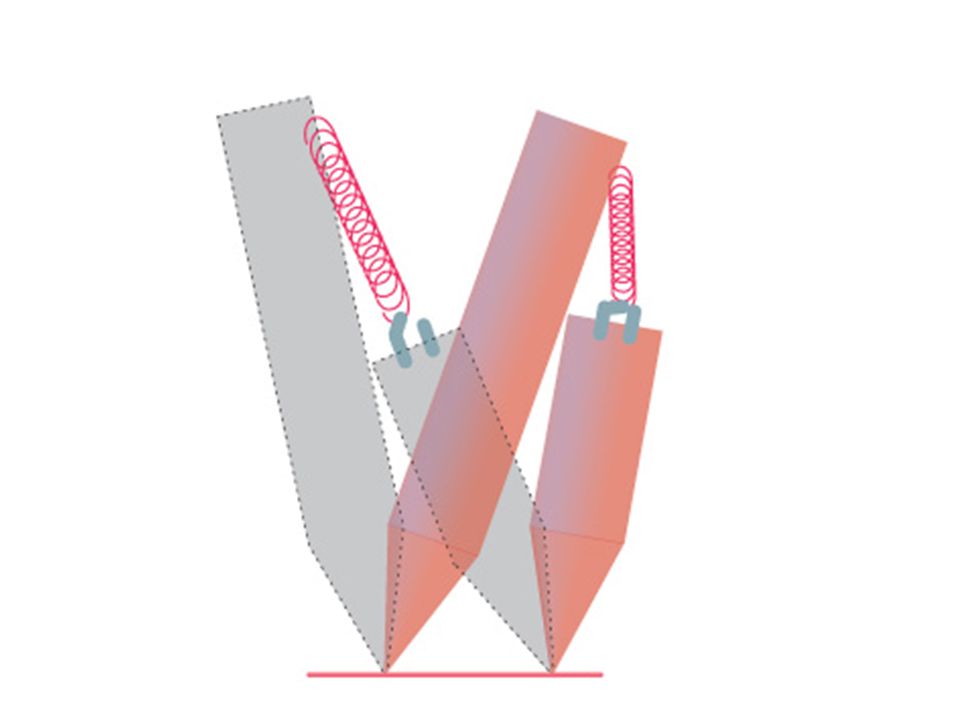
30
The motion on the basilar membrane causes shearing of the cilia on hair cells and thereby causes the hair cells to depolarize and hyperpolarize in response to sound

31
Organ of Corti Basilar membrane

32
Organ of Corti

33
basilar membrane shearing of stereocillia

36
Why are there two types of hair cells?

38
98% of the fibers that project into the central auditory system are innervated by inner hair cells!! 98%

39
What are the outer hairs doing? Answer: they act as amplifiers of the mechanical motion of the basilar membrane generated by sound

40
hyperpolarization ----- depolarization release of transmitter Hi K + K+K+

41
Evoked mechanical responses of isolated cochlear outer hair cells. Electromotility: OHC can change length in response to voltage change Direct evidence of an active mechanical process in the organ of Corti depolarizedhyperpolarized + + + _ _ _

42
42 Dancing hair cell

43
Outer hair cells are the only cells in the body that express prestin. Even inner hair cells do NOT have prestin. + + + + + + + +

44
Show movie of how Prestin works

46
Sound stimuli Basilar membrane motion Hair bundle deflection Membrane potential change Change in length of hair cells IHC Sensory signal transmission OHC Positive feedback loop

47
Normal response with cochlear amplifier response without cochlear amplifier base Apex base Apex Basilar membrane

48
How motion of basilar membrane generates tuning curves in auditory nerve fibers and thereby imparts frequency selectivity to auditory nerve fibers

50
6 kHz 7 kHz 8 kHz 9 kHz 10 kHz 10 678 9 11 Frequency ( kHz) 5 20 30 40 50 60 Intensity (dB SPL) baseapex 50 dB SPL
 Intensity (dB SPL) baseapex 50 dB SPL")
51
6 kHz 7 kHz 8 kHz 9 kHz 10 kHz 10 678 9 11 Frequency ( kHz) 5 20 30 40 50 60 Intensity (dB SPL) Tuning Curve The most basic feature of an auditory neuron best frequency 30 dB SPL baseapex
 Intensity (dB SPL) Tuning Curve The most basic feature of an auditory neuron best frequency 30 dB SPL baseapex")
53
frequency low high Sound intensity low high tuning curves in normal animalstuning curves in animals with no outer hair cells or in animals without prestin gene

54
How is the tonotopic organization that was first established on the basilar membrane preserved in in the central auditory system?

55
Inferior colliculus Inferior colliculus Medial geniculate Superior olive Cochlear nucleus Auditory cortex Cochlea Auditory nerve Flow of Information Along the Central Auditory Pathway

57
Inferior colliculus Inferior colliculus Medial geniculate Superior olive Cochlear nucleus Auditory cortex Cochlea Auditory nerve cochlear nucleus superior olive Inferior colliculus medial geniculate auditory cortex The Frequency Representation on the Cochlea is Preserved in Every Nucleus of the Central Auditory System, and thus the Auditory System is Tonotopically Organized

58
Inferior colliculus Inferior colliculus Medial geniculate Superior olive Cochlear nucleus Auditory cortex Cochlea Auditory nerve The Frequency Representation on the Cochlea is Preserved in Every Nucleus of the Central Auditory System, and thus the Auditory System is Tonotopically Organized

Similar presentations


 Physical properties of sound>")


