Download presentation
Presentation is loading. Please wait.

1
Nervous Systems

2
IB Assessment Statement
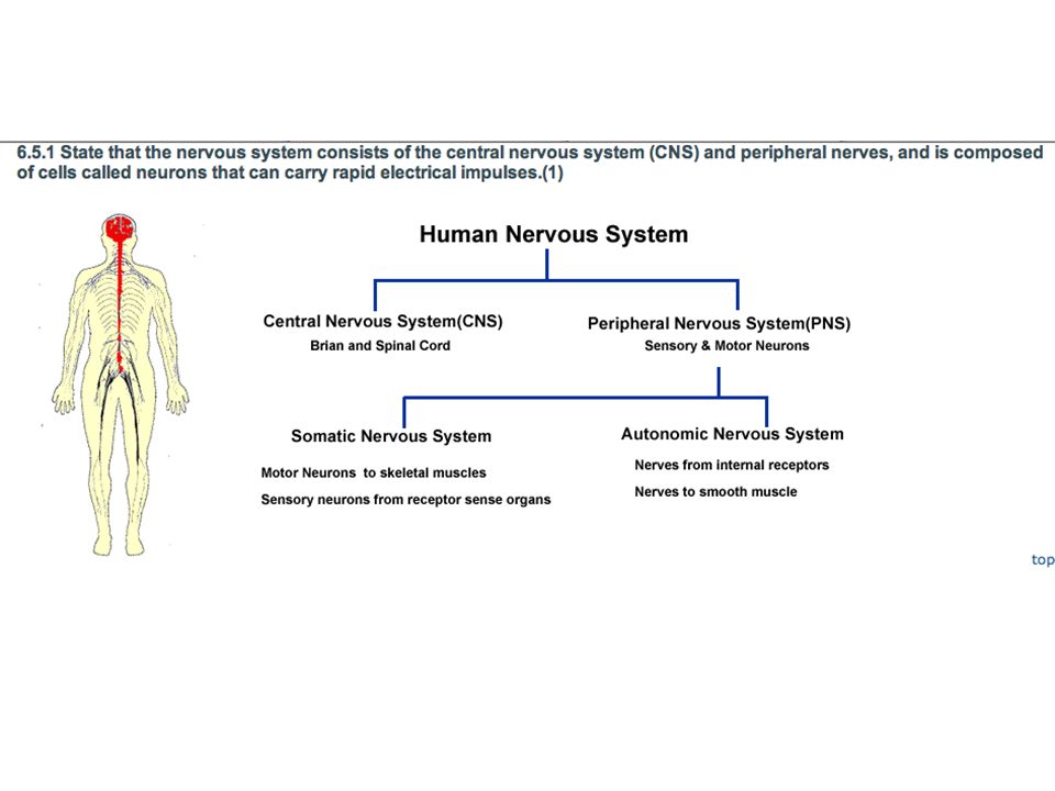
State that the nervous system consists of the central nervous system (CNS) and peripheral nerves, and is composed of cells called neurons that can carry rapid electrical impulses. Crash Course Video on the Nervous System
 and peripheral nerves, and is composed of cells called neurons that can carry rapid electrical impulses. Crash Course Video on the Nervous System. v=x4PPZCLnVkA.")
3
Organization of the Body
Nervous System Structures: Brain, spinal cord, peripheral nerves Function: Recognizes and coordinates the body’s response to changes in its internal and external environments

4
Overview: Command and Control Center
The human brain contains about 100 billion nerve cells, or neurons

5
Functional magnetic resonance imaging is a technology that can reconstruct a three-dimensional map of brain activity Brain imaging and other methods reveal that groups of neurons function in specialized circuits dedicated to different tasks

6
Parts of the Nervous System
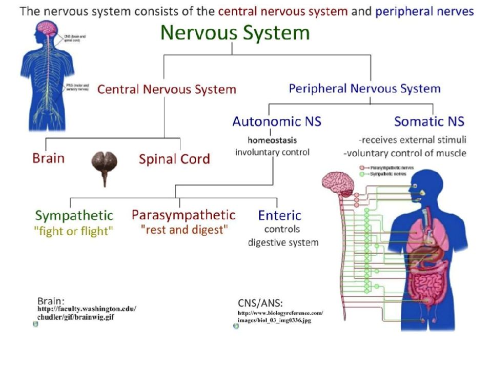
The nervous system consists of : the central nervous system (CNS) Brain and spinal cord and peripheral nerves (PNS), Nerves they run to all parts of the body. and is composed of cells called neurons that can carry rapid electrical impulses.
 Brain and spinal cord. and peripheral nerves (PNS), Nerves they run to all parts of the body. and is composed of cells called neurons that can carry rapid electrical impulses.")
7
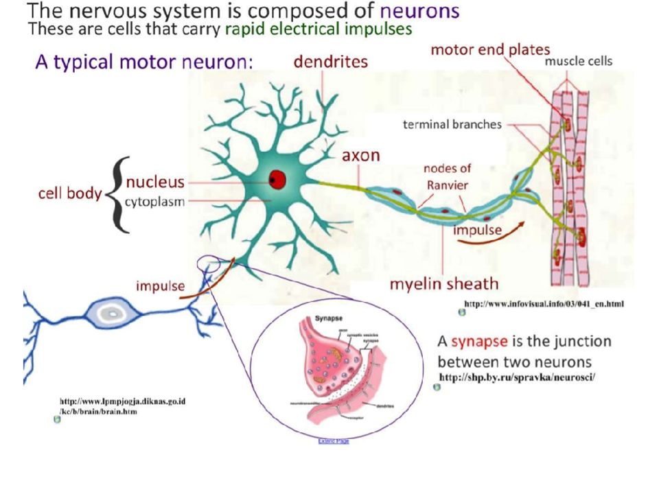
Neuron Cell Structure Neuron Cells have the following structures:
Cell Body dendrites axon myelin sheath Node of Ranvier Motor end plates Include dendrites, cell body with nucleus, axon, myelin sheath, nodes of Ranvier and motor end plates.

11
IB Assessment Statement
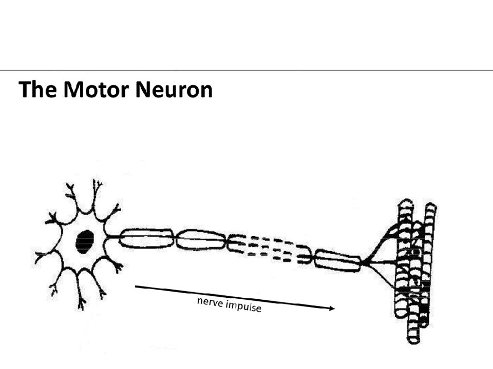
Draw and label a diagram of the structure of a motor neuron. Include dendrites, cell body with nucleus, axon, myelin sheath, nodes of Ranvier and motor end plates.

12
Cell Body The largest part of a typical neuron is the cell body.
It contains the nucleus and much of the cytoplasm. Cell body

13
Dendrites Dendrites extend from the cell body
and carry impulses from the environment toward the cell body. Dendrites

14
Axon The axon is the long fiber that carries impulses away from the cell body. Axon

15
Motor end plates The axon ends in motor end plates. Motor end plates

16
Myelin Sheath The axon is surrounded by an insulating lipid membrane called the myelin sheath. Myelin sheaths have high electrical resistance Myelin sheath Nodes of Ranvier

17
Myelin Sheath There are gaps in the myelin sheath, called nodes of Ranvier, where the membrane is exposed. Electrical Impulses jump from one node of Ranvier to the next. Myelin sheath Nodes of Ranvier

18
Neurons Structures of a Neuron Nucleus Dendrites Motor end plates
Cell body Myelin sheath The nervous system controls and coordinates functions throughout the body. The basic units of the nervous system are neurons. Axon Node of Ranvier

19
Draw and label a diagram of the structure of a motor neuron. Include
OK YOUR TURN Draw and label a diagram of the structure of a motor neuron. Include dendrites, cell body with nucleus, axon, myelin sheath, nodes of Ranvier and motor end plates

20
IB Assessment Statement
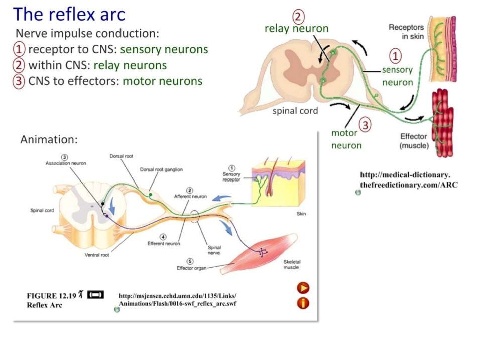
State that nerve impulses are conducted from receptors to the CNS by sensory neurons, within the CNS by relay neurons, and from the CNS to effectors by motor neurons.

21
The Path of Nerve Impulses
There are various receptor around the body such as skin and the eye. Stimuli (think of them as energy forms) are detected by the receptors and turned into an nerve impulse (chemical energy). Nerve impulses from sensory nerves are conducted to the central nervous system along sensory neurons. The impulse is sent to the relay neurons that move it around inside the central nervous system (brain and spine). Motor neurons take the relayed nerve impulse to the effectors (often muscles) which then produce the response.
 are detected by the receptors and turned into an nerve impulse (chemical energy). Nerve impulses from sensory nerves are conducted to the central nervous system along sensory neurons. The impulse is sent to the relay neurons that move it around inside the central nervous system (brain and spine). Motor neurons take the relayed nerve impulse to the effectors (often muscles) which then produce the response.")
22
Path of Nerve Impulses This is a cross section through the vertebrate spinal column. The receptor is deep in the biceps muscle. Sensory neuron conducts nerve impulses from the receptor to the central nervous system. The relay nerve conduct the impulse through the spinal cord and in a reflex back to the motor neuron. The motor neuron connects to the effector which in this case is the biceps muscle.

24
Reflex Arc Video Reflex Arc Animation

25
IB LEARNING Objective Define resting potential and action potential (depolarization and repolarization).
.")
26
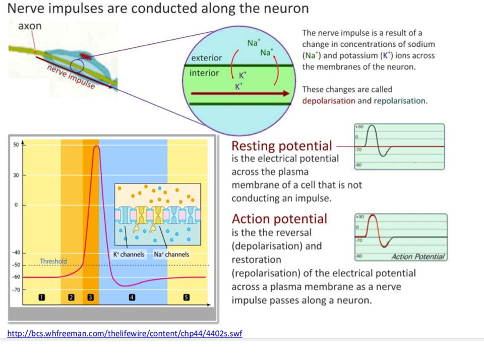
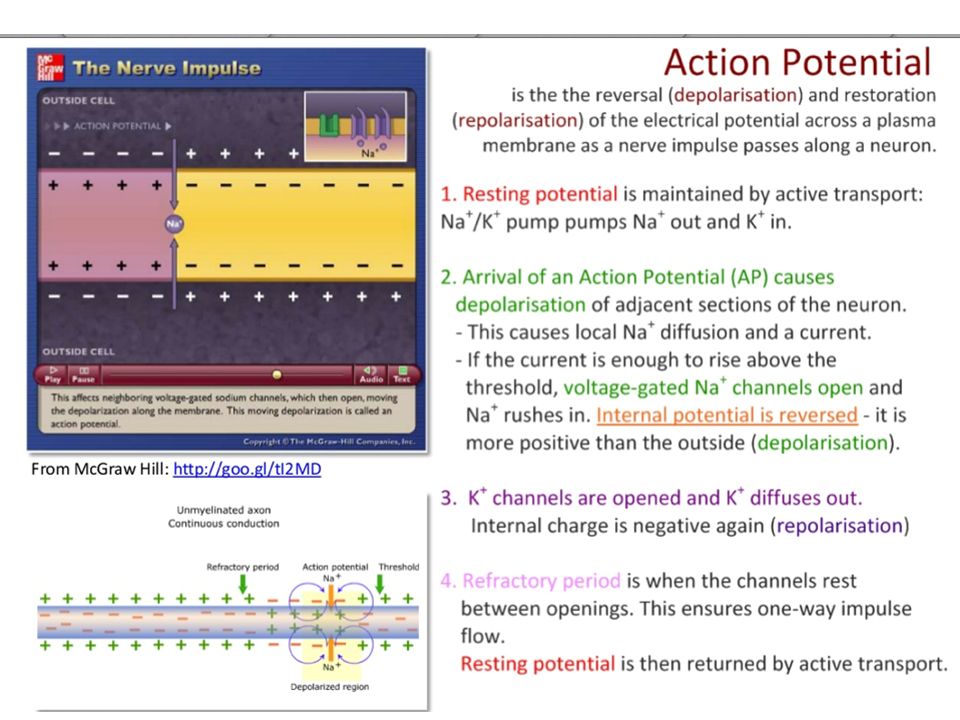
Definitions: Resting potential is the negative charge registered when the nerve is at rest and not conducting a nerve impulse. Action potential is the positive electrochemical charge generated at the nerve impulse. Normally this is seen as the 'marker' of the nerve impulse position. Depolarisation is a change from the negative resting potential to the positive action potential. Re-polarisation is the change in the electrical potential from the positive action potential back to the negative resting potential.

28
IB Assessment Statement
Explain how a nerve impulse passes along a non-myelinated neuron. Include the movement of Na+ and K+ ions to create a resting potential and an action potential.

30
How Neuron cells transmit an impulse
Neurons transmit information in the form of electrical impulses An electrical impulse is transmitted along nerve fibers. Dendrites Cell body Nucleus Axon Signal direction Myelin sheath Synaptic terminals Synapse State that nerve impulses are conducted from receptors to the CNS by sensory neurons, within the CNS by relay neurons, and from the CNS to effectors by motor neurons.

31
Electrical Potential Differences
An impulse is a change in electrical potential difference in the membrane of a neuron cell. A potential difference is when there are more positive ions (+) on one side of a membrane than negative ions(-) Positive Ion examples: Sodium (Na+), Potassium (K+), Calcium (Ca2+) Negative Ion examples: Chlorine (Cl-)
 on one side of a membrane than negative ions(-) Positive Ion examples: Sodium (Na+), Potassium (K+), Calcium (Ca2+) Negative Ion examples: Chlorine (Cl-)")
32
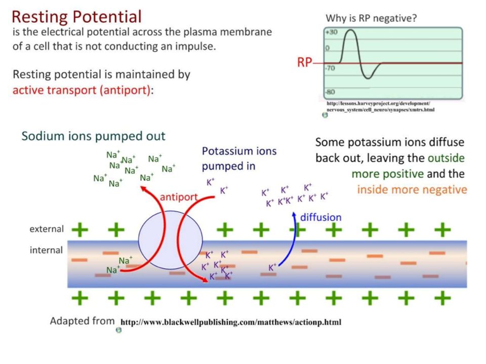
Resting Potential Normally cells has a rest potential of -70 millivolts (mV) This means a neuron cell will have more negative ions on the inside of it means than the outside. This is called its resting potential.

34
Resting Potential where
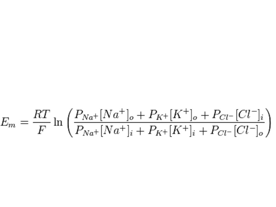
Em is the membrane potential, measured in volts EX is the equilibrium potential for ion X, also in volts PX is the relative permeability of ion X in arbitrary units (e.g. siemens for electrical conductance) Ptot is the total permeability of all permeant ions, in this case PK+ + PNa+ + PCl-
 Ptot is the total permeability of all permeant ions, in this case PK+ + PNa+ + PCl-")
35
An electrical Impulse in neuron cells
An electrical impulse is a momentary reversal in electrical potential difference in the neuron cell membrane Between conduction of one impulse the cell is said to be ‘resting.’ The ‘resting’ neuron membrane maintains the electrical potential difference between the inside and outside of the cell using active transport.

36
Resting Potential is maintained by TWO processes
Active transport of potassium ions (K+) in across the membrane and sodium (Na+) out across the membrane. Facilitated Diffusion of potassium ions (K+) out, and sodium ions (Na+) back in.
 in across the membrane and sodium (Na+) out across the membrane. Facilitated Diffusion of potassium ions (K+) out, and sodium ions (Na+) back in.")
37
LE 48-9 Microelectrode –70 mV Voltage recorder Reference electrode

38
Active Transport across Neuron Cell Membranes
The sodium-potassium pump in the nerve cell use ATP and active transport to pump two ions: Pumps 3 sodium (Na+) ions out of the cell 2 potassium (K+) ions into the cell by means of active transport. As a result, the inside of the cell contains more K+ ions and fewer Na+ ions than the outside.
 ions out of the cell. 2 potassium (K+) ions into the cell by means of active transport. As a result, the inside of the cell contains more K+ ions and fewer Na+ ions than the outside.")
39
Sodium-Potassium Pump
The sodium-potassium pump in the neuron cell membrane uses the energy of ATP to pump sodium out of the cell and, at the same time, to pump potassium in. This ongoing process maintains resting potential.

40
Sodium-Potassium ATPase -- Protein Pump
This uses the energy from ATP splitting to simultaneously pump 3 sodium ions out of the cell and 2 potassium ions in. If this was to continue unchecked there would be no sodium or potassium ions left to pump, but there are also sodium and potassium ion channels in the membrane. These channels are normally closed, but even when closed, they “leak”, allowing sodium ions to leak in and potassium ions to leak out, down their respective concentration gradients. The combination of the Na +K +ATPase pump and the leak channels cause a stable imbalance of Na + and K + ions across the membrane. This imbalance causes a potential difference across all animal cell membranes, called the membrane potential. The membrane potential is always negative inside the cell, and varies in size from –20 to –200 mV in different cells and species.

41
[Na+] 150 M [K+] 150 M Inside Cell Outside Cell [Na+] 15 M [K+] 5 M
LE 48-10 Inside Cell Outside Cell [Na+] 15 M [Na+] 150 M [K+] 150 M [K+] 5 M M = Concentration Plasma membrane
![[Na+] 150 M [K+] 150 M Inside Cell Outside Cell [Na+] 15 M [K+] 5 M](http://slideplayer.com/slide/8427301/26/images/41/%5BNa%2B%5D+150+M+%5BK%2B%5D+150+M+Inside+Cell+Outside+Cell+%5BNa%2B%5D+15+M+%5BK%2B%5D+5+M.jpg "LE Inside Cell. Outside Cell. [Na+] 15 M. [Na+] 150 M. [K+] 150 M. [K+] 5 M. M = Concentration. Plasma. membrane.")
42
Facilitated diffusion across cell membranes
Because K+ ions are in higher concentration inside than outside the cell, they slowly diffuse OUT across the membrane via facilitated diffusion Because Na+ ions are in higher concentration outside the cell they do not diffuse out. This produces a negative charge on the inside and a positive charge on the outside. The electrical charge across the cell membrane of a neuron at rest is known as the resting potential.

43
The Moving Impulse An impulse begins when a neuron is stimulated by another neuron or by the environment.

44
The Nerve Impulse At the leading edge of the impulse, gates in the sodium channels open allowing positively charged Na+ ions to flow inside the cell membrane. An impulse begins when a neuron is stimulated by another neuron. At the leading edge of an action potential, gates in the sodium channels open, allowing Na+ ions to flow into the cell. This flow of ions causes the action potential to move.

45
Sodium Channels Sodium Channels/ Gates –
Globular proteins that are imbedded in the neuron cell membrave They have a central pore that they can open and close Only sodium can diffuse through them This is facilitated diffusion.

46
The Nerve Impulse 2. The inside of the membrane temporarily becomes more positive than the outside, reversing the resting potential. This is called depolarization. An impulse begins when a neuron is stimulated by another neuron. At the leading edge of an action potential, gates in the sodium channels open, allowing Na+ ions to flow into the cell. This flow of ions causes the action potential to move.

47
Depolarization & Action Potential
Action potential causes local & temporary depolarization of the neuron. Depolarization - positive ions (sodium Na+) flow back into the cell via sodium channels. Sodium will passively diffuse down it electrochemical gradient (from high concentration to low concentration) This depolarizes the neuron from -70mV (resting potential) to +40mV (action potential)
 flow back into the cell via sodium channels. Sodium will passively diffuse down it electrochemical gradient (from high concentration to low concentration) This depolarizes the neuron from -70mV (resting potential) to +40mV (action potential)")
48
The Nerve Impulse 3. This reversal of charges f is called a nerve impulse, or an action potential. An impulse begins when a neuron is stimulated by another neuron. At the leading edge of an action potential, gates in the sodium channels open, allowing Na+ ions to flow into the cell. This flow of ions causes the action potential to move.

49
The Nerve Impulse 4. As the action potential passes, gates in the potassium channels open, allowing K+ ions to flow out restoring the negative potential inside the axon. At the trailing edge of an action potential, gates in the potassium channels open, allowing positive ions to flow out and restoring the resting potential of the neuron.

50
Potassium Channels Potassium channels/ gates
Similar to sodium channels/ gates Globular proteins that are imbedded in the membrane of the neuron cell. All only potassium to diffuse in and out of cell via facilitated diffusion.

51
The Nerve Impulse 5. Then the gates in the potassium channels CLOSE, and the resting potential is re-established by sodium/ potassium pumps and facilitated diffusion. This is called repolarize. At the trailing edge of an action potential, gates in the potassium channels open, allowing positive ions to flow out and restoring the resting potential of the neuron.

52
Repolarize Repolarize means to return back to resting potential.
Resting potential -70mV More negative ions on the inside than out of neuron More potassium (K+) inside of cell More sodium (Na+) outside of cell
 inside of cell. More sodium (Na+) outside of cell.")
53
The Nerve Impulse 6. Meanwhile, the impulse continues to move along the axon. An impulse at any point of the membrane causes an impulse at the next point along the membrane. At the trailing edge of an action potential, gates in the potassium channels open, allowing positive ions to flow out and restoring the resting potential of the neuron.

54
Nerve Impulse More Summary
LE 48-14c An action potential is generated as Na+ flows inward across the membrane at one location. Na+ Action potential Axon K+ The depolarization of the action potential spreads to the neighboring region of the membrane, re-initiating the action potential there. To the left of this region, the membrane is repolarizing as K+ flows outward. The depolarization-repolarization process is repeated in the next region of the membrane. In this way, local currents of ions across the plasma membrane cause the action potential to be propagated along the length of the axon. Nerve Impulse More Summary Action potential is generate Na+ flows in cell. Depolarizing the cell. Action potential spreads to neighboring cell and depolarizes this cell. Initial cell undergoes repolarization as K+ flows outward. Depolarization & Repolarization process is repeated in the next region of the membranes.

55
Nerve Impulse Animations/ Tutorials

58
IB LEARNING OBJECTIVE Explain the principles of synaptic transmission.
Include the release, diffusion and binding of the neurotransmitter, initiation of an action potential in the post-synaptic membrane, and subsequent removal of the neurotransmitter.

59
LE 48-15 Depolarized region (node of Ranvier) Cell body Myelin sheath
Axon

60
The Synapse The Synapse
At the end of the neuron cell , the impulse reaches the motor end plates. At this site, one neuron cell makes contact with another neuron cell. The neuron passes the impulse along to the second cell. The location at which a neuron can transfer an impulse to another neuron cell is called a synapse.

61
LE 48-5 Dendrites Cell body Nucleus Synapse Signal direction
Axon hillock Axon Presynaptic cell Motor end plates Myelin sheath Postsynaptic cell

63
The Synapse Pre-synaptic neuron
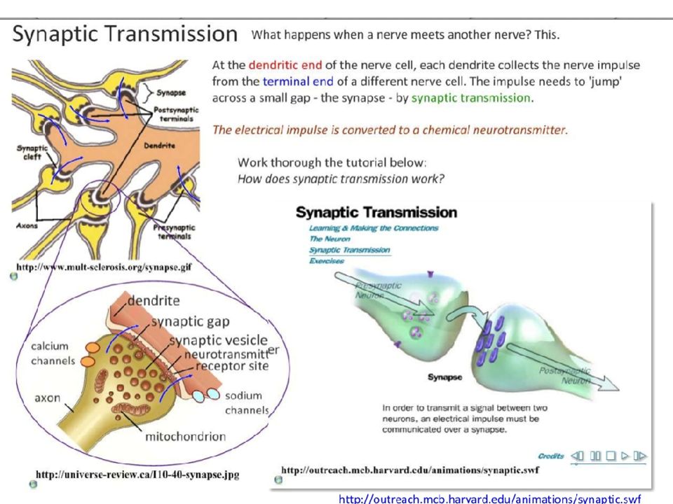
A Synapse is a link between two neuron cells. A cell carrying and transmitting an impulse is called the pre-synaptic neuron When an impulse reaches the end of the axon of one neuron, neurotransmitters are released into the synaptic cleft. The neurotransmitters bind to receptors on the membrane of an adjacent dendrite.

64
The Synapse Pre-synaptic neuron
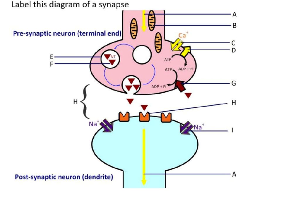
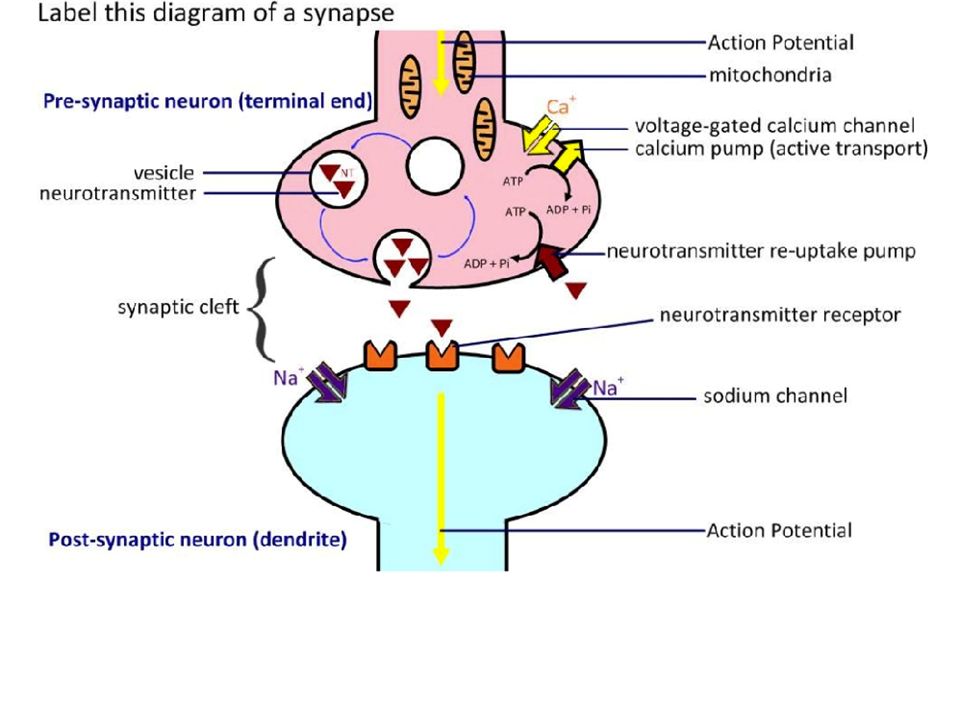
The synaptic cleft is a gap that separates the motor end plates from the dendrites of the adjacent cell. Pre-synaptic neuron opens up calcium channels and calcium moves into the cell Synaptic cleft

65
The Synapse Pre-synaptic neuron
Calcium causes vesicles of neurotransmitters to fuse with pre-synaptic membrane. Vesicle Synaptic cleft

66
The Synapse Neurotransmitters are chemicals used by a neuron to transmit an impulse across a synapse to another cell. Vesicle Neurotransmitter

67
The Synapse As an impulse reaches the motor end plates, vesicles send neurotransmitters into the synaptic cleft. These diffuse across the cleft The cell membrane receiving the impulse is called the post-synaptic membrane. Receptor

68
The Synapse Sodium ions then rush across the membrane, stimulating a nerve impulse (action potential/ depolarization) in the next cell, the postsynaptic cell. Once an action potential has been generated in the post-synaptic cell membrane an enzyme inactivates the neurotransmitter. The inactivated neurotransmitter is packaged for re-use later by the Golgi apparatus.
 in the next cell, the postsynaptic cell. Once an action potential has been generated in the post-synaptic cell membrane an enzyme inactivates the neurotransmitter. The inactivated neurotransmitter is packaged for re-use later by the Golgi apparatus.")
69
Inactivation of Neurotransmitter
The neurotransmitter must be rapidly broken down to prevent continuous synaptic transmission of impulse. Calcium ions will started to be pumped out of the pre-synaptic neuron into the synaptic cleft.

70
LE 48-17 Postsynaptic cell Presynaptic cell Na+ Neuro- K+ transmitter
Synaptic vesicles containing neurotransmitter K+ Presynaptic membrane Postsynaptic membrane Sodium ion channel Voltage-gated Ca2+ channel Postsynaptic membrane Ca2+ Neuro- transmitter Na+ Synaptic cleft Sodium ion channels

71
LE 48-5 Dendrites Cell body Nucleus Synapse Signal direction
Axon hillock Axon Presynaptic cell Synaptic terminals Myelin sheath Postsynaptic cell

74
Synaptice Transmission Videos / Tutorials

75
Postsynaptic neuron Synaptic terminals of pre- synaptic neurons 5 µm
LE 48-16 Postsynaptic neuron Synaptic terminals of pre- synaptic neurons 5 µm

76
LE 48-7 50 µm

Similar presentations


The nervous system>")


 Nervous system functions Structure of a neuron Sensory, motor, inter- neurons Membrane potential Sodium.>")


 use of active transport>")


