Download presentation
Presentation is loading. Please wait.

1
Jalal Jalal Shokouhi M.D. – Radiologist – Jam-e-jam - Ali Akbar Ameri M.D – Radiologist – Jam-e- jam - Mohsen Shamsolahrar – M.S- Radiology - Ali Saeidi – B.S. Radiology - Rasoul Sa'adat – B.S. Radiology

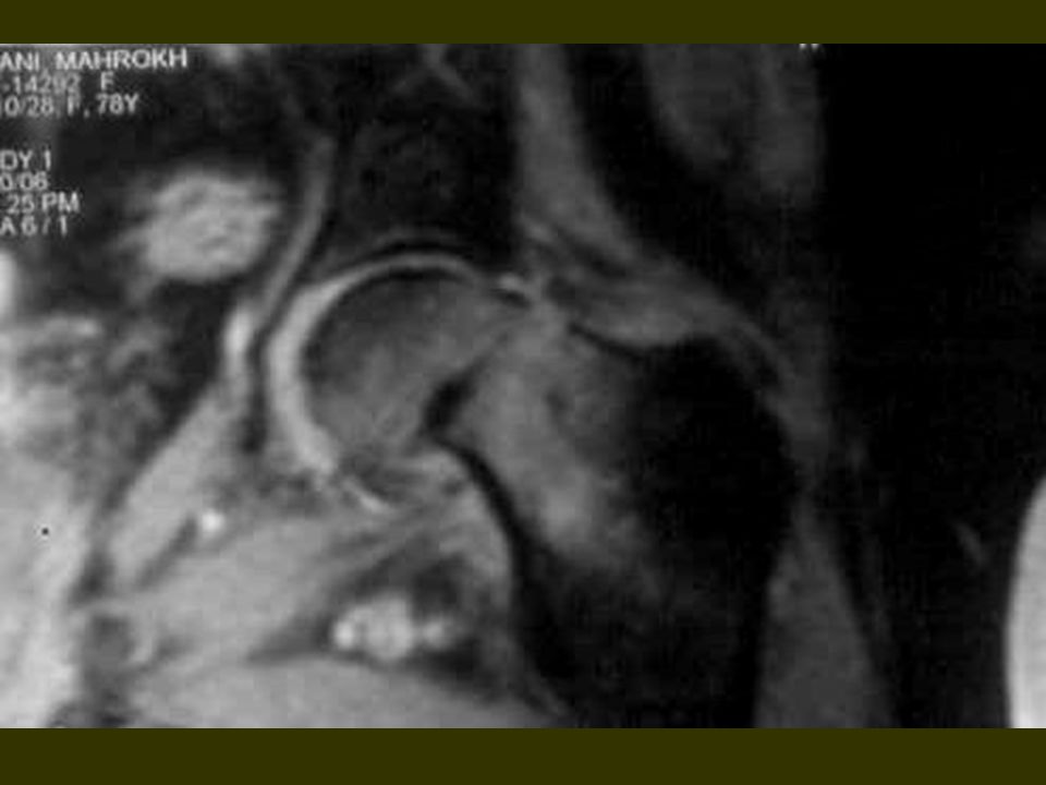
2
Demonestration of occult fractures by MRI

3
Abstract Bakground / objectives : We assessed the utility of MRI in diagosis of occult bone fractures and compared with x – rays. T1 and fat saturation images was the best for this diagnosis " few patients, x – ray CT SCANS, are out of study ".

4
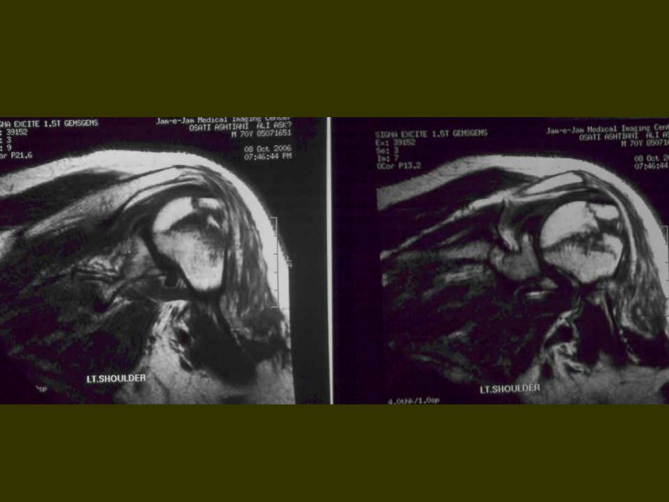
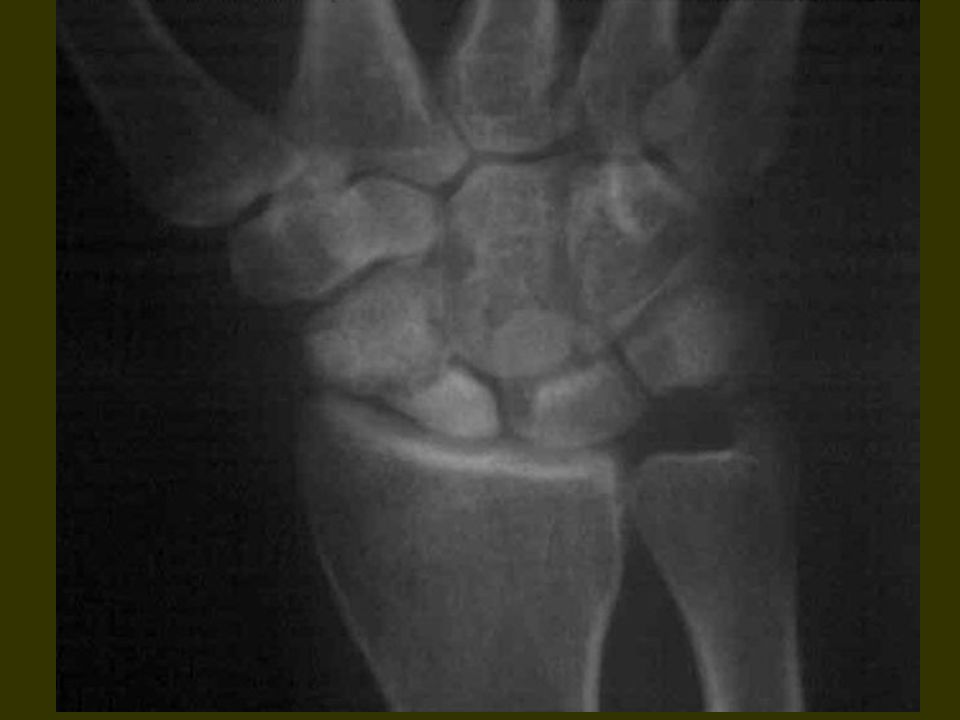
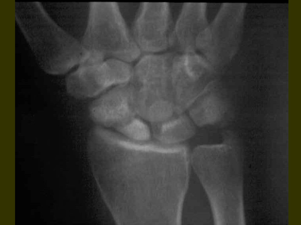
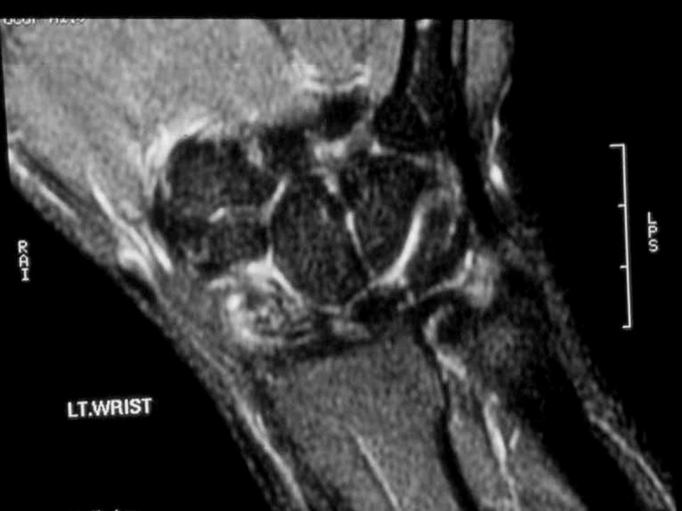
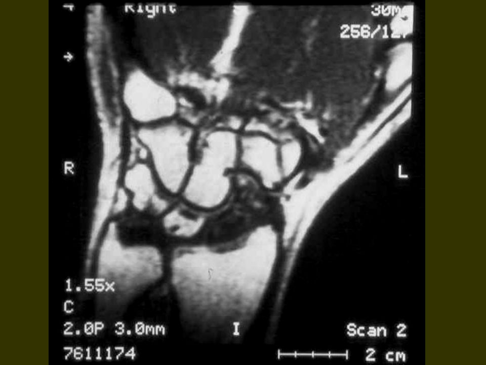
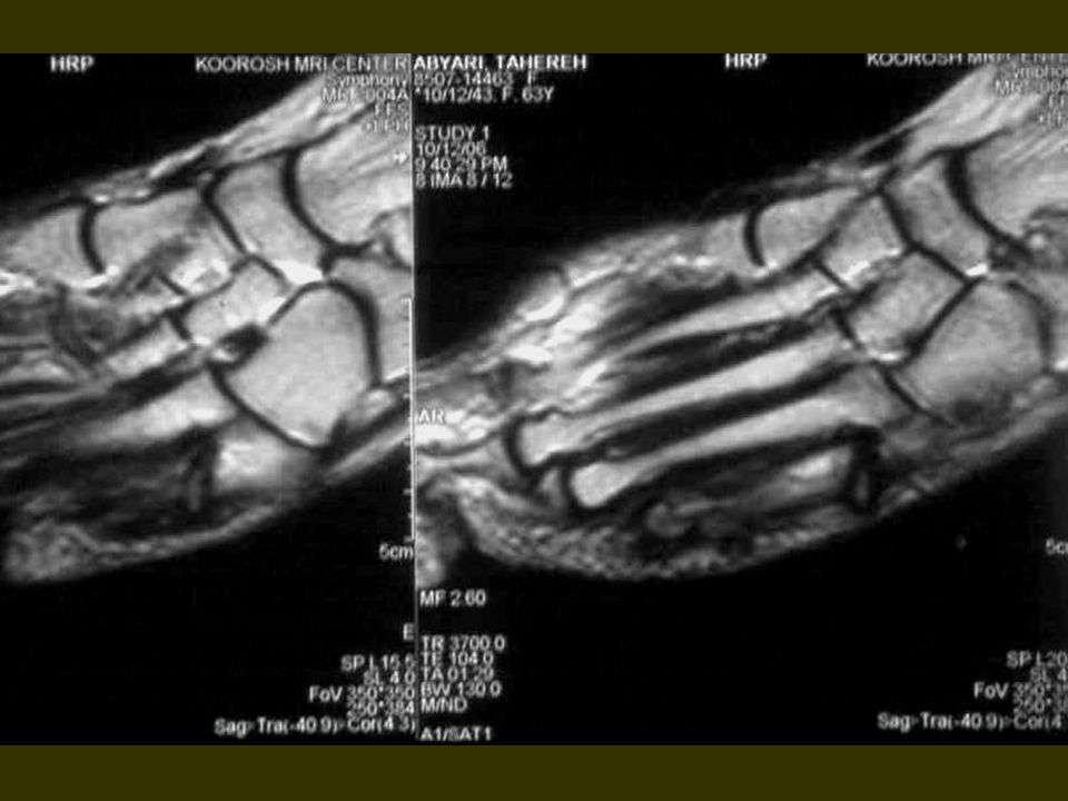
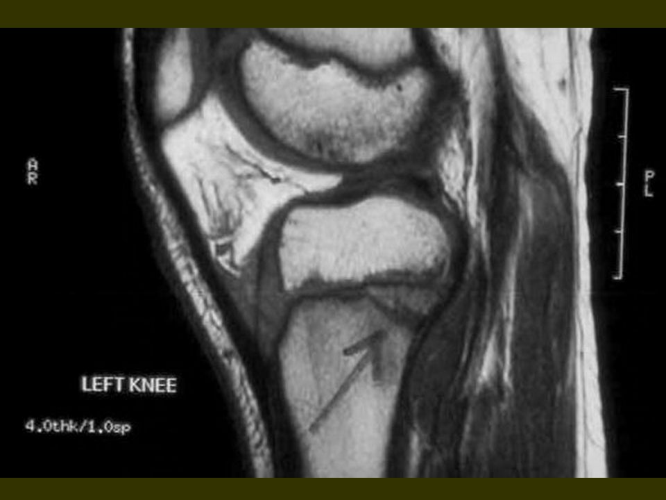
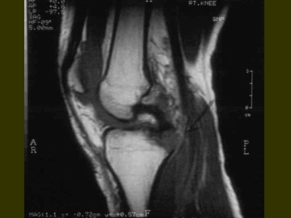
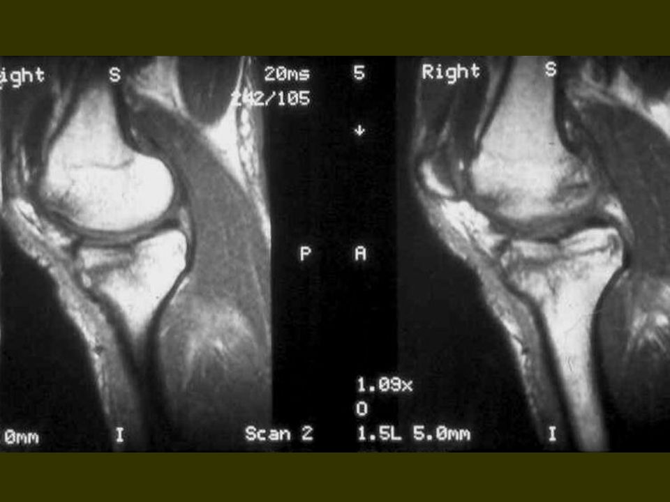
subjects consisted 75 patients with MRI and x – ray and x – ray CT " few of them fixed in cast or splint " Result : MRI was so helpful in determining bone marrow signal and minimum edema in the bright T1 signal. MRI can't see compact bone or cortex but because of muscle and bone marrow interfaces FX line can be demonstrated by MRI as a bright line. "compact bone as a dark line "

5
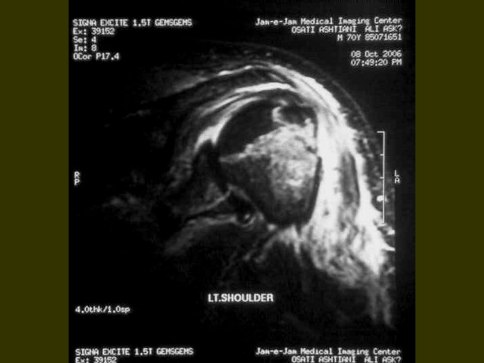
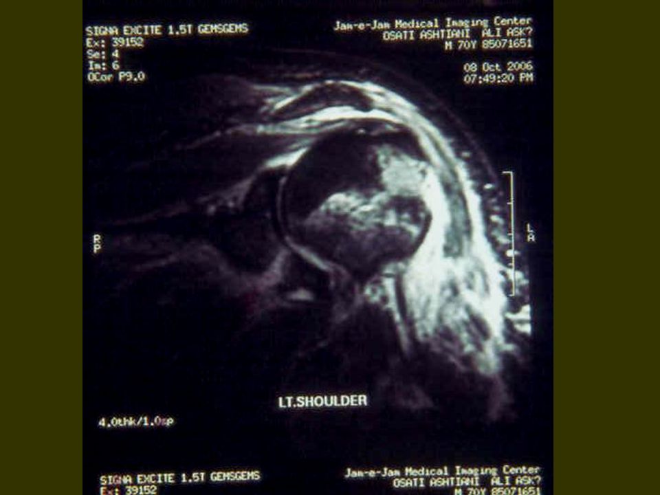
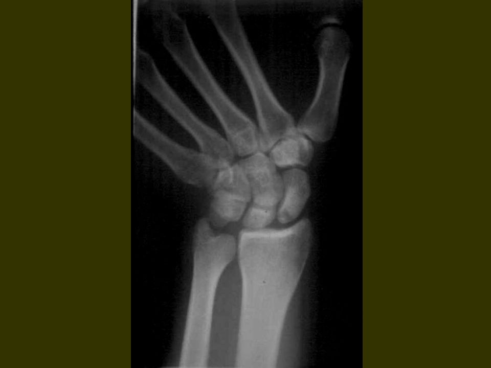
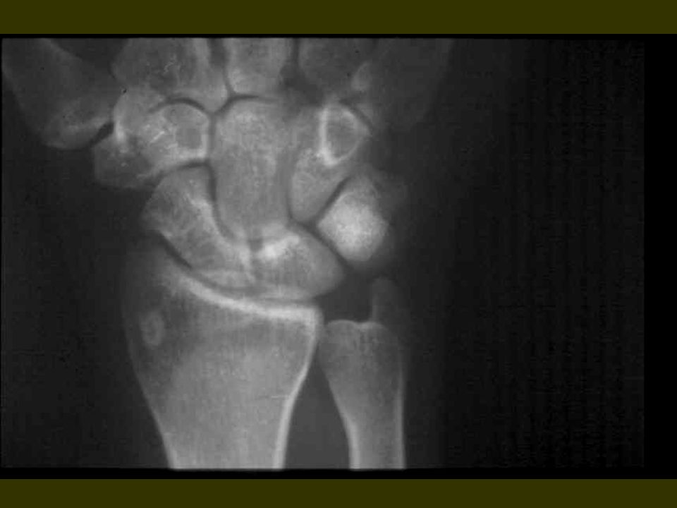
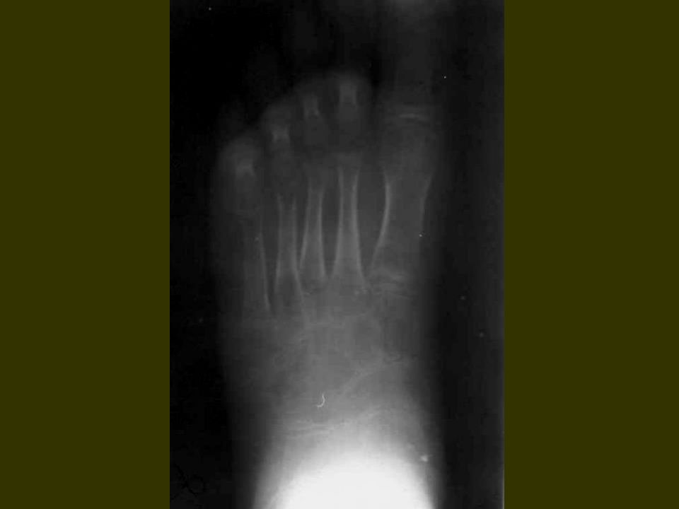
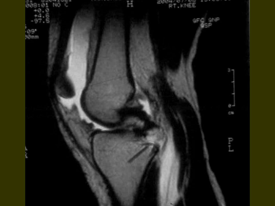
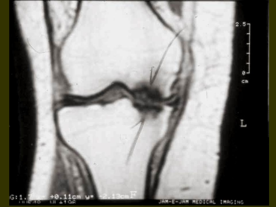
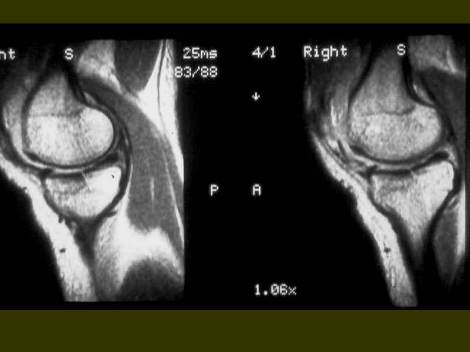
Conclusion : Because of bone fractures without replasment and diastasis they can't detect by x – ray but MRI is helpful and specifically diagnostic to show delicate fracture line and other complications like adjacent bone marrow edema and fat – fluid level in the adjacent joint spaces. " hemorrhage " MRI also can show other lesions beside fracture, " Tendon, menisc " and …. Lesions ".

6
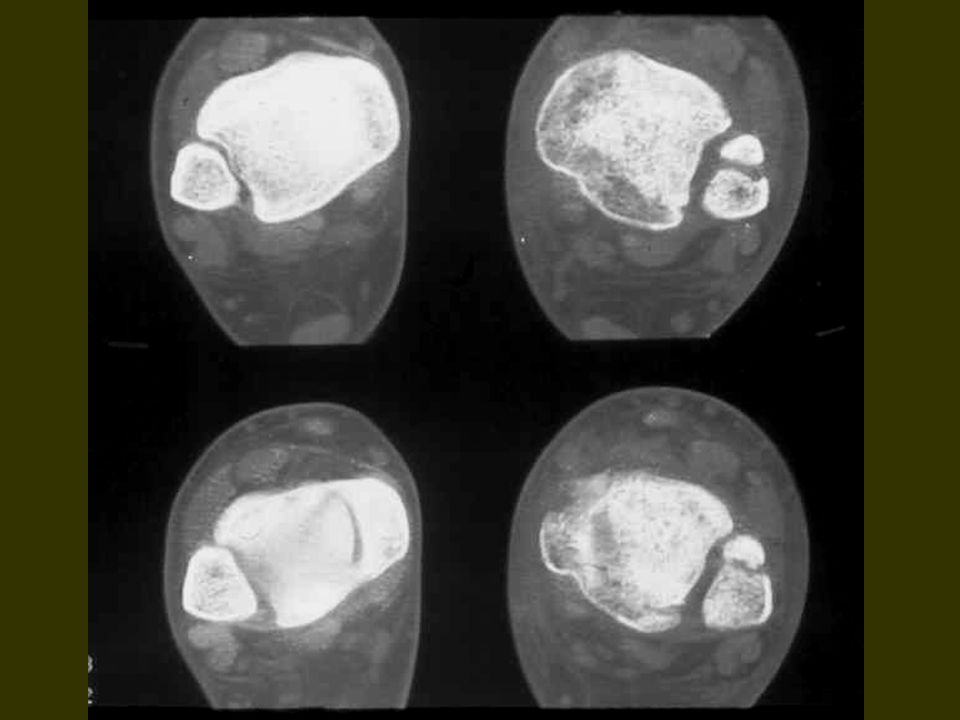
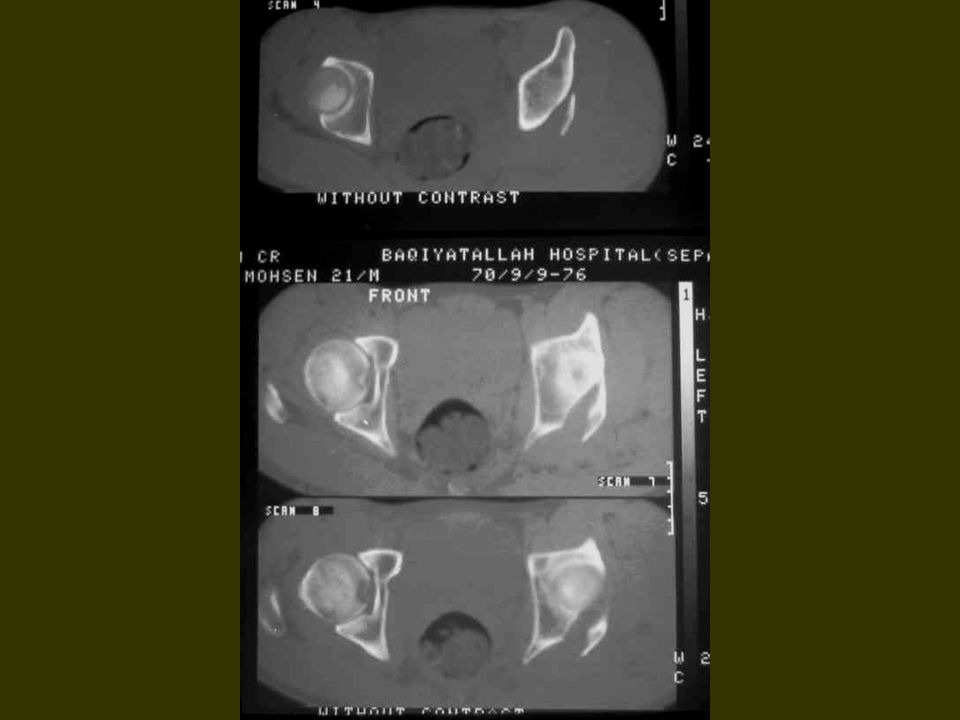
MRI is reliable and method of choice for assessing fracture line in the bone and complicated condition in the adjacent joint. Other modalities like x – ray, spiral CT – SCAN can show chip bones in case of replacement and could be also negative for FX. Line but can show fat – fluid in the joint spaces " an indirect sign of bone marrow opening in to the joint space " hidden fracture ". MRI also show soft tissue contusion, dirty fat, hematoma, capsule and tendon damages.

7
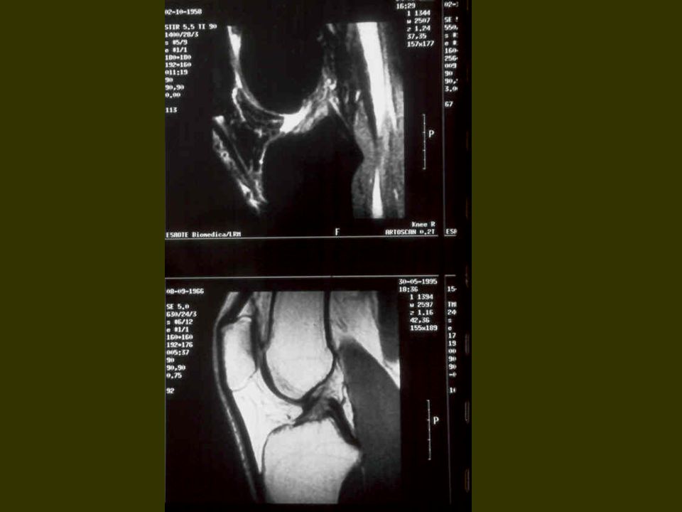
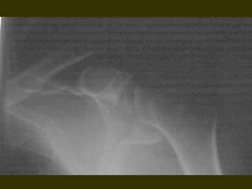
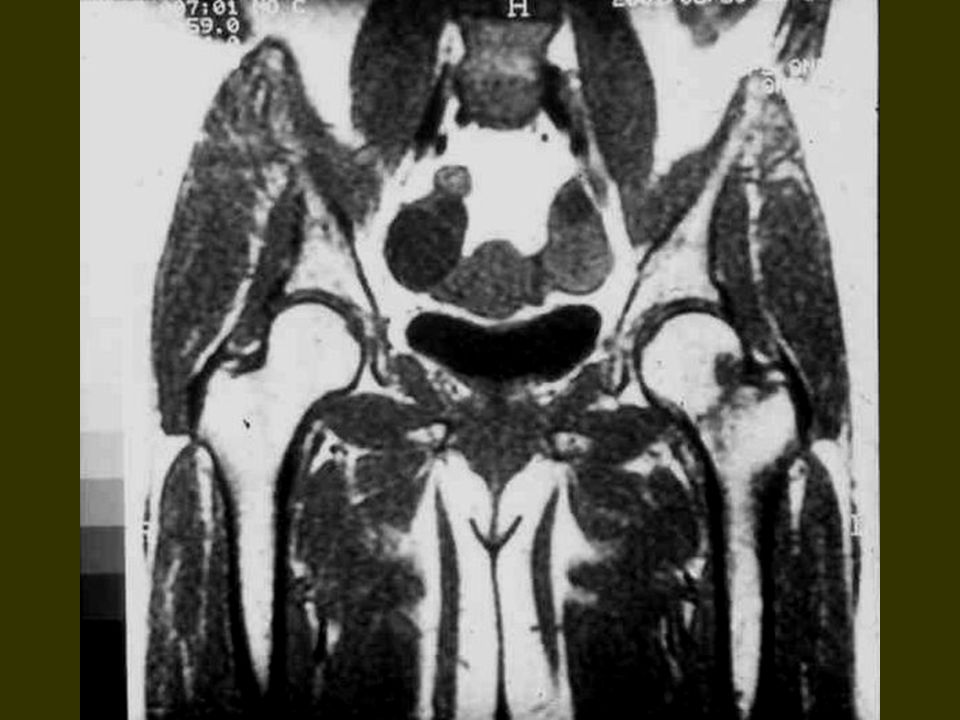
75 traumatic patients are addmited, 52 patient was male and 23 patient was female. From all patients, 57 traumatized knee joint bones, 10 shoulder joints, 4 hip joints, 3 long bones are imaged.

8
Different parts was 5 cases. Positive X – ray for FX observed in 11 cases. CT demonstrated lesion was 3 cases " and CT SCAN is not a modality in our study " There was no negative MRI but negative x – ray observed in 64 cases. Slice thickness was 5 to 8 mm. Comparison was drawn between MRI images and x – ray and few cases of x – ray spiral CT " main porpoise was comparing plain x – ray with MRI ".

9
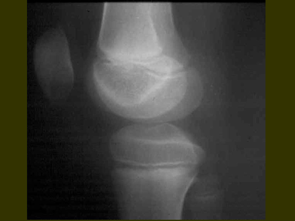
Assessted anatomic areas was shoulder, knee, hip and long bones. 57 cases was assessted at knee joint area specialy in proximal tibia " tibial plateau. Main cause was trauma and second was osteoporosis. Our study mainly occurred in young age group because of random admission.

10
Bone marrow is brigh and white in T1 images, by standard T2 images it could be white, bright gray and by fat saturation, it should be dark and black. Mild or small amount of edema in bone marrow looks like a small spot on white doctors wearing and named dirty fat.

11
MRI has 100 % specificity and sensitivity for occult fractures of bone by T1 and fat sat.images.

12
Because of high trauma incidence in this century, specially in our country and equipped centers and hospitals with trauma and MRI center and high sensitivity of MRI to detect small changes in bone marrow signal and water content, traumatized bone with normal x – ray and unusual clinical complains could be check by MRI for occult or hidden fracture line.

13
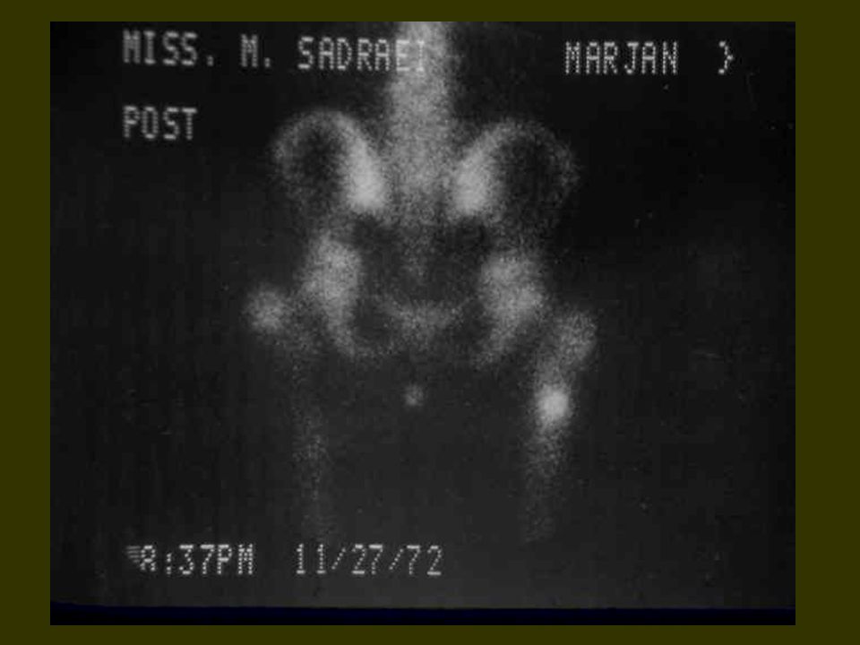
Use of spiral CT with reformatted images in multiple plans also useful for bone chip detection. RN SCAN is not used in emergency periods but could be helpful in subacute and chronic phases. In all cases MRI should be positive.

Similar presentations