Download presentation
Presentation is loading. Please wait.

1
Muscle Types There are 3 types of muscles Skeletal muscle – skeletal movement Cardiac muscle – heart movement Smooth muscle – peristalsis (pushes substances through hollow tubes)
")
2
Skeletal Muscles (focus) Major Functions: 1. movement 2. maintain posture 3. stabilize joints 4.generate heat 5.facial expressions

3
Characteristics: 1. excitability (irritability): ability to respond to a stimulus (usually a neurotransmitter) 2. contractility: the ability to shorten when adequately stimulated 3. extensibility: the ability to be stretched; muscles can be stretched beyond their normal resting length when relaxed 4. elasticity: the ability of a muscle fiber to resume its resting length after being stretched
: ability to respond to a stimulus (usually a neurotransmitter) 2. contractility: the ability to shorten when adequately stimulated 3. extensibility: the ability to be stretched; muscles can be stretched beyond their normal resting length when relaxed 4. elasticity: the ability of a muscle fiber to resume its resting length after being stretched.")
4
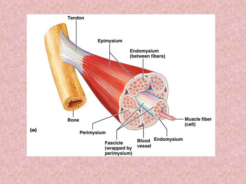
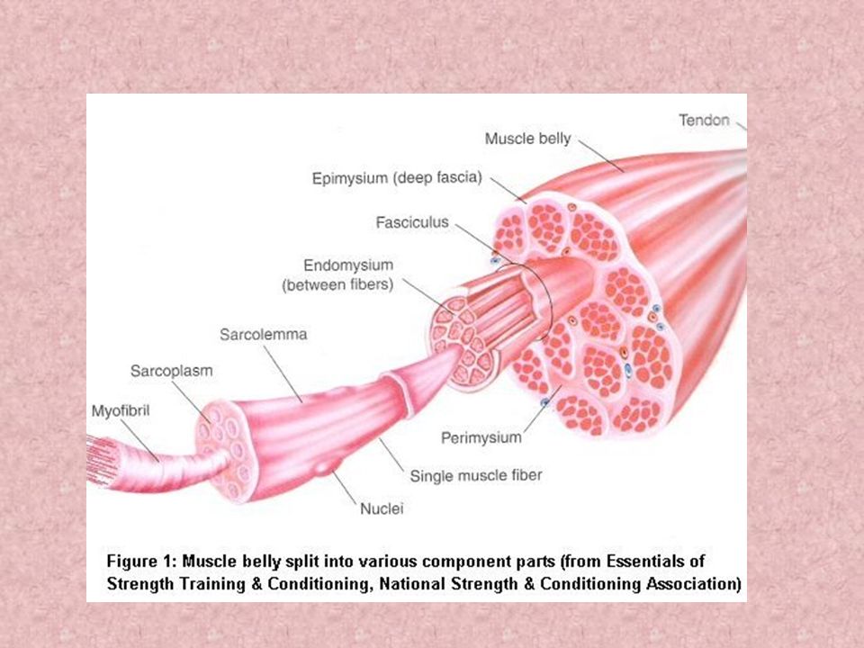
Connective Tissue Wrappings (external to deep) 1. epimysium: layer of connective tissue that surrounds the entire muscle; blends into tendons 2.perimysium: connective tissue extending inward from epimysium and separates muscle tissue into compartments called fascicles 3.fascicles: groups of muscle fibers (cells) 4. endomysium: a sheet of connective tissue that surrounds each muscle fiber (myofiber)
 4. endomysium: a sheet of connective tissue that surrounds each muscle fiber (myofiber).")
7
Nerve and Blood Supply 1. Each skeletal muscle fiber is innervated 2. Muscle tissue is vascular

8
Parts of Muscle Fibers - Muscle Cell Organelles a. sarcolemma – plasma membrane of muscle cell; surrounds myofibers b. sarcoplasm – similar to cytoplasm; contains large amounts of glycogen for energy

9
c. sarcoplasmic reticulum – similar to endoplasmic reticulum of cells; stores and releases Ca 2+ on demand when muscles are ready to contract d.transverse tubules (t-tubules) – channels that carry nerve impulses (action potentials) deep into the muscle cell e.mitochondria - muscle cells have many for energy
 – channels that carry nerve impulses (action potentials) deep into the muscle cell e.mitochondria - muscle cells have many for energy.")
10
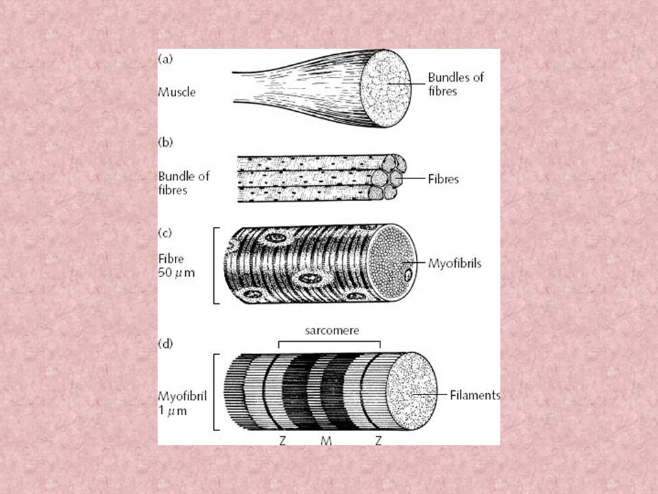
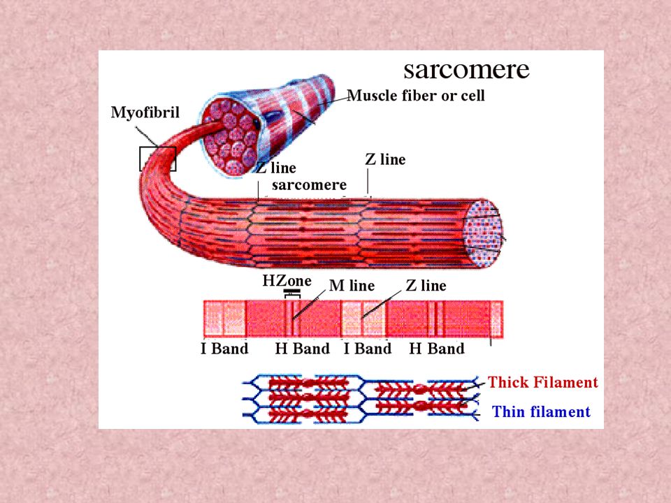
Myofibrils: rod-like fibers that run parallel to the muscle cell Hundreds to thousands in a muscle fiber composed of striations: repeating series of dark and light bands What causes those striations? The arrangement of myofilaments!

11
Structure of Myofilaments The thick filaments are primarily made of myosin. The thin filaments primarily contain actin. Thin filaments also contain tropomyosin & troponin.

12
View of myofilaments

13
Striation Pattern: A band: dark appearance, made of primarily of thick filaments (myosin) & some thin filaments (actin) I band: light appearance, made of thin filaments Z line (disc): connects each myofibril to the next throughout the width of the muscle fiber; Z line to Z line is one sarcomere – the smallest functional unit of a muscle contraction H Zone: holds thick filaments together, only visible when the muscle fiber is relaxed
 & some thin filaments (actin) I band: light appearance, made of thin filaments Z line (disc): connects each myofibril to the next throughout the width of the muscle fiber; Z line to Z line is one sarcomere – the smallest functional unit of a muscle contraction H Zone: holds thick filaments together, only visible when the muscle fiber is relaxed")
14
Muscle (organ) Fascicles Muscle cell Myofibril Myofilaments (Arranged From Largest to Smallest)
 Fascicles Muscle cell Myofibril Myofilaments (Arranged From Largest to Smallest)")
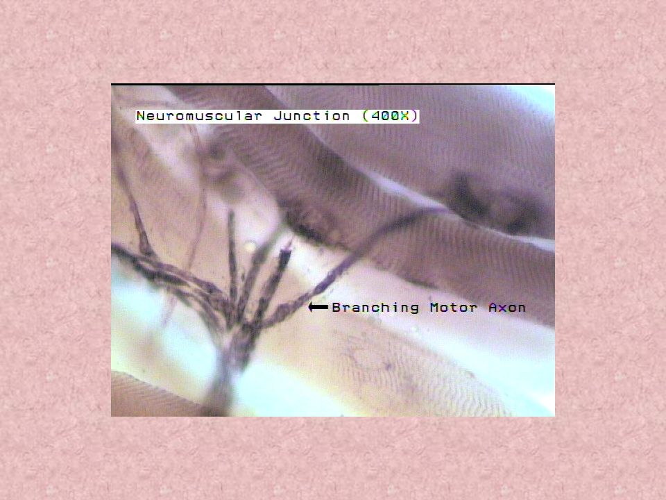
19
Contraction of Skeletal Muscle Cells One motor neuron (nerve cell) may stimulate a few muscle cells or hundreds. A motor neuron + all the myofibers = a motor unit * When an axon of a nerve cell reaches a muscle cell it branches into a number of axon terminals. Each axon terminal forms junctions with sarcolemma of different muscle cells – (neuromuscular junction - NMJ). * Although they are very close the axon and sarolemma do not touch directly this is a synaptic cleft.
. * Although they are very close the axon and sarolemma do not touch directly this is a synaptic cleft..")
22
A Blast from the past...Active Transport! Sodium / potassium pump, maintains cell resting potential at -70mV (charge inside a cell); a change is this charge 30mV will cause an action potential (nerve impulse) 2 K+ in for every 3 Na + out
; a change is this charge 30mV will cause an action potential (nerve impulse) 2 K+ in for every 3 Na + out.")
23
Actions Occurring At NMJ – Web animations

24
An action potential travels along an axon to an axon terminal. This stimulates the release of the neurotransmitter – acetylcholine or ACh from synaptic vesicles. ACh diffuses across the synaptic cleft and attaches to ACh receptors on the sarcolemma of a muscle fiber – this region is known as the motor end plate. The ACh receptors open channels on the motor end plate which allows Na+ to diffuse into the muscle cell. This change in charge in the muscle cell allows for the action potential to continue and move deep into the muscle cell via the transverse tubules.

25
An action potential passes into the muscle cells via t-tubules (transverse tubules) This stimulates the release of Ca 2+ from the sarcoplasmic reticulum Ca 2+ binds to the troponin / tropomyosin complex and allows myosin heads to bond to actin and push thin filaments toward the center of the sarcomere This process requires ATP
 This stimulates the release of Ca 2+ from the sarcoplasmic reticulum Ca 2+ binds to the troponin / tropomyosin complex and allows myosin heads to bond to actin and push thin filaments toward the center of the sarcomere This process requires ATP")
27
Contraction of a Skeletal Muscle Muscle fiber contraction is “all or none” Not all fibers may be stimulated during the same interval Different combinations of muscle fiber contractions differing responses

28
Graded responses – different degrees of skeletal muscle shortening

29
Types of Graded Response: Twitch –Single, brief contraction –Not a normal muscle function

30
Types of Graded Response: Unfused (incomplete) tetanus –Some relaxation occurs between contractions –The results are summed
 tetanus –Some relaxation occurs between contractions –The results are summed")
31
Types of Graded Response: Fused (complete) tetanus –No evidence of relaxation before the following contractions –The result is a sustained muscle contraction
 tetanus –No evidence of relaxation before the following contractions –The result is a sustained muscle contraction")
32
Muscle Response to Strong Stimuli Muscle force depends upon the number of fibers stimulated More fibers contracting results in greater muscle tension Muscles can continue to contract unless they run out of energy

33
Types of Muscle Contractions Isotonic contractions –Myofilaments are able to slide past each other during contractions –The muscle shortens Isometric contractions –Tension in the muscles increases –The muscle is unable to shorten

34
Effects of Exercise on Muscle Aerobic or Endurance: result in stronger muscles due to increase blood supply Muscle fibers increase mitochondria and oxygen storage Muscle becomes more fatigue resistant Heart enlarges to pump more blood to body Does not increase skeletal muscle size

35
Resistance or Isometric Exercises Results of increased muscle use from resistance training Individual muscle cells make more contractile filaments & connective tissue increases –Increase in muscle size –Increase in muscle strength

36
How do all of our muscles get the energy they need for contractions?

37
Energy for Muscle Contractions Initially, muscles used stored ATP for energy –Bonds of ATP are broken to release energy –Only 4-6 seconds worth of ATP is stored by muscles Then, other pathways must be utilized to produce ATP

38
1. Direct Phophorylation –Muscle cells contain creatine phosphate (CP) –After ATP is depleted, ADP is left –CP transfers energy to ADP, to regenerate ATP –CP supplies are exhausted in about 20 seconds
 –After ATP is depleted, ADP is left –CP transfers energy to ADP, to regenerate ATP –CP supplies are exhausted in about 20 seconds.")
39
2. Aerobic (Cellular) Respiration – Occurs in the mitochondria –Glucose is broken down to carbon dioxide and water, releasing energy –Slower reaction that requires continuous oxygen
 Respiration – Occurs in the mitochondria –Glucose is broken down to carbon dioxide and water, releasing energy –Slower reaction that requires continuous oxygen.")
40
3. Anaerobic Glycolysis –Reaction that breaks down glucose without oxygen –Glucose is broken down to pyruvic acid to produce some ATP –Pyruvic acid is converted to lactic acid – Not as efficient, but is fast Huge amounts of glucose are needed Lactic acid produces muscle fatigue

41
Muscle Fatigue When a muscle is fatigued, it is unable to contract The common reason for muscle fatigue is oxygen debt –Oxygen must be “repaid” to tissue –Oxygen is required to get rid of accumulated lactic acid Increasing acidity (from lactic acid) & lack of ATP causes the muscle to contract less
 & lack of ATP causes the muscle to contract less")
Similar presentations