Download presentation
Presentation is loading. Please wait.

1
Lab Diagnosis of Viruses Dr Syed Suhail Ahmed College of Medicine Qassim University

2
Laboratory Diagnosis of Viral Infections : Clinical specimen: 1.Throat or nasopharyngeal swab or aspirate 2.Bronchial and bronchoalveolar washes 3.Rectal swabs and stool specimens 4.Urine 5.Sterile body fluids and Blood 6.Bone marrow 7.Tissue 8.Serum for antibody testing

3
5 approaches for the diagnosis of viral infections A.Microscopic examination : viruses can be detected and identified by direct microscopic examination. 3 different procedures can be used 1.Light microscopy can reveal characteristic inclusion bodies or multinucleated giant cells.Cytological examination for inclusions Herpes infection eg tzanck smear, which shows herpes – induced multinucleated giant cells in vesicular skin lesions. Rabies virus inclusions in brain tissue are called Negri bodies. Cytology are less sensitive than culture but are especially helpful for viruses that are difficult or dangerous to isolate in the lab such as rabies virus. 2. UV microscopy is used for fluorescent antibody staining of the virus in infected cells. Direct and indirect fluorescent antibody staining

4
Inclusion bodies Electron Microscopy Fluorescent technique

5
3.Electron Microscopy (EM) : EM detects virus particles, which can be characterized by their size and morphology : EM is most useful for detection of viruses that do not grow readily in cell culture and works best if the titer of virus is at least 10 6 to 10 7 particles per ml. Immune EM allows visualization of virus particles in numbers too small for easy direct detection. The addition of specific antiserum causes the virus particles to form antibody-bound aggregates, which are more easily detected than are single virus particle. Eg detection of viruses causing gastroenteritis like rotavirus.

6
B. Detection of viral antigen: viral antigens can be detected in patients blood or body fluids by various tests but most often by an ELISA. Tests for Rota virus & HBs Ag. RIA and latex agglutination test are additional techniques to detect viral antigen.

7
C. Isolation of virus A. Inoculation into animals B. Egg inoculation c. Cell culture. A. Inoculation into animals : monkeys, mice etc were initially used for viruses and the growth is indicated by death, disease or visible lesions

8
2. Egg inoculation : egg offers several sites for the cultivation of viruses CAM – pox viruses yolk sac- Amniotic cavity – influenza.

9
3.Cell culture : this type of culture is routinely employed for growing viruses. As viruses replicate only in living cells, the growth of viruses require cell culture. Virus growth in cell culture can be detected by the following methods

10
1.Cytopathic effect : many viruses cause morphological changes in cultured cells in which they grow. These changes can be readily observed by microscopic examination of the cultures. Eg measles virus produce syncytium formation. 2. Metabolic inhibition : In normal cell culture the medium turns acid due to cellular metabolism. When viruses grow in cell culture, cell metabolism is inhibited and there is no acid production. this can be checked by the color of the indicator phenol red. 3.Haemadsorption : when hemagglutinating viruses such as influenza & parainfluenza grow in cell

11
Cytopathic effect of viruses

12
culture,there presence can be indicated by the addition of guinea pig erythrocytes to the culture. If the viruses are multiplying in the culture, the erythrocytes will adsorb onto the surface of cells. This is known as hemadsorption. 4. Interference : the growth of non- cytopathogenic viruses in cell culture can be tested by the subsequent challenge with a known cytopathogenic virus. The growth of the first will inhibit infection by the second virus by interference. Fluorescent – antibody assay Shell vial culture :rapid modification of conventional cell culture. Virus is detected more quickly.

13
D.Serological procedures : A increase in titer of antibody to the virus can be used to diagnose current infection. a serum sample is obtained as soon as viral etiology is suspected (acute phase), and a second sample is obtained 10- 14 days later(convalescent phase). If the antibody titer in the convalescent phase serum is at least 4 fold higher than the titer in the acute phase serum sample, the patient is considered to be infected. In certain viral infections, the presence of IgM antibody is used to diagnose current infection
, and a second sample is obtained days later(convalescent phase). If the antibody titer in the convalescent phase serum is at least 4 fold higher than the titer in the acute phase serum sample, the patient is considered to be infected. In certain viral infections, the presence of IgM antibody is used to diagnose current infection.")
14
Many serological methods are used to detect anti- viral antibody like A.Complement fixation test B. ELISA : Enzyme Linked Immuno Sorbent Assay C. Indirect Immunofluorescence D. Western immunoblotting

15
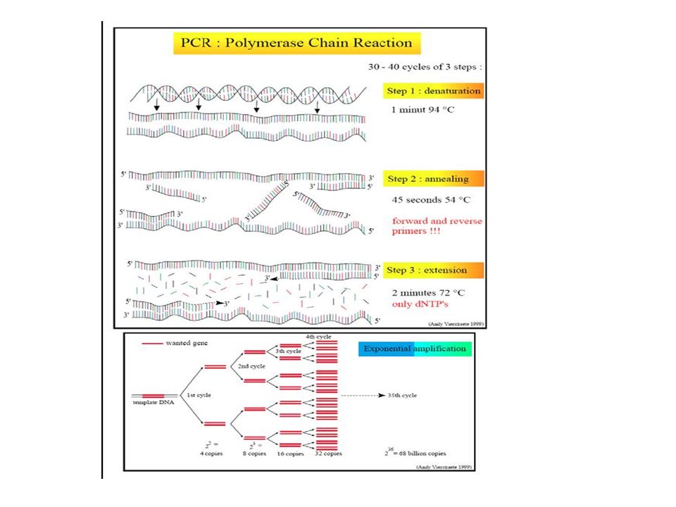
E.Molecular technique : They detect viral genome. 1.Nucleic acid probes : they are short segments of DNA that are designed to hybridize with complementary viral DNA or RNA segments. The probe is labelled with fluorescent or chromogenic tag that allows detection if hybridization occurs.nucleic acid probes are more useful in situations were amount of virus is abundant. 2.PCR (polymerase chain reaction): is a method that duplicates short DNA segments thousand to million fold and detect them.
: is a method that duplicates short DNA segments thousand to million fold and detect them..")
17
References: 1.Diagnostic Microbiology by Bailey & Scotts. 2.Medical Microbiology by Levinson

Similar presentations


 Or to precipitate.>")


 that polioviruses could be cultured tissue, cell culture has become a very.>")


