Download presentation
Presentation is loading. Please wait.

2
intro-VIRUSES Virus NamePDB ID HUMAN PAPILLOMAVIRUS 161DZL BACTERIOPHAGE GA1GAV L-A virus1M1C SATELLITE PANICUM MOSAIC VIRUS1STM SATELLITE TOBACCO NECROSIS2BUK CANINE PARVOVIRUS2CAS Human hepatitis B virus2G34 RYEGRASS MOTTLE VIRUS2IZW BACTERIOPHAGE MS22MS2 CARNATION MOTTLE VIRUS1OPO BACTERIOPHAGE PP71DWN

3
LIST OF RESIDUE NAMES/NUMBER #/Code Name Hydropathy 1 R+ ARG Arginine-4.5 2 K+ L YS Lysine -3.9 3 D- ASP Aspartic Acid -3.5 4 E- GLU Glutamic Acid -3.5 5 N ASN Asparagine -3.5 6 Q GLN Glutamine -3.5 7 H+ HIS Histidine -3.2 8 P PRO Proline -1.6 9 Y TYR Tyrosine -1.3 10 W TRP Trytophan -0.9 11 S SER Serine -0.8 12 T THR Threonine -0.7 13 G GLY Glycine -0.4 14 A ALA Alanine 1.8 15 M MET Methionine 1.9 16 C CYS Cysteine* (CYX*) 2.5 17 F PHE Phenylalanine 2.8 18 L LEU Leucine 3.8 19 V VAL Valine 4.2 20 I ILE Isoleucine 4.5
 F PHE Phenylalanine L LEU Leucine V VAL Valine I ILE Isoleucine 4.5")
4
intro, R2R STATS 11 viruses are used. Residue 2 residue statistics are collected if any non-hydrogen atoms within each residue are within 3.5 Å of each other. Statistics are ONLY collected for interactions of residues within the same protein of the viral capsid. All viruses used have icosahedral symmetry. A protein was used to collect statistics if it was a protein within the “identity” subunit.

5
Distances, Cα If two residues are neighbors then the distance between their Cα atoms was calculated. These distances were tabulated by residue- residue type and their average and standard deviation were found.

6
Distances, Cα: AVERAGE nmin = 23 nmax = 178

7
Distances, Cα: AVERAGE

8
Distances, Cα: NSET tot # of data points =14827

9
Distance, Cα: STANDARD DEV

10
intro, SASA 11 viruses are used. The SASA is calculated for every atom using the LCPO method (J Comp Chem, 22, 2, 217-230, 1999). The LCPO method occasionally gives a negative SASA, if this occurred the SASA for that atom was set to zero. Each residue is then assigned a SASA value based on the sum of the SASA’s of the atoms in that residue. These values are then tabulated into averages, and standard deviations for each residue type.
. The LCPO method occasionally gives a negative SASA, if this occurred the SASA for that atom was set to zero. Each residue is then assigned a SASA value based on the sum of the SASA’s of the atoms in that residue. These values are then tabulated into averages, and standard deviations for each residue type..")
11
intro, SASA Side Note: When calculating the neighbor map for the LCPO algorithm on atom i only atoms that were on the same molecule of atom i were used as neighbors of atoms i. This has the effect of finding not whether an atom is buried inside a viral capsid but rather it is buried within its own protein.

12
SASA: Average

13
SASA: # of amino acids

14
SASA: Standard Deviation

15
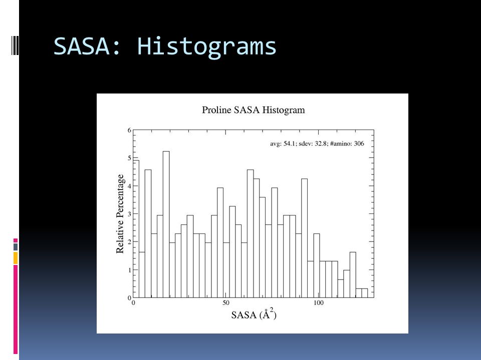
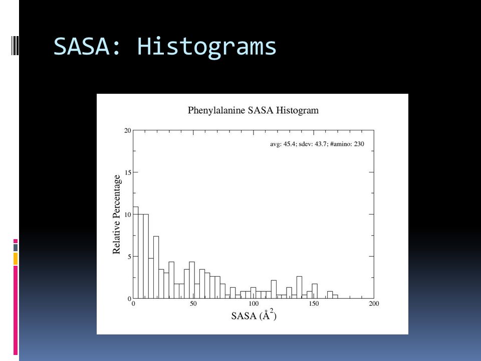
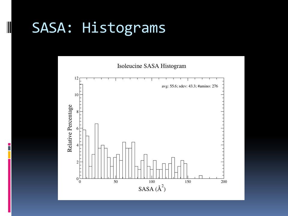
SASA: Histograms

19
SASA: Conclusion The general trend of the average seems to imply that the more hydrophilic an amino acid is the higher its SASA is going to be. However, from the large standard deviations and the evidence given by the histograms this trend should probably not be taken as an absolute.

Similar presentations


 Leucina (Leu) Isoleucina (Ile) Fenilalanina (Phe) Metionina (Met)>")


 1’ C 5’ PO4 (free) DNA is a linear polymer of nucleotide subunits joined together by phosphodiester bonds - covalent bonds between.>")


 Urea cycle and reactions that feed amino groups into the cycle. The enzymes catalyzing these reactions (named in the text) are distributed.>")


