Download presentation
Presentation is loading. Please wait.

1
Final Diagnosis Intralobar Pulmonary Sequestration

2
8/15/2005

5
History of recurrent pneumonia

6
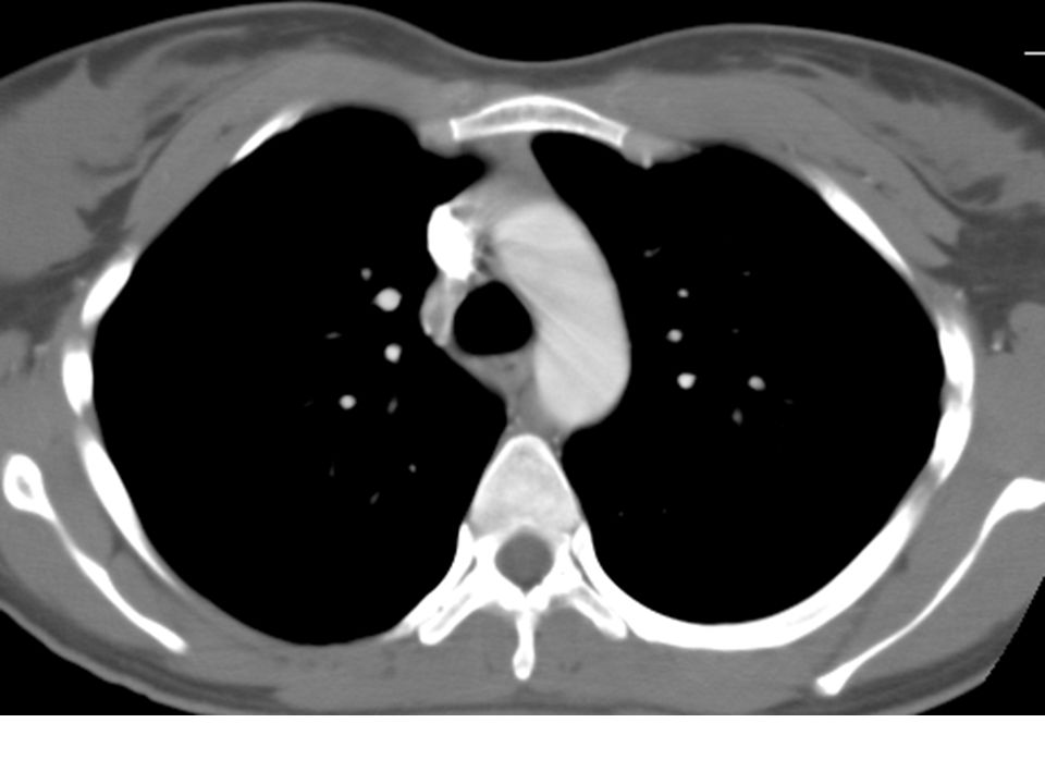
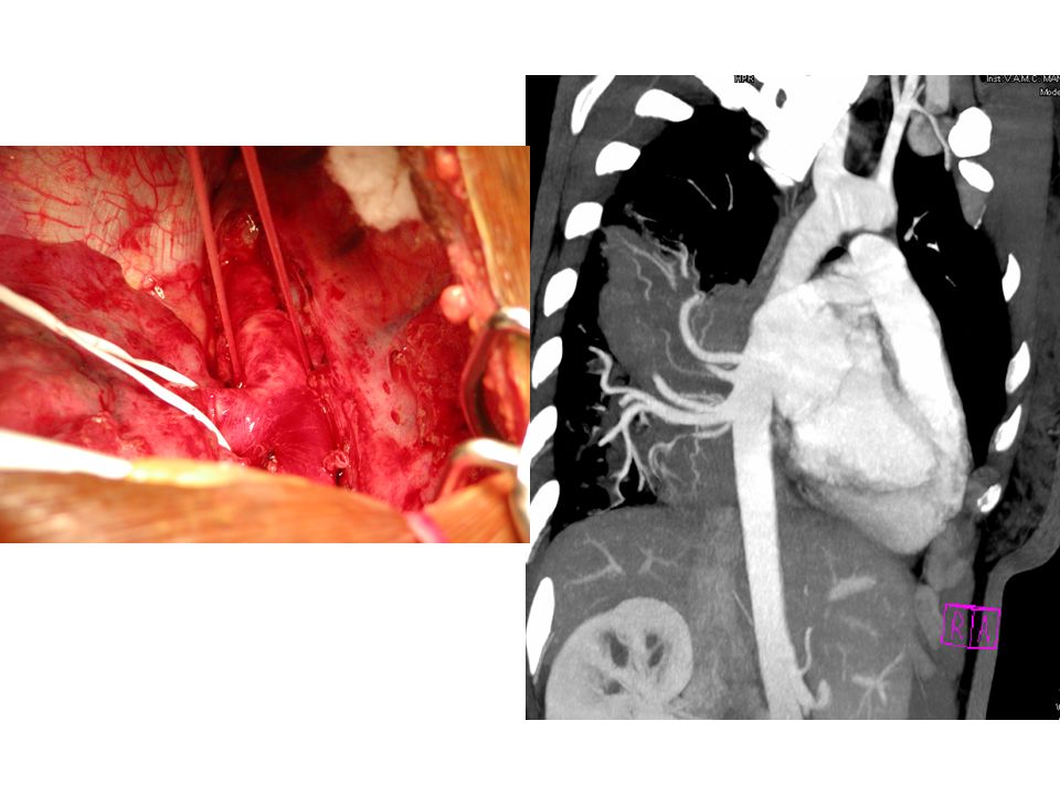
Surgical Report Intralobar Pulmonary Sequestration, treated with right lower lobectomy Two major arteries from the thoracic aorta Large vein draining sequestered lobe

13
Bronchopulmonary Sequestration Definition Non-functioning lung tissue Separated from normal bronchial tree Vascularized by a systemic artery Two Forms Intralobar (ILS): within the visceral pleura Extralobar (ELS): separated from the lung by its own pleura
: within the visceral pleura Extralobar (ELS): separated from the lung by its own pleura")
14
Intralobar Pulmonary Sequestration (ILS) Characteristics More common type May present at any age Generally as recurrent infection No sexual predominance Almost exclusively affects lower lobe Arterial supply: descending aorta Venous drainage: pulmonary veins
 Characteristics More common type May present at any age Generally as recurrent infection No sexual predominance Almost exclusively affects lower lobe Arterial supply: descending aorta Venous drainage: pulmonary veins")
15
Etiology of Bronchopulmonary Sequestration Accessory bud that forms caudal to lung buds Traction theory Postnatal formation

16
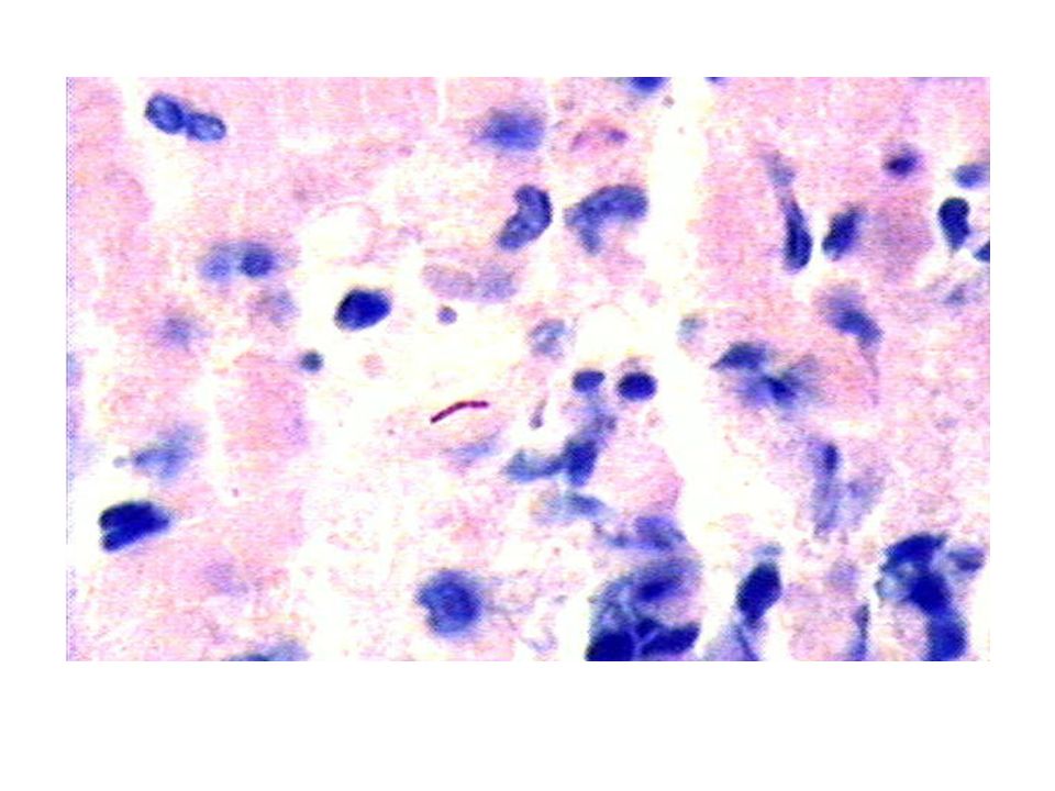
Pulmonary Sequestration Complications Recurrent infection Heart failure Intralobar malignancy

17
Treatment of Pulmonary Sequestration Symptomatic Disease Surgery: lobectomy or segmental resection Arterial embolization

18
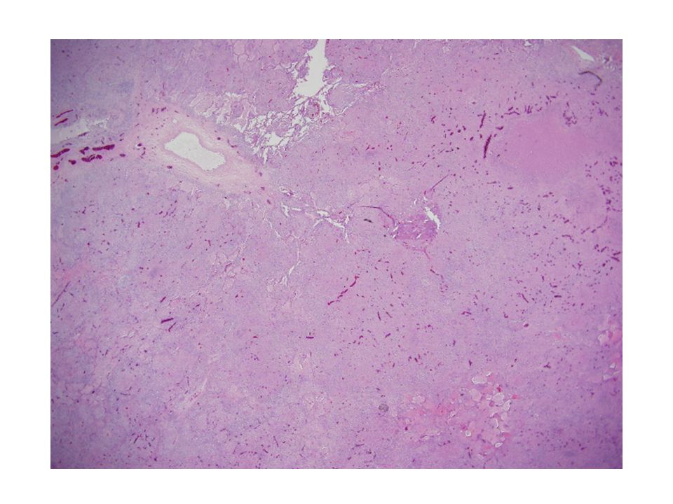
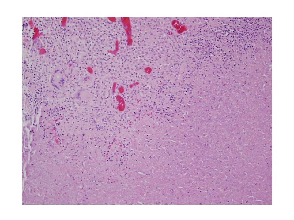
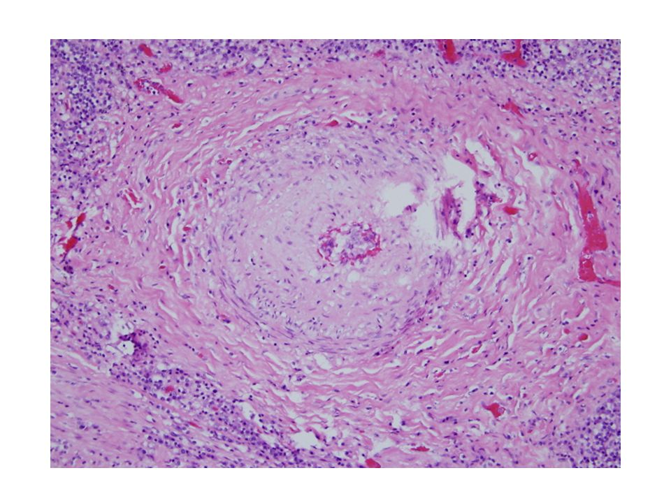
Patient Follow-up Given granulomas and acid fast bacilli, sputum was sent for culture. Three sputum samples were negative for acid fast bacilli. A ppd was placed and was also negative. Patient was treated empirically with IRPE for four months.

Similar presentations


 LCDR.>")


