Download presentation
Presentation is loading. Please wait.

1
Mitosis Is a process of cell division which results in the production of two daughter cells from a single parent cell. The daughter cells are identical to one another and to the original parent cell. Mitosis can be divided into four stages: 1.Prophase. 2.Metaphase. 3.Anaphase. 4.Telophase.

3
Prophase 1.The chromatin, condenses into chromosomes. 2. Each chromosome has duplicated and now consists of two sister chromatids. 3. The nuclear membrane disappear. Metaphase 1.The chromosomes align at the equatorial plate. {in this stage the chromosomes are very dense and best seen}.

4
Anaphase 1.The centromeres divide. 2. Sister chromatids separate and move toward the corresponding poles. Telophase 1.Daughter chromosomes arrive at the poles. 2. The condensed chromatin expands. 3. The nuclear envelope start to reappears. 4.The cytoplasm divides, the cell membrane pinches inward ultimately producing two daughter cells (phase: Cytokinesis).
..")
5
Chromosome Analysis

6
It was possible to visualize chromosomes under microscopes as early as the mid-1800s, it was quite difficult to observe individual chromosomes. It was thus hard to count the number of chromosomes in a cell or to examine structural abnormalities. Beginning in the 1950s, several techniques were developed that improved our ability to observe chromosomes. These included : (1)The use of spindle poisons, such as colchicine and colcemid, that arrest dividing somatic cells in metaphase, when chromosomes are maximally condensed and easiest to see. (2) The use of hypotonic (low salt) solution, which causes swelling of cells, rupture of the nucleus, and better separation of individual chromosomes. (3) The use of staining materials that are absorbed differentially by different parts of chromosomes, thus producing the characteristic bands that help to identify individual chromosomes.
The use of spindle poisons, such as colchicine and colcemid, that arrest dividing somatic cells in metaphase, when chromosomes are maximally condensed and easiest to see. (2) The use of hypotonic (low salt) solution, which causes swelling of cells, rupture of the nucleus, and better separation of individual chromosomes. (3) The use of staining materials that are absorbed differentially by different parts of chromosomes, thus producing the characteristic bands that help to identify individual chromosomes..")
7
Karyotyping Is a test to examine chromosomes in a sample of cells, which can help identify genetic problems as the cause of a disorder or disease. This test can: Count the number of chromosomes. Look for structural changes in chromosomes.

8
Picture 1. Chromosomes as they appear under a Microscope.

9
Picture 2. The chromosomes arranged according to size; chromosome 1 is the largest. The last two chromosomes are the sex chromosomes.

10
How The Test is Performed Sample Collection Chromosomes are analyzed by collecting a living tissue (usually blood). Karyotype will be done on the white blood cells which are actively dividing (a state known as mitosis), fetal skin cells (from amniotic fluid or the placenta) and occasionally bone marrow cells. Transport to the Laboratory karyotype is a specialized test that is done in a specific laboratory called a cytogenetics lab.
, fetal skin cells (from amniotic fluid or the placenta) and occasionally bone marrow cells. Transport to the Laboratory karyotype is a specialized test that is done in a specific laboratory called a cytogenetics lab..")
11
Growing the Cells In order to have enough cells to analyze, the dividing cells are grown in special media or a cell culture. This media contains chemicals and hormones that enable the cells to divide and multiply. This process of “culturing” the cells can take 3 to 4 days for blood cells, and up to a week for fetal cells. Synchronizing Cells The cells are treated with a chemical ”colcemid” which stops cell division at the metaphase where the chromosomes are the most compact and best seen.

12
harvesting the cells, placing the cell sediment on a slide. Rupturing the cell nucleus with a hypotonic saline solution. Staining with a designated nuclear stain. Photographing the metaphase “spread” of chromosomes on the slide. The image of the 22 pairs of autosomes are arranged according to the length, with the sex chromosomes in the right hand corner.

13
This ordered display of chromosomes is termed a Karyotype. Currently, computerized image analyzers are usually used to display chromosomes. Three parameters in identifying chromosomes: 1.Size 2.Banding pattern. 3.Centromere position.

14
Karyotype key

16
Chromosome Banding Early Karyotype were useful in counting the number of chromosomes, but structural abnormalities, such as balanced rearrangement or small chromosomal deletions, were often undetectable. Staining techniques were developed in the 1970s to produce the chromosome bands characteristic of modern Karyotype. chromosome banding helps greatly in the detection of deletions, duplications, and other structural abnormalities, and it facilitates the correct identification of individual chromosomes.

17
Several chromosome-banding techniques are used in cytogenetic laboratories Quinacrine banding (Q-banding). Fluorescent stain, to identify specific chromosomes and structural rearrangements. Giemsa banding (G-banding). Giemsa staining. Reverse banding ( R banding). C banding. staining heterochromatin
. Giemsa staining. Reverse banding ( R banding). C banding. staining heterochromatin.")
18
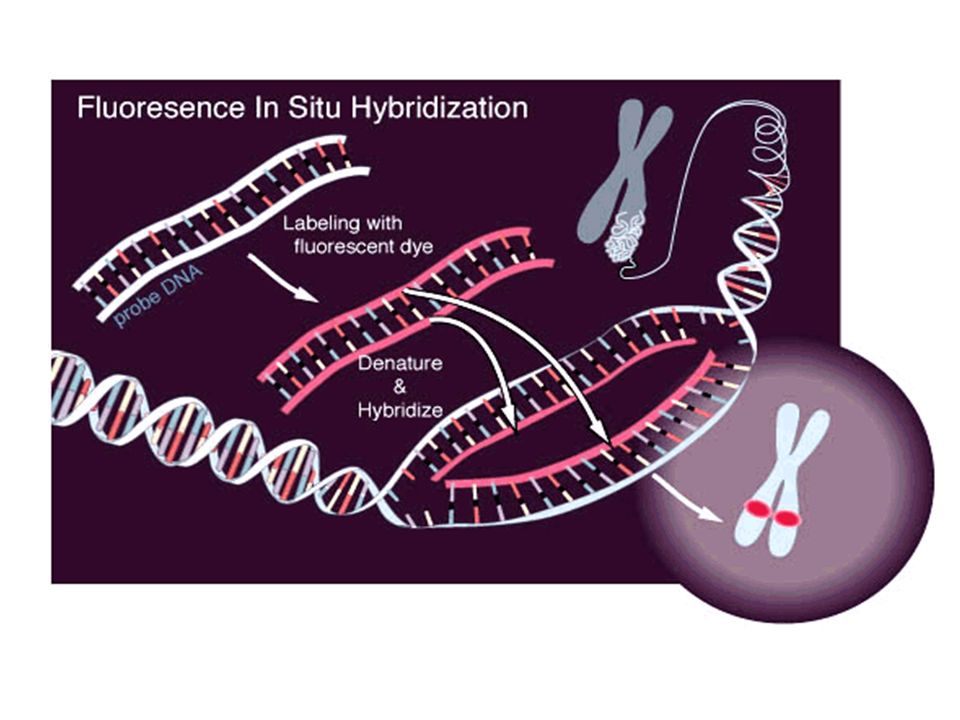
FLUORESENCE IN SITU HYBRIDISATION Cytogenetic technique used to detect and localize the presence or absences of specific DNA sequences on the chromosomes. FISH uses fluorescent probes that bind only to those part of chromosomes with which they show a high degree of similarity. Fluorescence microscopy can be used to find out where the fluorescent probe bound to the chromosomes.

21
The chromosomes can be seen in blue. The chromosome that is labeled with green and red spots (upper left) is the one where the wrong rearrangement is
 is the one where the wrong rearrangement is.")
22
Barr body A microscopic feature of female cells due to the presence of two X chromosomes in the female. One of these X chromosomes is inactive and is crumpled up to form the Barr body.

Similar presentations