Download presentation
Presentation is loading. Please wait.

1
The Arthropods: Blueprint for Success
Chapter 14 Zoology

2
What is the most abundant animal on Earth?
Answer: Copepods, a type of marine Arthropod. 1-2 mm planktonic crustacean Basis of marine food webs. One of the smallest creatures is food to one of the largest – blue whales.

3
Number of Species by Phylum
Chordata Mollusca Platyhelminthes Nematoda Arthropoda Porifera Annelida Echinodermata Sarcomastigophora Apicomplex Ciliophora

4
Arthropoda - Jointed Legs
What is an Arthropod? Arthro – joint + podas – foot Crayfish, lobsters, spiders, mites, scorpions, and insects. + 1 million species described. Estimated to be 30 – 50 million undescribed species. Most successful Phylum in the Animal Kingdom. Arthropoda - Jointed Legs

5
Arthropods - Most Successful Animals
Number of species Diversity Distribution Longevity

6
Reasons for Arthropod Success
Versatile exoskeleton Segmentation Oxygen piped directly to cells (terrestrial) Highly developed sensory organs (compound eye) Complex behavior Metamorphosis
 Highly developed sensory organs (compound eye) Complex behavior. Metamorphosis.")
7
General Characteristics of Arthropods
Triploblastic Protostome Bilateral Symmetry Metamerism – segmented body with tagmatization Jointed Appendages Exoskeleton Ecdysis or molting Ventral Nervous System Open Circulatory System Complete Digestive Tract Metamorphosis

8
Phylogeny of Arthropods
Arthropoda Annelids (worms) Onychophorans (worms w/legs) Chelicerates (spiders) Crustaceans (lobsters) Insects (butterflies) Trilobites (extinct) Worm-like Ancestor
 Onychophorans. (worms w/legs) Chelicerates. (spiders) Crustaceans. (lobsters) Insects. (butterflies) Trilobites. (extinct) Worm-like. Ancestor.")
9
Metamerism and Tagmatization
Metamerism – segmented body (like Annelida) but specialization of body regions for specific functions – Tagmatization. Segmented externally, but no internal septa to divide each segment; organ systems are not divided. Internal metamerism is not needed; exoskeleton replaces the hydrostatic compartments of annelids for support.
 but specialization of body regions for specific functions – Tagmatization. Segmented externally, but no internal septa to divide each segment; organ systems are not divided. Internal metamerism is not needed; exoskeleton replaces the hydrostatic compartments of annelids for support.")
10
Tagmatization Body regions are called tagmata - specialized for feeding, locomotion, and visceral functions. Examples are: head, thorax, and abdomen Most internal organs are not segmented.

11
The Exoskeleton Arthropods enclosed in an external, jointed skeleton – exoskeleton or cuticle. Provides: structural support, protection, prevents water loss, attachment points for muscle movement. It is a nonliving mixture of lipids, proteins and a tough polysaccharide called – chitin. Made by a deeper layer of tissue – epidermis or hypodermis.

12
Two Layers of The Exoskeleton
Epicuticle – waxy, outermost layer; impermeable to water, bacteria, and pesticides. Procuticle – deeper, thicker layer of chitin and protein; outer portion may darken and harden by sclerotization (proteins) or with calcium carbonate. Hard, but flexible armor.
 or with calcium carbonate. Hard, but flexible armor.")
13
Hardened Procuticle Softer Procuticle Fig

14
Modifications of Exoskeleton
Copyright © The McGraw-Hill Companies, Inc. Permission required for reproduction or display. Modifications of Exoskeleton Soft and flexible at joints for movement Invaginations for gas exchange From A Life of Invertebrates, Copyright © 1979, W. D. Russell-Hunter.

15
Growth with an Exoskeleton
Growing arthropods must periodically shed the exoskeleton by molting – ecdysis. Triggered by hormones (ecdysone) – which causes the old procuticle to breakdown and separate from the epidermis. A new epicuticle and procuticle are made underneath by the epidermis. Old exoskeleton splits open as the animal stretches with air or water intake and wriggles out of old exoskeleton. See a crab do ecdysis
 – which causes the old procuticle to breakdown and separate from the epidermis. A new epicuticle and procuticle are made underneath by the epidermis. Old exoskeleton splits open as the animal stretches with air or water intake and wriggles out of old exoskeleton. See a crab do ecdysis.")
16
How do Arthropods grow?

17
Ecdysis in my tarantula! They grow up so fast!
Old Exoskeleton New Exoskeleton and Animal Crawling Out

18
How big can something with an exoskeleton get?

19
The Hemocoel An internal cavity for the open circulatory system of arthropods; results from no internal segmentation. Internal organs are bathed by body fluids (hemolymph) for exchanges of nutrients, wastes and gases. Because of an exoskeleton, the coleom is no longer used as a hydorstatic skeleton.
 for exchanges of nutrients, wastes and gases. Because of an exoskeleton, the coleom is no longer used as a hydorstatic skeleton.")
20
Review of Circulatory Systems
Open Circulatory System Closed Circulatory System Ostia

21
Metamorphosis Changes in body form and physiology as arthropods grow and develop. Immature stages (larvae) often are radically different than adult forms. These differences reduce competition between adult and young (different body forms, behaviors, and habitats). Many types of metamorphosis (discussed more with insects).
 often are radically different than adult forms. These differences reduce competition between adult and young (different body forms, behaviors, and habitats). Many types of metamorphosis (discussed more with insects).")
22
Metamorphosis

23
Metamorphosis in a Monarch Butterfly
Larva – Caterpillar eats leafy vegetation Chrysalis Adult – Butterfly eats nectar

24
Phylum Arthropoda Groups
Subphylum Trilobitomorpha (trilobites) Subphylum Chelicerata Class Merostomata (horseshoe crabs) Class Arachnida (spiders,scorpions,ticks) Class Pycnogonida (sea spiders) Subphylum Crustacea Class Malacostraca (lobsters, crabs, shrimp) Class Branchiopoda (brine shrimp, water fleas) Class Maxillopoda (barnacles, copepods) Subphylum Hexapoda (insects) Subphylum Myriapoda (millipedes and centipedes)
 Subphylum Chelicerata. Class Merostomata (horseshoe crabs) Class Arachnida (spiders,scorpions,ticks) Class Pycnogonida (sea spiders) Subphylum Crustacea. Class Malacostraca (lobsters, crabs, shrimp) Class Branchiopoda (brine shrimp, water fleas) Class Maxillopoda (barnacles, copepods) Subphylum Hexapoda (insects) Subphylum Myriapoda (millipedes and centipedes)")
25
Subphylum Trilobitomorpha
Trilobites, dominant life form that went extinct 345 million years ago. Oval, flattened bodies divided into 3 tagmata (head, thorax, pygidium). All body parts could roll up (like a rollie-pollie). Appendages – two lobes or branches (biramous) – one as a walking leg and the other for digging or as a gill). Crawled on the bottom feeding on annelids, molluscs, and decaying matter.
. All body parts could roll up (like a rollie-pollie). Appendages – two lobes or branches (biramous) – one as a walking leg and the other for digging or as a gill). Crawled on the bottom feeding on annelids, molluscs, and decaying matter.")
26
Subphylum Chelicerata
chele – “claw”; spiders, mites, horshoe crabs,etc. Two Tagmata: Prosoma (Cephalothorax) - “head” for sensory, feeding, locomotion; often eyes but NEVER antennae. Opisthosoma – “abdomen” for digestion, reproduction, excretion, and respiration.
 - head for sensory, feeding, locomotion; often eyes but NEVER antennae. Opisthosoma – abdomen for digestion, reproduction, excretion, and respiration.")
27
Prosoma has all the appendages!
First two pairs of appendages for feeding: Chelicerae – first pair that are pincerlike (chelate) used in feeding; maybe hollow fangs. Pedipalps – second pair that are for sensory, feeding, movement, or reproduction. Paired walking legs are posterior to the pedipalps; number of legs varies.
 used in feeding; maybe hollow fangs. Pedipalps – second pair that are for sensory, feeding, movement, or reproduction. Paired walking legs are posterior to the pedipalps; number of legs varies.")
28
Prosoma or Cephalothorax Opisthosoma or Abdomen
Chelicerata Legs 1st Pair - Chelicerae 2nd Pair - Pedipalps Prosoma or Cephalothorax Posterior Pairs –Walking Legs Opisthosoma or Abdomen

29
Class Merostomata Ancient group of 4 species of horseshoe crabs and giant water scorpions (extinct 280 mya). Horseshoe crabs are benthic scavengers and predators. Hard, horseshoe-shaped carapace covers the prosoma. Has cheilicerae, pedipalps and first three pairs of walking legs are chelate – used in walking and feeding. Last pair of legs for walking and digging – clams, annelids, and other invertebrates.

30
Fig. 18.2a Dorsal

31
Fig. 18.2b Ventral

32
Opisthosoma of Horseshoe Crabs
Long, unsegmented tail – telson – used to self-right the animal. First Pair of Appendages – genital opercula - cover genital pores. The remaining Five Pairs of Appendages – book gills – platelike gills used in gas exchange between blood and water.

33
Horseshoe Crab Horseshoe crab info
Live up to 19 years; they are dioecious, may take 9-12 years to sexually mature. Mates during spring and summer full and new moons, onto ocean beaches. Females lay up to 30 thousand eggs, which males fertilize before burying them in the sand These eggs provide a major food source for migrating birds along the Atlantic coast. Those that are not eaten hatch during the next high tide, and the tiny larvae are carried away to sea.

34
Class Arachnida arachne – “spider”; includes mites, ticks and scorpions. Some of the first terrestrial animals – exoskeleton helps retain water; many other adaptations for land. Have 4 pair of walking legs. 2 body segments (prosoma and opisthosoma) Except mites & ticks All are dioecious.
 Except mites & ticks. All are dioecious.")
35
Digestion Most are carnivores.
Some inject enzymes into prey with their chelicerae (venomous) to help digest food. Suck digested material into pharynx. Gut tract is in three parts: foregut, midgut, and hindgut- lined with cuticle so the lining is shed as the exoskeleton.
 to help digest food. Suck digested material into pharynx. Gut tract is in three parts: foregut, midgut, and hindgut- lined with cuticle so the lining is shed as the exoskeleton.")
36
Excretory Organs for Terrestrial Arachnids
Fig. 18.5 Excretory Organs for Terrestrial Arachnids

37
Excretion Arachnids use coxal glands and/or Malpighian tubules to excrete waste and reabsorb nutrients. Coxal Glands – paired, thin-walled sacs to remove nitrogenous wastes; leave via excretory pores at base of posterior appendages. Uric acid is the main nitrogenous product of land arachnids.

39
Malpighian Tubules In insects and other terrestrial arthropods, Malpighian tubules Remove nitrogenous wastes from hemolymph and function in osmoregulation Digestive tract Midgut (stomach) Malpighian tubules Rectum Intestine Hindgut Salt, water, and nitrogenous wastes Feces and urine Anus tubule Reabsorption of H2O, ions, and valuable organic molecules HEMOLYMPH
 Malpighian. tubules. Rectum. Intestine. Hindgut. Salt, water, and. nitrogenous. wastes. Feces and urine. Anus. tubule. Reabsorption of H2O, ions, and valuable. organic molecules. HEMOLYMPH.")
40
Malpighian Tubules Na+/K+-ATPase K+ Hemolymph Water and K+ K+
Water and waste K+ Na+/K+-ATPase Conc. Waste Hindgut

41
Respiration To reduce water loss, some arachnids have book lungs – paried, leaf-like, infoldings on the ventral surface. Air enters the book lung through slit opeing and circulates between lamellae. Other arachnids have a tracheal system for gas exchange. Exchange gases with the hemolymph fluid. Remember…no blood vessels!

42
Copyright © The McGraw-Hill Companies, Inc
Copyright © The McGraw-Hill Companies, Inc. Permission required for reproduction or display. Arachnid Book Lung

44
Tracheal Systems in Insects
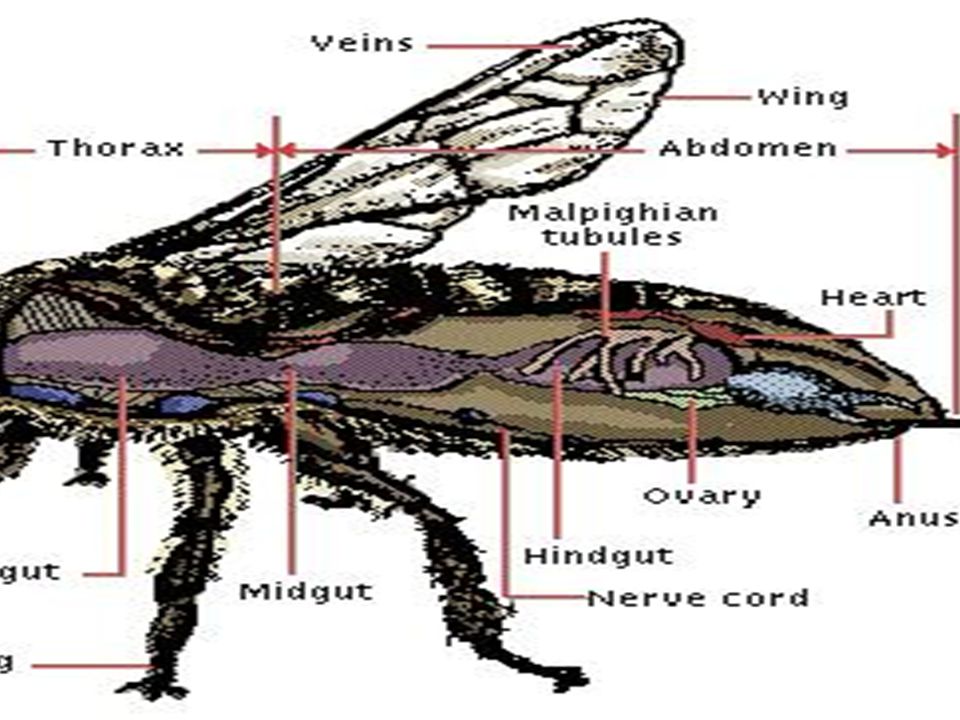
The tracheal system of insects Consists of tiny branching tubes that penetrate the body Tracheae Air sacs Spiracle (a) The respiratory system of an insect consists of branched internal tubes that deliver air directly to body cells. Rings of chitin reinforce the largest tubes, called tracheae, keeping them from collapsing. Enlarged portions of tracheae form air sacs near organs that require a large supply of oxygen. Air enters the tracheae through openings called spiracles on the insect’s body surface and passes into smaller tubes called tracheoles. The tracheoles are closed and contain fluid (blue-gray). When the animal is active and is using more O2, most of the fluid is withdrawn into the body. This increases the surface area of air in contact with cells.
 The respiratory system of an insect consists of branched internal tubes that deliver air directly to body cells. Rings of chitin reinforce the largest tubes, called tracheae, keeping them from collapsing. Enlarged portions of tracheae form air sacs near organs that require a large supply of oxygen. Air enters the tracheae through openings called spiracles on the insect’s body surface and passes into smaller tubes called tracheoles. The tracheoles are closed and contain fluid (blue-gray). When the animal is active and is using more O2, most of the fluid is withdrawn into the body. This increases the surface area of air in contact with cells.")
45
The tracheal tubes Supply O2 directly to body cells. Body cell Air sac
Tracheole Tracheoles Mitochondria Myofibrils Body wall (b) This micrograph shows cross sections of tracheoles in a tiny piece of insect flight muscle (TEM). Each of the numerous mitochondria in the muscle cells lies within about 5 µm of a tracheole. 2.5 µm Air
 This micrograph shows cross sections of tracheoles in a tiny piece of insect flight muscle (TEM). Each of the numerous mitochondria in the muscle cells lies within about 5 µm of a tracheole. 2.5 µm. Air.")
46
Generic Arthropod Nervous System
Ladder-like nervous system designed for a segmented body plan – similar to annelids. Ventral longitudinal nerve cord.

47
NERVOUS SYSTEM Lateral view of body showing relative position of circulatory (yellow), digestive (green), and nervous (blue) systems.
, digestive (green), and nervous (blue) systems.")
48
Sense Organs Sensilla - modified extensions of the exoskeleton act as receptors (mechano- and chemoreceptors). May be slits or membraneous drums.

49
Photoreceptors Arachnids have one or more pairs of eyes.
Detect movement, light intensity or images (hunting spiders). Two Types: Simple or Pigment – cup Ocelli – median eyes. Compound Eyes – lateral eyes.
. Two Types: Simple or Pigment – cup Ocelli – median eyes. Compound Eyes – lateral eyes.")
50
Pigment – cup Ocelli highly variable within the arthropods.
all the receptor units (retina cells) share a common lens. concavity of the cup is oriented toward the light source. pigment layer prevents the entry of light from any other direction.
 share a common lens. concavity of the cup is oriented toward the light source. pigment layer prevents the entry of light from any other direction.")
51
Compound Eyes Most have one pair of compound eyes.
Each compound eye is made of thousands of light-receiving units called ommatidia. Ommatidium is the individual, self-contained, independent light-detecting unit. Each includes focusing system (cuticular cornea and crystalline cone)
")
52
Compound Eye Ommatidia Lens Crystalline cone Pigment cells Facet
Visual cells Nerve fibers from visual cells Optic nerve Ommatidia

53
Compound Eye Ommatidium Anatomy

54
Arachnid Reproduction
All are dioecious Genital openings are ventral; sperm usually transferred indirectly. Male may package sperm in a spermatophore which he either places into the female or the female crawls over the spermatophore and picks it up. May use modified pedipalps to transfer sperm. (spiders)
")
55
Order Scorpionida Scorpions Tropical and temperate climates
All are predatory Prosoma fused into hard carapace Small chelicerae, but enlarged pedipalps Opisthosoma is divided into preabdomen and postabdomen Sting and venomous gland located on postabdomen

56
Scorpion Anatomy

57
3 Types of Birth in Scorpions
Oviparous – females lay eggs that develop outside of body. Ovoviviparous – young develop in large, yolky eggs held internally in body, then are born, fully developed. Viviparous – mother provides nutrients to embryos; eggs develop in special chambers close to female digestive tract. After birth, young crawl onto mother’s back for about 1 month.

58
Viviparous Scorpion Seeing is believing!

59
Order Araneae +34,000 species spiders; largest group of arachnids
Chelicerae are modified with poison and fangs. Pedipalps are leg-like; males are modified for sperm transfer. 6-8 eyes Spinnerettes at the posterior end that make silk using silk glands.

60
Fig. 18.4

61
Fig. 18.5

62
What kinds of prey can they eat?
Tarantula Feeding What kinds of prey can they eat?

63
Latrodectus mactans Black widow spider Neurotoxin

64
Loxosceles reclusa Fiddle back spider Necrotoxin- causes tissue death.
Brown recluse Brown Violin Necrotoxin- causes tissue death. Warning!!! The following pictures may be distrubing!

65
Loxosceles reclusa Necrosis of tissue

66
Day 3

67
Day 4

68
Day 5

69
Day 6

70
Day 9

71
Day 10

72
Order Acarina Mites and ticks
Many are ectoparasites, others are free-living. Most serious to human health Prosoma and opisthosoma are fused and covered by a single carapace. Chelicerae and pedipalps are variously modified for piercing, biting, etc. and have 4 pair of walking legs.

73
Rocky Mountain Spotted Fever
Ticks are vector High fever Headache Muscle pain Rash BEGINS ON EXTREMETIES 25% fatal without antibiotics

74
Dermacentor variabilis
Dog tick Dermacentor andersoni Wood tick

75
Fig

76
Dermatophagoides Dust mite Allergies to fecal products
1 gram of dust holds 250,000 droppings

77
Trombicula Chigger mite Larvae feed on skin Dermatitis
Adults are feed on insect eggs.

78
Sarcoptes scabiei, the human itch mite
See it in action!

79
Class Pycnogonida Sea Spider
Marine arthropods- found especially in the Mediterranean and Caribbean Seas, as well as the Arctic and Antarctic Oceans 1300 known species Walking legs is usually eight (four pairs), but species with five and six pairs exist. small size and slender body and legs, no respiratory system is necessary for the Sea Spiders, with gases moving by diffusion.
, but species with five and six pairs exist. small size and slender body and legs, no respiratory system is necessary for the Sea Spiders, with gases moving by diffusion.")
80
Subphylum Crustacea Lobsters, shrimp, crayfish, copepods, barnacles
Have two pairs of antennae Biramous appendages - two distal projections Medial ramus – endopodite Lateral ramus - exopodite

81
Class Malacostraca Crabs, lobsters, shrimp; largest class of crustraceans. Possess mandibles and maxillae – modified appendages for chewing and grinding food.

82
Fig. 19.1

83
Dorsal Fig. 19.2a

84
Ventral Fig. 19.2b

85
Internal Anatomy of Crustaceans
Excretory Organs Fig. 19.5

86
Crayfish

87
Fig. 19.6

88
Who cares about crustaceans?
The Krill

89
Some of the biggest aquatic crustaceans!

90
Order Isopoda Pillbugs- A terrestrial crustacean.

91
A Strange Isopod Parasite!
Cymothoa exigua See it in action!

92
Class Branchiopoda Branchio – gill + podos –foot; fairy and brine shrimp, water fleas; freshwater. Flattened appendages for respiration, filter-feeding, and swimming. Females may reproduce parthenogenetically.

93
Brine Shrimp

94
Class Maxillopoda Subclass Copepoda – copepods; kope – oar + poda – foot; most abundant crustacean; marine and freshwater. Cylindrical body and one compound eye. First antennae are modified for swimming and no other appendages. Most are planktonic and filter feeding.

95
Copepods

96
Class Maxillopoda Subclass Thecostraca – species of barnacles; sessile marine crustaceans. Most are monecious. A larval form develops a bivalved carapace and first antennae become used for attachement. Gut tract becomes U-shaped and thoracic appendages used for filter feeding. Attach to rocks, ships, whales Some are parasitic

97
Barnacle Cirri Testis Penis Anus Mouth Stomach Ovary Cement gland

98
Barnacles

99
The End?

100
Fig a Fig a

Similar presentations


 is found in Australia. They are herbivores who cannot bite or sting in defense, but.>")


