Download presentation
Presentation is loading. Please wait.

2
Practical Part Microscopic Examination of Microorganisms Experiments Identification of MOs Different Staining Techniques

3
Experimental Microbiology

4
Pour Plate Technique 1 Distribution of Microorganisms in the Environment 2 Techniques for Isolation of Pure Cultures “ Streaking” 3 Determination of Viable Count of Bacteria

5
Objectives : To enumerate and count viable cells of a given culture. Determination of Viable Count of Bacteria

6
Principle: Viable count is defined as the number of living cells per 1 ml of solution. Determination of viable cells depends on the fact that each colony represents one cell, when the cell was allowed to grow on a solid medium.

7
Because bacterial population is usually growing in a huge number, a serial dilution is performed to facilitate counting of bacteria. Viable Count (VC) = Number of Colony Forming Unit (CFU) X dilution factor / Inoculum size
 = Number of Colony Forming Unit (CFU) X dilution factor / Inoculum size.")
8
Materials : Bacterial suspension in a test tube. 3 tubes, each containing 9-ml sterile saline. 2 sterile 1- ml pipettes. Sterile plates (9 plates). Nutrient agar tubes.
. Nutrient agar tubes..")
9
Procedure

10
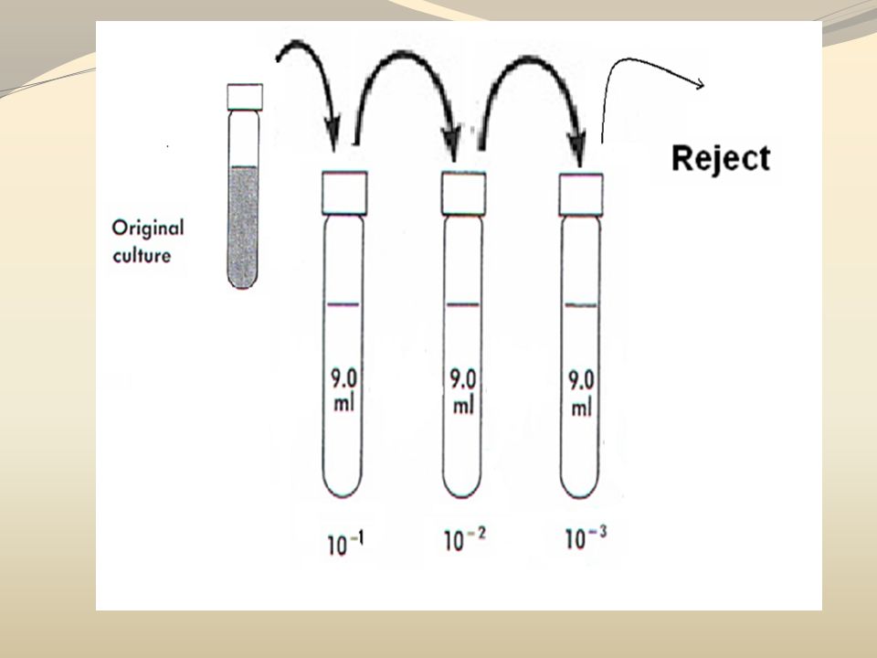
Mark the saline tubes as 10 -1, 10 -2 and 10 -3 (1)
")
11
Divide the petri dishes into 3 groups and mark each group the same manner you follow for the saline tubes. Thus each group containing 3 plates will be marked as follow: 10 -1, 10 -2 and 10 -3. (2)
.")
13
Shake the tube vigorously using vortex. Then transfer 1ml aseptically from the original suspension to the first tube. Mix well repeat the same step by transferring one ml from the first tube to the second tube and then from the second tube to the third one. (3)
.")
14
Start

15
After good mixing, transfer aseptically one ml from the third tube (10 -3 ) to each of three plates marked as 10 -3. Repeat the same step by transferring 1 ml from the tube marked 10 -2 and 10 -1 to their corresponding plates. Aseptically pour the agar in each plate and mix well. Make sure that the agar is cooled to 55 ○ C to avoid killing of microorganism. (4)
.")
16
Results of Experiment After incubation, count the colonies in plates in which the CFU range between (30-300). The detected colonies are counted as colony forming unit (CFU).
..")
17
Plate number Colony count at dilution 10 -1 10 -2 10 -3 1 2 3 Mean

18
Number of colonies = Dilution Factor = 1/Dilution = Number of viable cells per ml Viable Count (VC) = Number of colonies x dilution factor Inoculum size (1)
 = Number of colonies x dilution factor Inoculum size (1)")
19
Example

20
Plate number Colony count at dilution 10 -1 10 -2 10 -3 1 250 150 50 2 250 125 50 3 250100 50 Mean 250 12550

21
Comment VC of 10 -1 = (250 x 10) / 1 = 2500 CFU/ml VC of 10 -2 = (125 x 100) / 1 = 12500 CFU/ml VC of 10 -3 = (50 x 1000) / 1 = 50000 CFU/ml
 / 1 = 2500 CFU/ml VC of = (125 x 100) / 1 = CFU/ml VC of = (50 x 1000) / 1 = CFU/ml")
22
Plate number Colony count at dilution 10 -1 10 -2 10 -3 1 400 (rejected) 150 50 2 500 (rejected) 125 50 3 600 (rejected) 100 50 Mean ------ 12550
 (rejected) (rejected) Mean")
23
Plate number Colony count at dilution 10 -1 10 -2 10 -3 1 400 (rejected) 400 (rejected) 50 2 500 (rejected) 420 (rejected) 50 3 600 (rejected) 400 (rejected) 50 Mean ------ -------50
 400 (rejected) (rejected) 420 (rejected) (rejected) 400 (rejected) 50 Mean")
Similar presentations