Download presentation
Presentation is loading. Please wait.

1
Development of the Heart 212 – 2004 – Week 6 Avinash Bharadwaj

2
Retrospect… Trilaminar embryo Three germ layers Head and tail folds

3
Cardiovascular System The Heart Blood vessels Abnormal development Correlation

4
Some Special Features Early development and function Requirements of the foetus Non-functional lungs Placental circulation Planning for postnatal life Right-left passage essential during foetal life, obliteration required immediately after birth. The phylogenetic sequence.

5
Earliest Development Angiogenic cell clusters or Blood Islands Blood cells and endothelium

6
Cardiogenic Area Flat embryo Position of cardiogenic area

7
Head Fold Cardiac area Gut formation Pericardium and heart tube

8
Later…

10
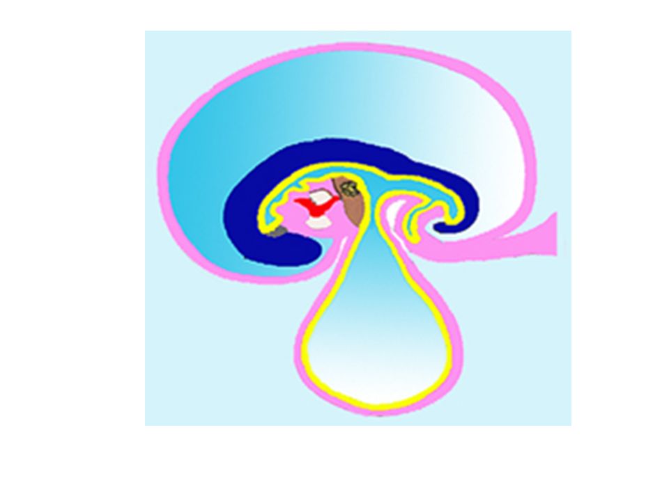
The Heart Tube Venous and arterial ends Regions defined Bulbus cordis Ventricle Atrium Sinus venosus Conus and Truncus…?

11
The Tube Bends SV A V B VD

12
The Chambers Left Front A AA V BVL

13
The Interior R and L atria Atrioventricular canal : common Outflow Aortic arches RA LA AVC A-Ar

14
Left – Right Partitioning Interatrial septum Interventricular septum Spiral (aortico-pulomonary septum Endocardial cushions (A-V cushions) Functional requirements There must always be a right to left passage!
 Functional requirements There must always be a right to left passage!")
15
Interatrial septum Partitioning Right to left passage Mechanism for closing the passage

16
A V

17
Septum Primum This is a sagittal section seen from the right. V AVC

18
Foramen Primum Foramen primum : Between the septum and the AV Cushions

19
Passage is a Must! Foramen secundum Foramen primum about to disappear

20
Septum Secundum To the right of primum Foramen primum has disappeared

21
Foramen ovale F. Ovale – In septum secundum Further…

22
The ‘Valve’ Two septa Two foramina

23
Sinus Venosus Originally a symmetrical structure Right and left “horns” Venous return more to the right Left horn becomes smaller Opening shifts to the right Later – part of right atrium

24
Left Atrium Four pulmonary veins Common opening “Absorption” of veins into atrium Rough part - auricle

25
The Ventricular Septum Three Parts Interventricular septum AV Cushions Spiral Septum

26
The Ventricular Septum R Membranous Muscular Spiral (Aorticopulmonary)
")
27
Foetal Circulation Very little pulmonary flow Placental Circulation Right to Left Passages

28
IVC : Blood from placenta Ductus venosus F. ovale Ductus arteriosus

29
Changes At Birth Closure of interatrial septum Closure of ductus arteriosus Closure of ductus venosus

30
Congenital Heart Disease Septal Defects – Atrial and Ventricular Endocardial cushion defects Aorticopulmonary defects PDA Others

Similar presentations


, UNSEPARATED VENTRICLE (18), LIVER (12), UMBILICAL VEIN (17), TRANSVERSE.>")


