Download presentation
Presentation is loading. Please wait.

1
Electrocardiography investigation of heart (ECG).
.")
2
Elecrtocardiogram It is the method of registration of heart bioelectrical potential from the chest of patient It is the method of registration of heart bioelectrical potential from the chest of patient

3
Electro gram of cardiac muscle

4
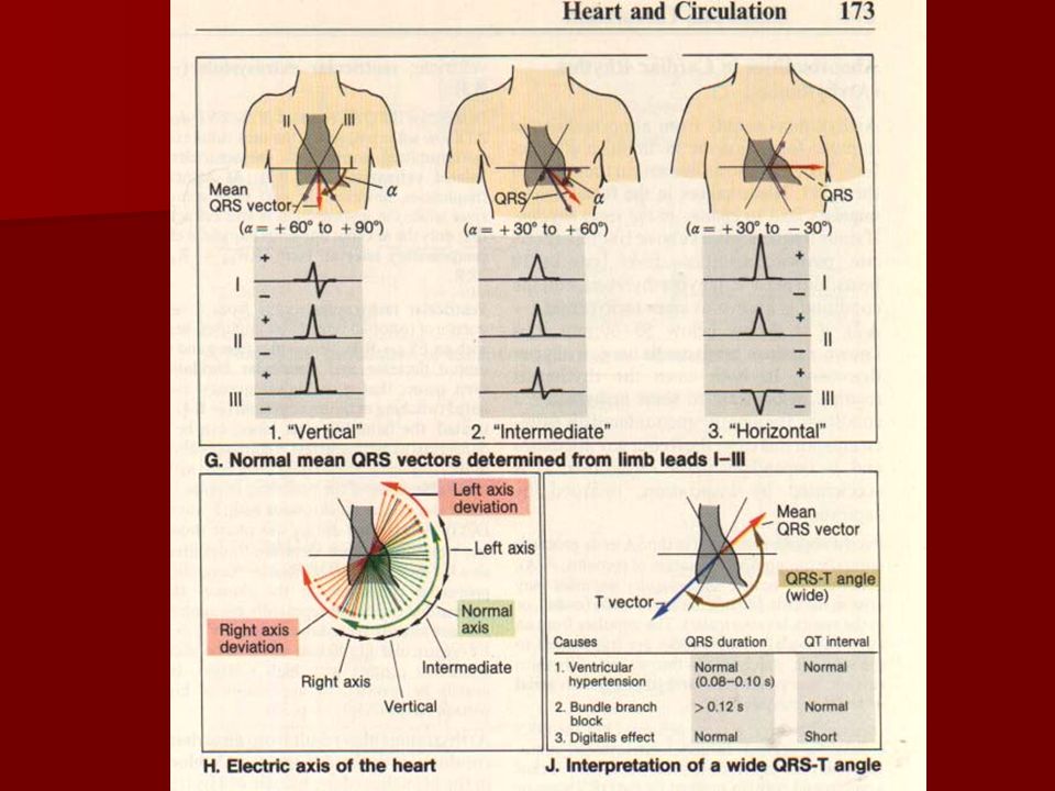
Vector direction

8
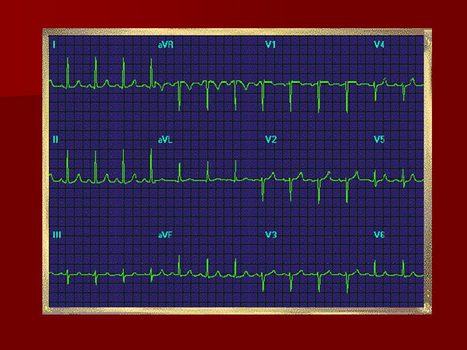
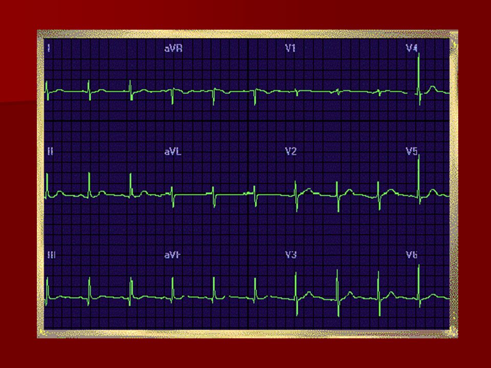
The electrocardiogram

9
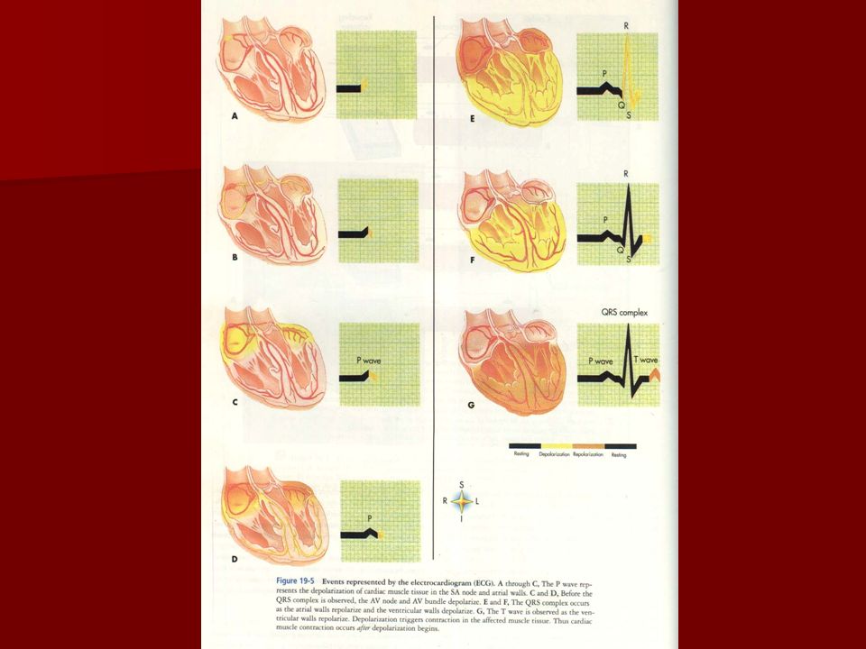
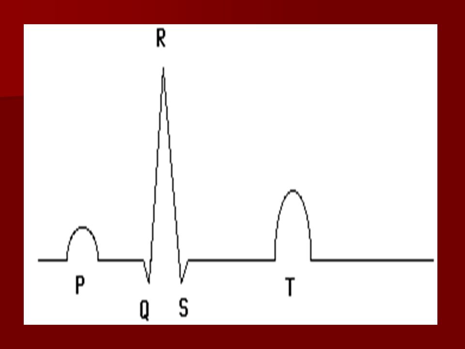
Waves of ECG 1. P wave – depolarization of atria, precedes atria systole 1. P wave – depolarization of atria, precedes atria systole 2. QRS wave is depolarization of ventricles, precedes ventricular systole 2. QRS wave is depolarization of ventricles, precedes ventricular systole 3. atria repolarization also occurs at QRS 3. atria repolarization also occurs at QRS 4. T wave indicates ventricular repolarization 4. T wave indicates ventricular repolarization

11
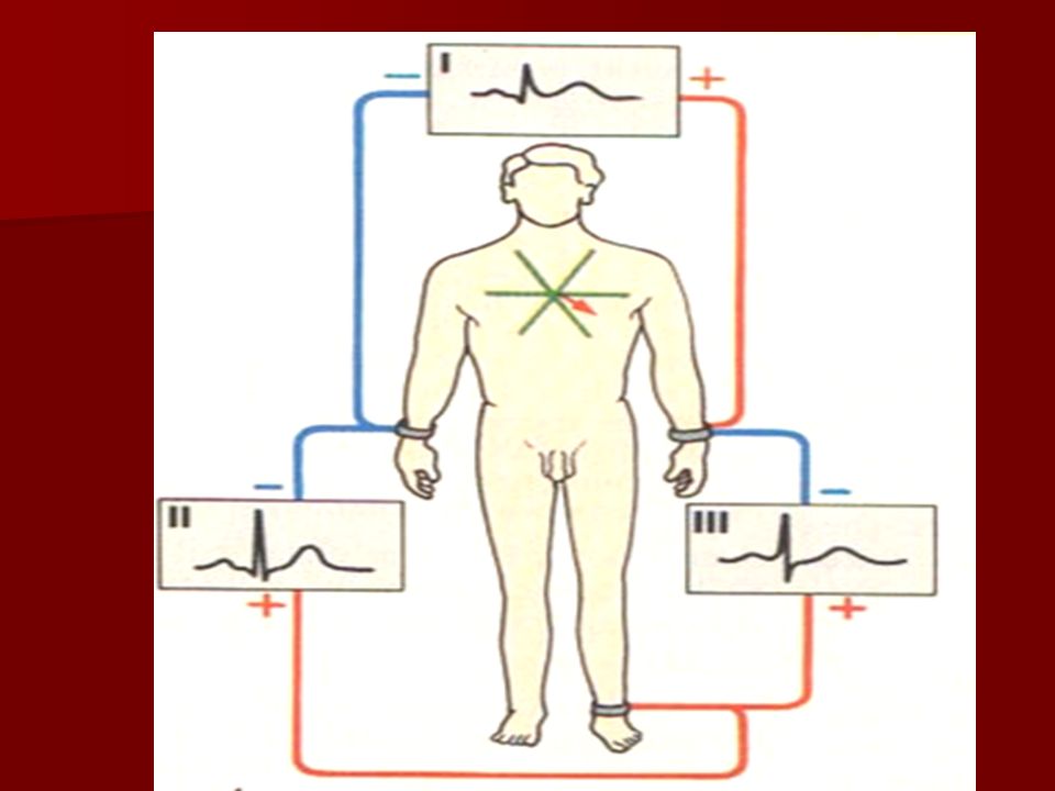
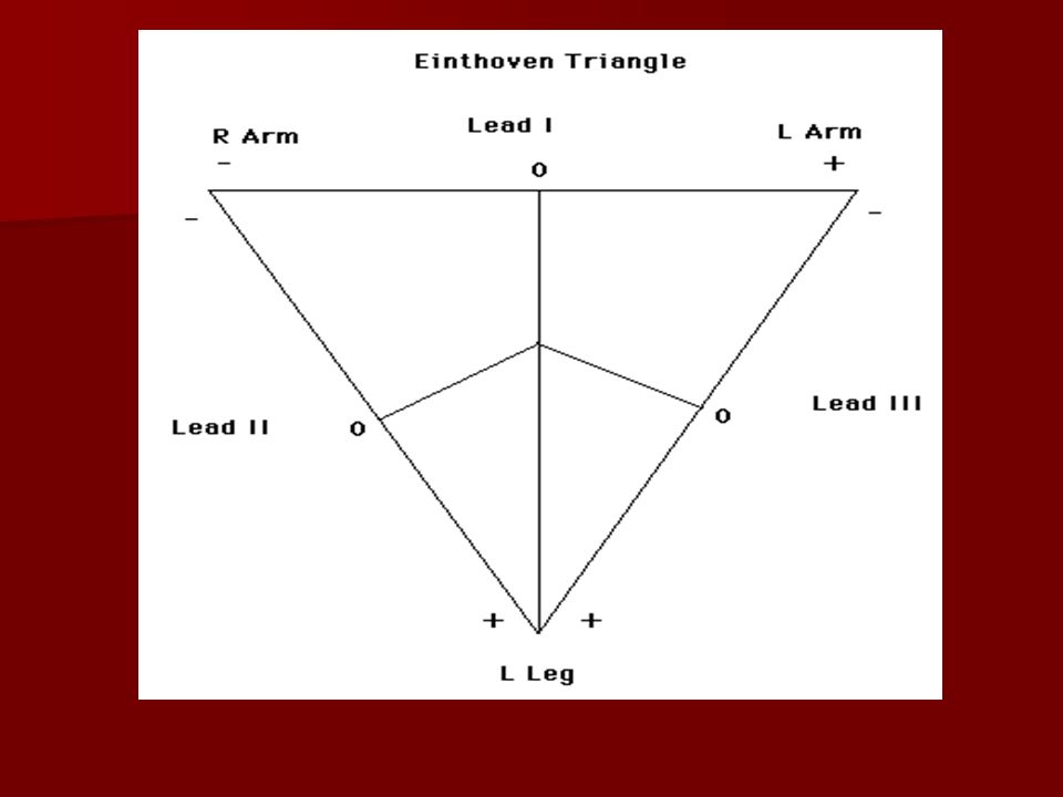
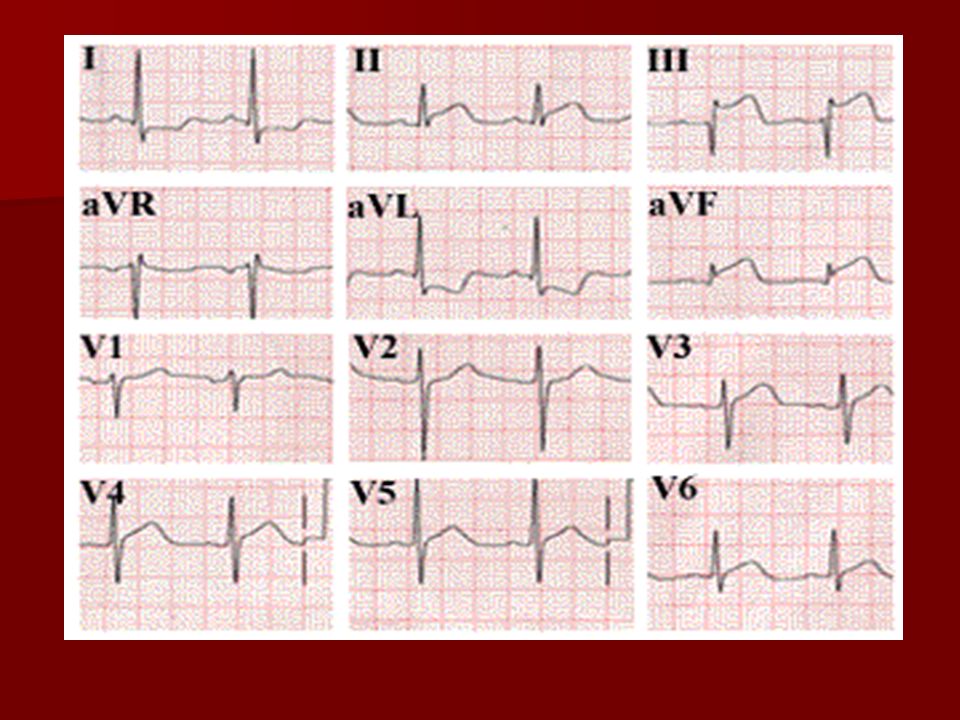
ECG leads a) Bipolar limb leads. The bipolar limb leads record the voltage between electrodes placed on the wrists and legs. These leads were proposed by Einthoven in 1913. a) Bipolar limb leads. The bipolar limb leads record the voltage between electrodes placed on the wrists and legs. These leads were proposed by Einthoven in 1913. I lead: left arm (+) - right arm (-); I lead: left arm (+) - right arm (-); II lead: left leg (+) - right arm (-); II lead: left leg (+) - right arm (-); III lead: left arm (+) - left leg (-). III lead: left arm (+) - left leg (-). For recording limb leads we put red electrode on right arm, yellow - on left arm, green - on left leg and black - on right leg. Black electrode has zero potential (ground). For recording limb leads we put red electrode on right arm, yellow - on left arm, green - on left leg and black - on right leg. Black electrode has zero potential (ground).
 Bipolar limb leads. The bipolar limb leads record the voltage between electrodes placed on the wrists and legs. These leads were proposed by Einthoven in I lead: left arm (+) - right arm (-); I lead: left arm (+) - right arm (-); II lead: left leg (+) - right arm (-); II lead: left leg (+) - right arm (-); III lead: left arm (+) - left leg (-). III lead: left arm (+) - left leg (-). For recording limb leads we put red electrode on right arm, yellow - on left arm, green - on left leg and black - on right leg. Black electrode has zero potential (ground). For recording limb leads we put red electrode on right arm, yellow - on left arm, green - on left leg and black - on right leg. Black electrode has zero potential (ground)..")
12
Bipolar limb leads

13
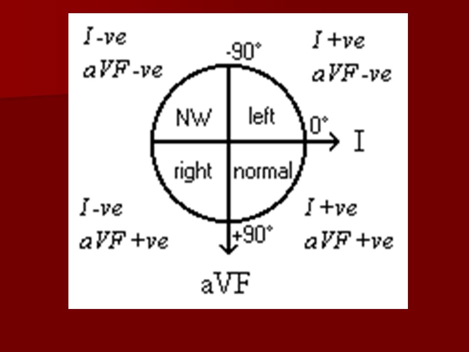
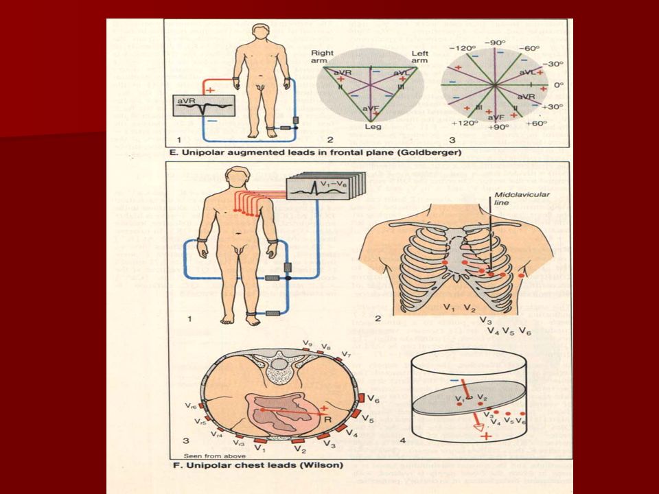
ECG leads The unipolar limb leads were proposed by Goldberger in 1942. They record voltage between single “exploratory electrode” fro one limb and zero joined electrode from two other limbs. So there are three leads AVR, AVL, AVF. In fact zero electrodes records middle voltage of two limbs. Bipolar limb leads and unipolar limb leads record electrical power in frontal projection. The unipolar limb leads were proposed by Goldberger in 1942. They record voltage between single “exploratory electrode” fro one limb and zero joined electrode from two other limbs. So there are three leads AVR, AVL, AVF. In fact zero electrodes records middle voltage of two limbs. Bipolar limb leads and unipolar limb leads record electrical power in frontal projection.

14
The unipolar limb leads

15
The unipolar chest leads The unipolar chest leads were suggested in 1934 by Wilson. One electrode, which is active, situated on the chest in six standard positions. They labeled V1 - V6. Joined zero electrode records middle potential of right arm, left arm and left leg. That is why every chest lead records voltage between active chest electrode and Vilson’s joined zero electrode. The unipolar chest leads were suggested in 1934 by Wilson. One electrode, which is active, situated on the chest in six standard positions. They labeled V1 - V6. Joined zero electrode records middle potential of right arm, left arm and left leg. That is why every chest lead records voltage between active chest electrode and Vilson’s joined zero electrode. Unipolar chest leads records changes of heart polarity in horizontal projection. Unipolar chest leads records changes of heart polarity in horizontal projection.

16
ECG leads V1 - in crossing right IV right intercostal space and parasternal line; V1 - in crossing right IV right intercostal space and parasternal line; V2 - in crossing left IV intercostal space and parasternal line; V2 - in crossing left IV intercostal space and parasternal line; V3 - between V2 and V4; V3 - between V2 and V4; V4 - in crossing V left intercostal space and medioclavicular line; V4 - in crossing V left intercostal space and medioclavicular line; V5 - in crossing V left intercostal space and anterior axilar line; V5 - in crossing V left intercostal space and anterior axilar line; V6 - in crossing V left intercostal space and middle axilar line. V6 - in crossing V left intercostal space and middle axilar line.

17
The unipolar chest leads

18
Algorithm of ECG registration Registration performs fare from electric motors and other electrical devices. Registration performs fare from electric motors and other electrical devices. Tested person may have rest before registration in 10-15 minutes. This procedure needs 2-hour interval after eating or worm procedures. Tested person may have rest before registration in 10-15 minutes. This procedure needs 2-hour interval after eating or worm procedures. For better contact between electrodes and skin use solution NaCl 5-10 % or special electrode past or electrode gel. Otherwise hindrances in ECG curve may occur. They will stand in the way of ECG analysis. For better contact between electrodes and skin use solution NaCl 5-10 % or special electrode past or electrode gel. Otherwise hindrances in ECG curve may occur. They will stand in the way of ECG analysis.

19
Algorithm of ECG registration ECG registration performs in quiet breathing in patients. ECG registration performs in quiet breathing in patients. Registration begins from standard voltage 1 mV from the electrocardiograph for regulation of amplitude in ECG. Usually standard voltage amplitude is 10 mm. Then continue registration of bipolar limb leads, the next - unipolar limb leads and afterwards - unipolar chest leads. Registration begins from standard voltage 1 mV from the electrocardiograph for regulation of amplitude in ECG. Usually standard voltage amplitude is 10 mm. Then continue registration of bipolar limb leads, the next - unipolar limb leads and afterwards - unipolar chest leads.

29
Holter Monitor

30
Thank you! Thank you!

Similar presentations


>")
>")


>")


 3 rd year UG, Department of Biological Sciences Electro Cardio Graphy Sandipan Dasgupta, Indian Institute of Science Education.>")
>")


