Download presentation
Presentation is loading. Please wait.

1
Thorax Intercostal Spaces

2
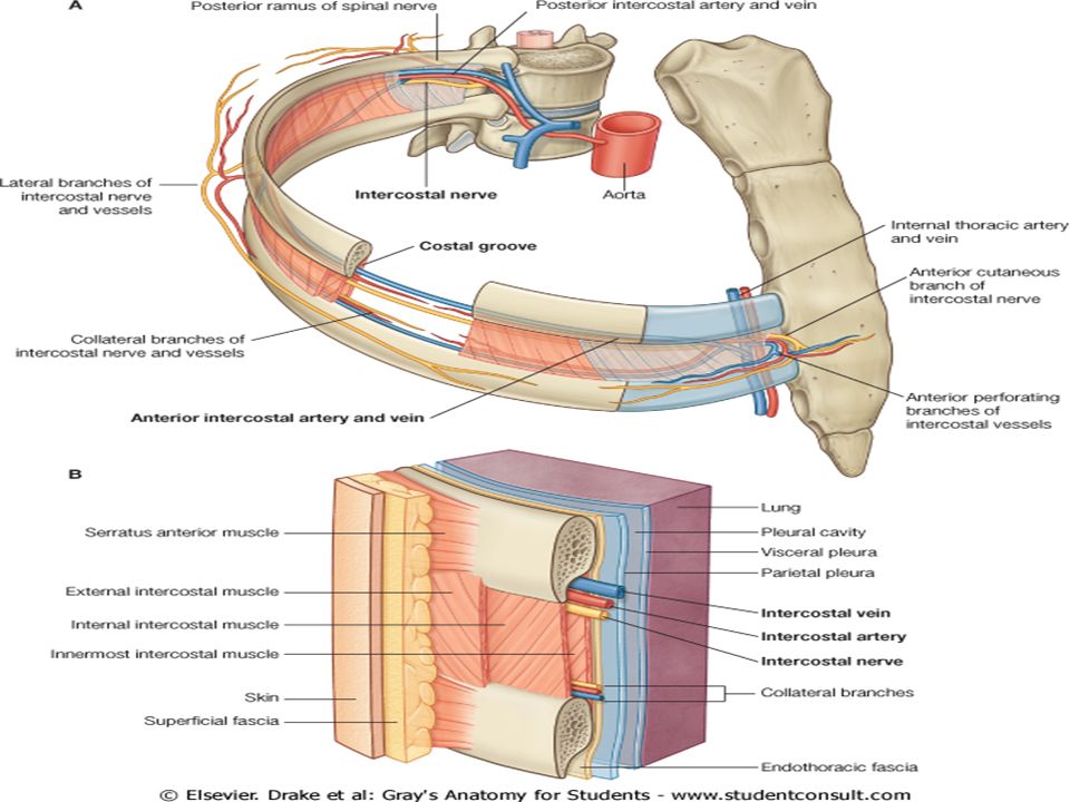
Intercostal spaces Separate the ribs and their costal cartilages from one another. The spaces are named according to the rib forming the superior border of the space(4th intercostal space lies between rib 4 and rib 5). Space below the 12th rib does not lie between ribs and thus is referred to as the subcostal space.
. Space below the 12th rib does not lie between ribs and thus is referred to as the subcostal space.")
3
Contents of Intercostal Spaces
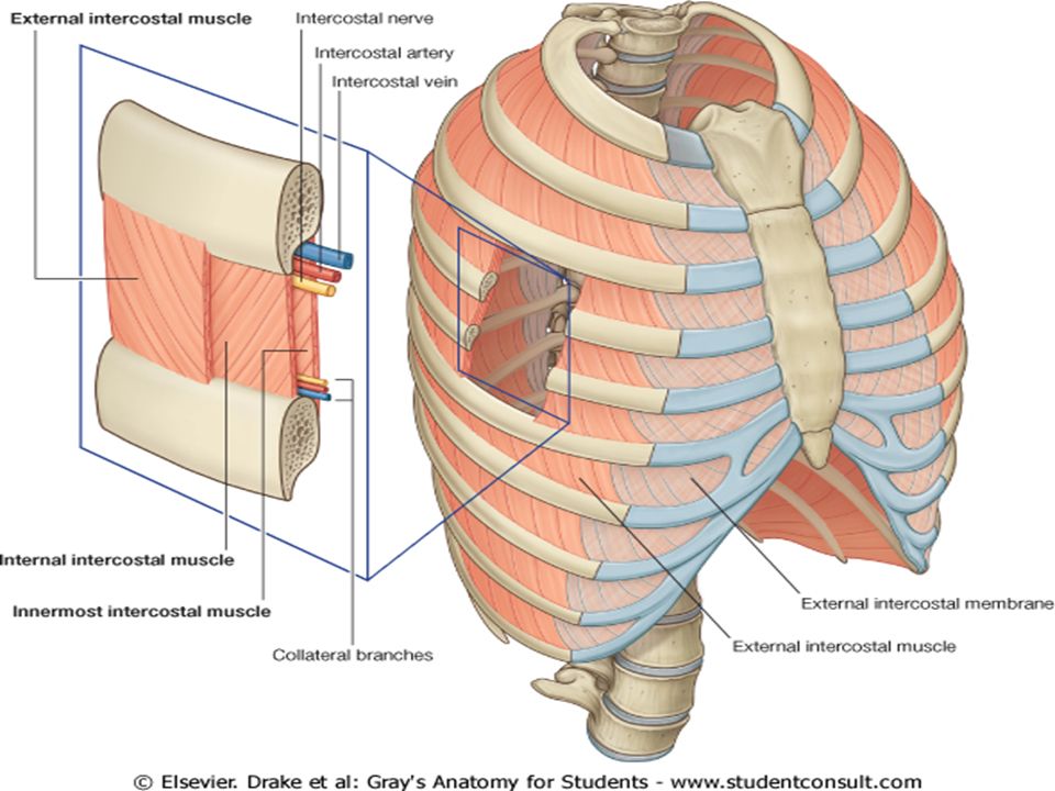
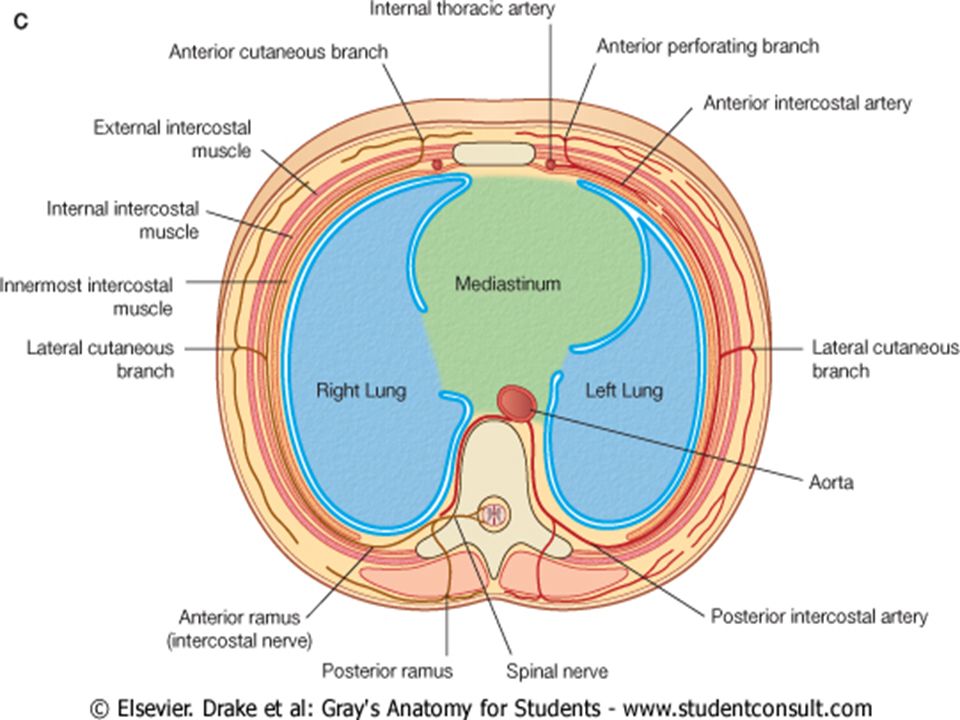
1. Intercostal Muscles: External intercostal, the internal intercostal, and the innermost intercostal muscle 2. The intercostal nerves and blood vessels run between the intermediate and deepest layers of muscles. They are arranged in the following order from above downward: intercostal vein, intercostal artery, and intercostal nerve (i.e., VAN).
.")
5
Muscles of the Thoracic Wall
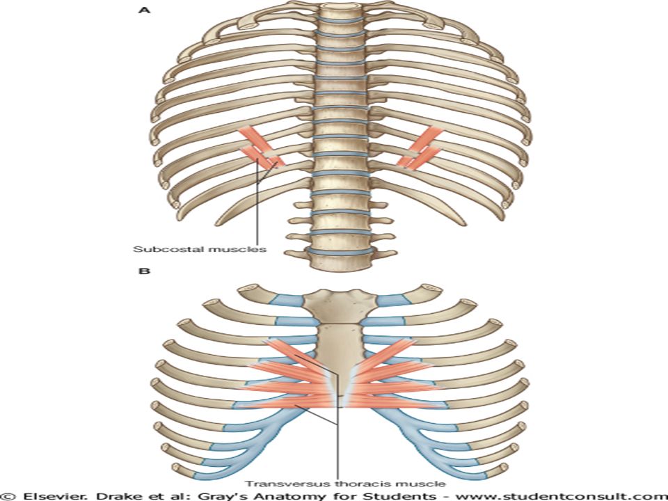
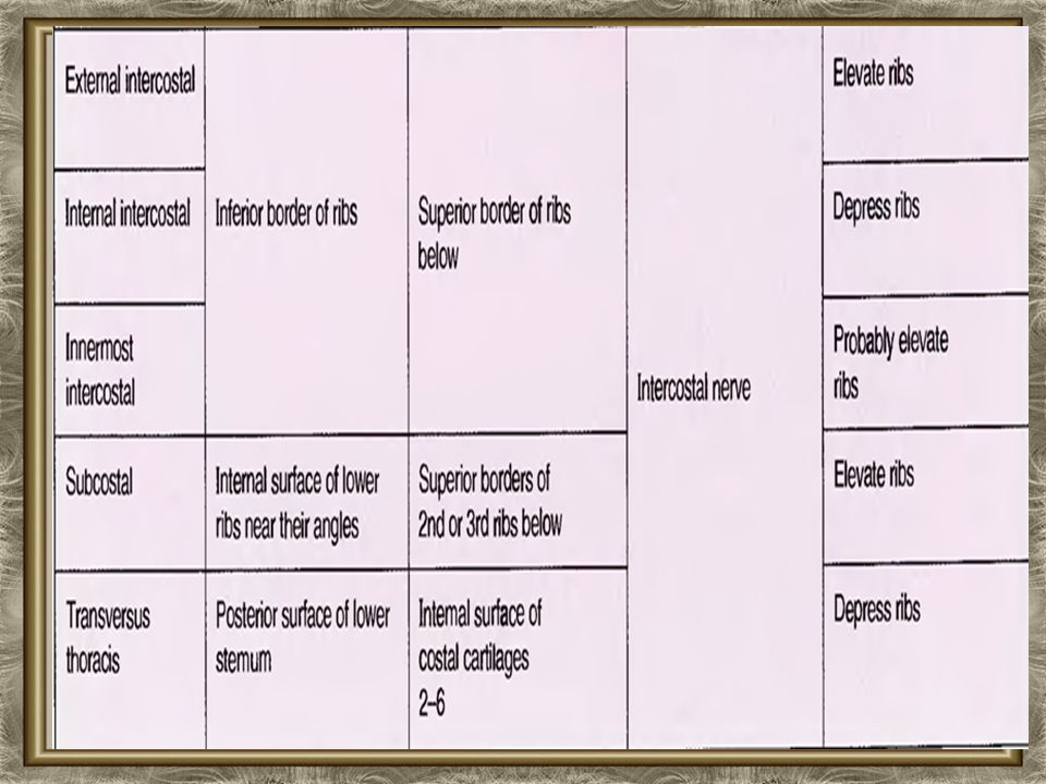
The intercostal muscles are arranged as three layers (external layer, internal layer and an incomplete innermost layer) between the ribs. The three layers of the intercostal muscles are: External layer -- External intercostal Internal layer -- Internal intercostal Innermost layer -- Transversus thoracic (anterior), Innermost (lateral) and subcostal (posterior)
 between the ribs. The three layers of the intercostal muscles are: External layer -- External intercostal. Internal layer -- Internal intercostal. Innermost layer -- Transversus thoracic (anterior), Innermost (lateral) and subcostal (posterior)")
9
A simple model of the action of the intercostal muscles

10
Vasculature of the Thoracic Wall

12
Arteries of the Thoracic Wall
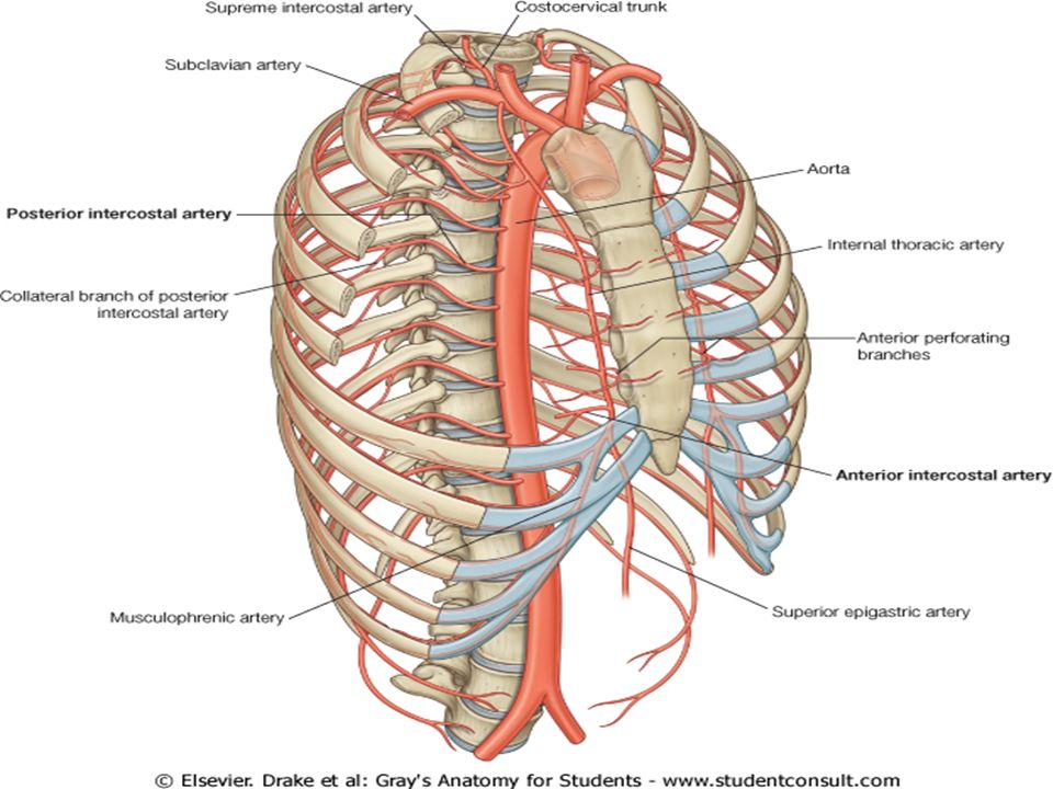
The arterial supply to the thoracic wall derives from the: 1. Thoracic aorta, through the posterior intercostal and subcostal arteries. 2. Subclavian artery, through the internal thoracic and supreme intercostal arteries.

13
The posterior intercostal arteries:
Of the 1st and 2nd intercostal spaces arise from the supreme (superior) intercostal artery, a branch of the costocervical trunk of the subclavian artery. Of the 3rd to 11th intercostal spaces (and the subcostal arteries of the subcostal space) arise posteriorly from the thoracic aorta.
 intercostal artery, a branch of the costocervical trunk of the subclavian artery. Of the 3rd to 11th intercostal spaces (and the subcostal arteries of the subcostal space) arise posteriorly from the thoracic aorta.")
14
Anterior intercostal arteries:
Supply the anterior parts of the upper 9 intercostal spaces. Of the 7 to 9th intercostal spaces derive from the musculophrenic arteries, also branches of the internal thoracic arteries. Are absent from the inferior two intercostal spaces; these spaces are supplied only by the posterior intercostal arteries and their collateral branches.

16
Veins of the Thoracic Wall
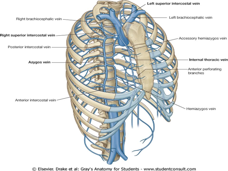
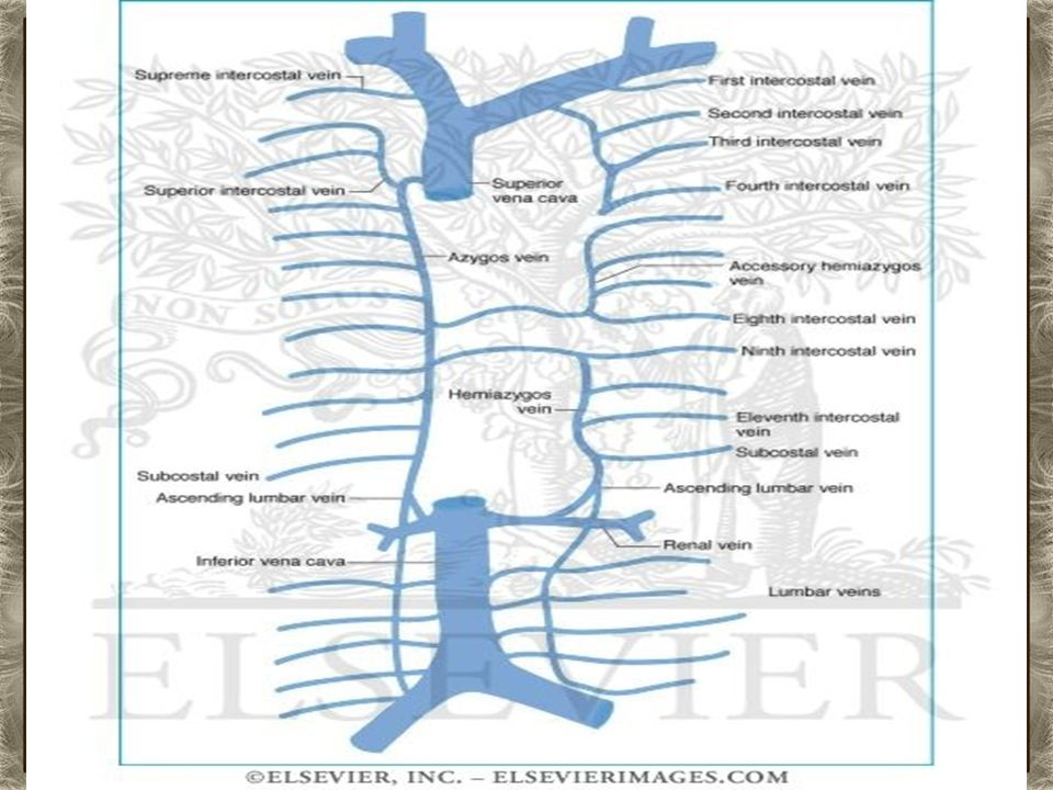
There are 11 posterior intercostal veins and one subcostal vein on each side. Most posterior intercostal veins (4 to11) end in the azygos/hemiazygos venous system, which conveys venous blood to the SVC.
 end in the azygos/hemiazygos venous system, which conveys venous blood to the SVC.")
19
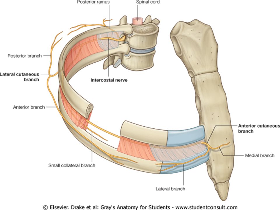
Nerve Supply The intercostal muscles are supplied by the corresponding intercostal nerves. The intercostal nerves are the anterior rami of the first 11 thoracic spinal nerves. The anterior ramus of the 12th thoracic nerve lies in the abdomen and runs forward in the abdominal wall as the subcostal nerve.

20
Branches 1. The lateral cutaneous branch reaches the skin on the side of the chest. It divides into an anterior and a posterior branch. 2. The anterior cutaneous branch, which is the terminal portion of the main trunk, reaches the skin near the midline. It divides into a medial and a lateral branch. 3. The collateral branch runs forward inferiorly to the main nerve on the upper border of the rib below.

22
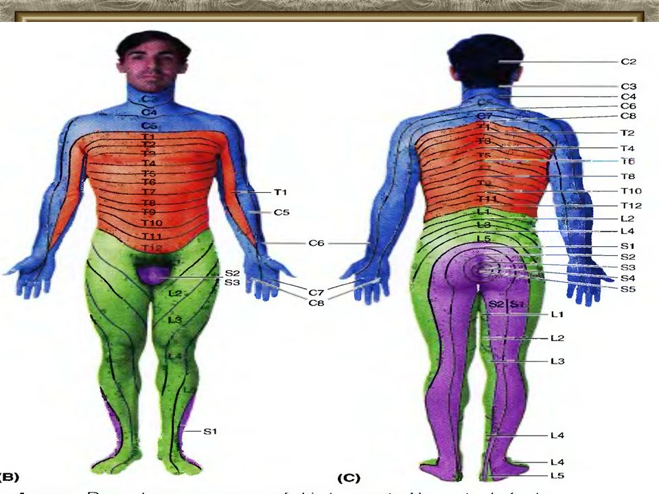
Segmental innervation (dermatomes) of thoracic wall.
Spinal nerve C5 supplies skin at the level of the clavicles . Anteriorly, the dermatome immediately inferior to the C5 dermatome is that of spinal nerve T1. Dermatomes C6 to C7 are located mostly in the upper limbs Dermatome T4 includes the nipple. Dermatome T10 includes the umbilicus.

24
Herpes Zoster Infection of the Spinal Ganglia
A herpes zoster infection causes a classic, dermatomally distributed skin lesion shingles. Herpes zoster is primarily a viral disease of spinal ganglia(varicella-zoster virus (VZV), or chickenpox virus). After invading a ganglion, the virus produces a sharp burning pain in the dermatome supplied by the involved nerve.
, or chickenpox virus). After invading a ganglion, the virus produces a sharp burning pain in the dermatome supplied by the involved nerve.")
25
Herpes Zoster Infection of the Spinal Ganglia

26
Intercostal Nerve Block
This procedure, an intercostal nerve block, involves infiltration of the anesthetic around the intercostal nerve trunk and its collateral branches. Indications Intercostal nerve block is indicated for repair of lacerations of the thoracic and abdominal walls, for relief of pain in rib fractures, and to allow pain-free respiratory movements.

Similar presentations