Download presentation
Presentation is loading. Please wait.

1
2. Basic Immunologic Procedures Terry Kotrla, MS, MT(ASCP)BB
BB")
2
This section of Unit 2 Contains Part 2 Measurement of Light Scattering Part 3 Passive Immunodiffusion Techniques Part 4 Electrophoretic Techniques

3
Introduction How are antigen antibody reactions detected? Multitude of test methodologies have been developed. Go from simple to complex. Can be used either as a screening test or a confirmatory test.

4
Measurement of Precipitation by Light Scattering Antigen-antibody complexes, when formed at a high rate, will precipitate out of a solution resulting in a turbid or cloudy appearance. Turbidimetry measures the turbidity or cloudiness of a solution by measuring amount of light directly passing through a solution. Nephelometry indirect measurement, measures amount of light scattered by the antigen- antibody complexes.

5
Precipitation/Flocculation When soluble antibody binds to soluble antigen (sensitization) there will come a point where lattice formation will occur resulting in precipitation occurring resulting in a visible reaction These immune complexes have fallen out of solution. The Ab at the bottom in the illustration at right is still in the soluble phase.

6
Turbidimetry Measures turbidity or cloudiness of a solution by measuring the amount of light PASSING THROUGH the solution. Soluble antigen and antibody join and once they join in sufficient amounts precipitate, results in cloudiness. The more cloudy the solution, the less light can pass through.

7
Turbidimetry The amount of substance being quantitated is calculated based on the results obtained on standards (aka calibrators) and controls. Calibrator is a substance of an EXACT amount, i.e. 50 mg/dL, used to create standard curve. Controls are substances similar to patient samples and have a range, i.e., 43-56 mg/dL Turbidimetry is very simple but not very sensitive.

8
Standard Curve

9
Nephelometry Widely used in clinical laboratories because it is relatively easily automated. Based on the principle that a dilute suspension of small particles will scatter light passed through it rather than simply absorbing it. The amount of scatter is determined by collecting the light at an angle (usually about 70 or 75 degrees).
..")
10
Nephelometry Can detect either antigen or antibody. Endpoint tests all Ag/Ab reaction to go to completion. If complexes get to large will fall out of solution. Causes falsely decreased results Kinetic tests add antigen and antibody then measure at a specific time. Rate of formation must be known. Calculate concentration based on standards.

11
Nephelometry Measures SCATTERED light bouncing off antigen- antibody complexes. The more light that is scattered the higher the concentration.

12
Turbidimetry versus Nephelometry Turbidimetry – measures light which PASSES through. Nephelometry – measures light which is SCATTERED.

13
Passive Immunodiffusion Reactions of antigens and antibodies in agar gel. Migrate towards each other and where they meet in optimal proportions form a precipitate. “Passive” because they are allowed to react to completion with no enhancements such as an electrical charge applied.

14
Factors Affecting Rate of Diffusion Size of the particles. Temperature Gel viscosity and hydration Interaction of reactants with gel

15
Four Methodologies Single diffusion, single dimension Single diffusion, double dimension Double diffusion, single dimension Double diffusion, double dimension

16
Ouidin Double Diffusion, Single Dimension

17
Oudin Precipitation Solution of antibody is carefully layered on top of a solution of antigen, such that there is no mixing between the two. With time at the interface where the two layers meet, antigen- antibody complexes form a visible precipitate. The other two tubes are negative controls, containing only antibody or only antigen plus an irrelevant protein in the second layer.

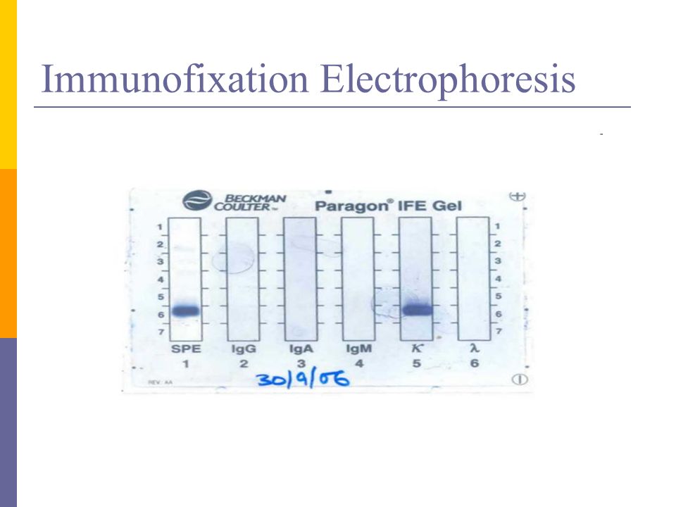
18
Radial Immunodiffusion Antibody mixed with agar poured into plate. Holes punched. Add standards, controls and patients to wells. Antigen will diffuse out and form precipitin ring. The diameter of the ring directly proportional to concentration. Create standard curve and read results.

19
Radial Immunodiffusion

20
Two methods Endpoint – allows reaction to go to completion. Kinetic – measurements taken at a specific time before zone of equivalence is reached. This is a QUANTITATIVE technique which will give the actual concentration.

21
Standard Curve Precipitin Rings A B C a b c Standards Samples Radial Immunodiffusion

22
Standard Curve

23
RID Sources of Error Over or under filling the well. Spilling sample onto the outside of the well. Nicking the well with the pipette tip. Improper incubation time or temperature. Incorrect measuring of the wells.

24
Ouchterlony Gel Diffusion Holes punched in agar. Known antibody or antigen added to center well. Known sample added to outer well. Unknown sample added to outer well next to unknown sample. Wait for bands to form. This is a QUALITATIVE technique, simply determines the presence NOT the concentration.

25
Ouchterlony Immunodiffusion

26
Ouchterlony - Identity Precipitation appears as a continuous line in the form of an arc between the two outer wells and the center well. There are no spurs at the angle and this type of reaction is termed a band of identity.

27
Ouchterlony – Partial Identity FIGURE 2: If a solution with antigens X and Y is placed in well 1, a solution with antigen X only is placed in well 2, and antiserum containing antibodies specific for both X and Y is placed in well 3, a reaction similar to that appearing in Fig. 2 will occur. Notice that there is a spur reaction towards the XY well. This indicates that the two antigenic materials in wells 1 and 2 are related, but that the material in well 1 possesses an antigenic specificity not possessed by the material in well 2. Such a reaction with spur formation indicates partial identity

28
Ouchterlony – Non-Identity If the material in wells 1 and 2 do not possess common antigens and the antiserum in well 3 possesses specificities for both materials, the reaction will appear as two crossed lines as in Fig. 3

29
Ouchterlony-Interpret Determine which interpretation fits for samples 1, 2 and 3.

30
Electrophoretic Techniques Immunodiffusion can be combined with electrical current to speed things up. Electrophoresis is a technique which separates molecules using electrical current. Small molecules move faster than large. For immunolectrophoresis antigen and antibody migrate through gel faster. Can be single or double diffusion.

31
Rocket Immunoelectrophoresis Adaptation of radial immunodiffusion (RID). Antibody incorporated (mixed) into the gel. Antigen added to wells. Apply electrical current and antigen will move forward and will bind to antigen. Dissolution and reformation occurs. Height of precipitin band related to concentration of antigen. Much faster than RID.
 into the gel. Antigen added to wells. Apply electrical current and antigen will move forward and will bind to antigen. Dissolution and reformation occurs. Height of precipitin band related to concentration of antigen. Much faster than RID..")
32
Rocket Immunoelectrophoresis Antigen is electrophoresed into gel containing antibody. The distance from the starting well to the front of the rocket shaped arc is related to antigen concentration.

33
Rocket Electrophoresis

34
Immunoelectrophoresis

35
Two step double diffusion technique. Electrophorese antigen. Antiserum added to trough parallel to line of separation. Incubate overnight. Diffusion occurs and bands of precipitate form. Most often used as a screening test.

36
Immunoelectrophoresis Two-dimensional immunoelectrophoresis. Antigens are separated on the basis of electrophoretic mobility. The second separation is run at right angles to the first which drives the antigens into the antiserum-containing gel to form precipitin peaks; the area under the peak is related to the concentration of antigen.

37
Immunoelectrophoresis -Antivenom Each antibody molecule can bind two separate sites on an antigen molecule (venom toxin), consequently antibodies have the ability to cross link many antigen molecules simultaneously. This cross-linking causes the antibody antigen-complex to become insoluble and precipitate out from the solution. The immunoelectrophoresis technique makes use of this capability of the antibodies to form giant insoluble complexes with their respective antigens. The antigen-antibody precipitate which forms can be visualized by specific staining techniques, or quantified by various means.

38
Immunofixation Electrophoresis Immunofixation Electrophoresis (IFE) combines electrophoresis with immunoprecipitation. This technique may be used to identify and characterize serum or urine proteins. In IFE, proteins of sample are first separated by electrophoresis on a support (agarose) according to their charge and after that the medium is overlaid with monospecific antisera reactive with specific protein - antigen. If the antigen is present a characteristic immunoprecipitin band will be formed.
 according to their charge and after that the medium is overlaid with monospecific antisera reactive with specific protein - antigen. If the antigen is present a characteristic immunoprecipitin band will be formed..")
39
Immunofixation Electrophoresis

41
Test is frequently ordered to identify moncolonal proteins. May be done on urine or serum depending upon what doctor suspects. Multiple myeloma Production of large amount of specific protein. Will be excreted in urine

42
Electrophoresis Sources of Error Applying current in wrong direction. Incorrect buffer pH Incorrect timing Incorrect current applied. Concentration of reactants must be appropriate.

43
End of this lecture.

Similar presentations